Performance Evolution of Various
Wavelets in Cervical Lesion Detection
P S RAMAPRABA*,
Research scholar,
Department of Electronics and Communication Engineering,, Sathyabama University, Chennai, Tamilnadu, India,
H. RANGANATHAN
Principal,
Rajiv Gandhi College of Engineering, Chennai, Tamil Nadu, India.
Abstract
Cervical cancer is one of most common cancers among women in the world caused by human papilloma virus infection. It develops in the tissue of cervix which connects upper body of the uterus to the vagina. The types of cancer are squamous cell carcinoma, adeno carcinoma and adeno squamous carcinoma based on location of cervix where cancer develops. In this paper, an automatic detection of squamous cell carcinoma in cervical images based on Discrete Wavelet Transform (DWT) and K-Nearest Neighbor (KNN) classifier is described. The energy features are extracted from DWT decomposed image of small area of cervical images. Then the features are fed into KNN classifier to classify whether the given area is normal or cancer affected region. The performance of the proposed system is evaluated by using three wavelets namely bi-orthogonal (bior3.7), Daubechies-8(db8) and Symlet (sym8). Experimental results show the performance of db8 with other wavelets that produces 97.22% average accuracy.
Keywords: DWT, cervical cancer, KNN.
1. INTRODUCTION
A domain-specific diagnostic feature in a probabilistic manner using conditional random fields to detect pre-cancerous and pre-cancerous lesions of the uterine cervix is presented [1]. Image regions corresponding to different tissue types are identified for the extraction of domain-specific anatomical features. The unique optical properties of each tissue type and the diagnostic relationships between neighboring regions are incorporated in the proposed conditional random field model.
An analysis of the Colposcopic images to help the expert to make a more robust diagnosis of precursor lesions of cervical cancer is reviewed [2]. This is a complete methodology to evaluate temporal changes of tissue color. The Colposcopic image segmentation based on integrated color and texture tools is described [3]. Principal component analysis and multidimensional histogram analysis are used for preprocessing.
In Cervigram, the lesions are of varying sizes and complex, non-convex shapes. A new methodology [4] that enables the segmentation of non-convex regions, thus providing a major step forward towards cervigram tissue detection and lesion description is presented. The framework transitions from pixels to a set of small coherent regions, which are grouped bottom-up into larger, non-convex, perceptually similar regions, utilizing a new graph-cut criterion and agglomerative clustering.
A cost-sensitive 2ν-SVM classification scheme to cervical cancer images to separate diseased regions from healthy tissue is proposed [5]. Multiplier classifier scheme is used instead of the traditional single classifier to test the NCI/NLM archive of 60000 images. Automatic lesion detection method in cervical images based on region based active contour segmentation is proposed [6]. The region based active contour segmentation method requires an initial curve or mask to segment the given image. To detect the lesion, the initial mask must be in the acetowhite region.
samples to characterize the attribute of contour. The spatial parameter is the number of the region around the edges and the frequency parameters are amplitude of first peak, frequency of the end of first peak, area under first peak and area under other peaks. Then the Principal Component Analysis is performed to test the parameters. Segmentation and classification of cervix lesions by pattern and texture analysis is presented in [8].
The spatial parameter used in lesion detection is the number of the region around the edges and the frequency parameters are amplitude of first peak, frequency of the end of first peak, area under first peak and area under other peaks. Then the Principal Component Analysis is performed to test the parameters is described [9] World Health Organization study reveals that every year 1, 32,082 women are diagnosed with cervical cancer and 74,118 die from the disease The growing risk of cervical cancer in women in India is 2.4% compared to 1.3% for the world. More than 90% of cervical cancer in India is squamous cell carcinoma type which develops in the ectocervix is described [10].
In this paper, an efficient lesion detection in cervix images based on DWT and KNN classifier is developed. The concept of DWT and KNN classifier are discussed in section 2. In section 3, the proposed method for lesion detection in cervix images is explained. The experimental results are evaluated in section 4 and finally concluded in section 5.
2. METHODOLOGY
The proposed lesion detection in cervical images is built based on Discrete Wavelet Transform and KNN classifier. In this section the theoretical background of Wavelet Transform and KNN classifier are discussed.
2.1 Wavelets
A wavelet is a wave-like oscillation with amplitude that starts out at zero, increases, and then decreases back to zero. It can typically be visualized as a "brief oscillation" like one might see recorded by a seismograph or heart monitor. Generally, wavelets are purposefully crafted to have specific properties that make them useful for signal processing. Wavelets can be combined, using a "shift, multiply and sum" technique called convolution, with portions of an unknown signal to extract information from the unknown signal.
As wavelets are a mathematical tool they can be used to extract information from many different kinds of data, including - but certainly not limited to - audio signals and images. Sets of wavelets are generally needed to analyze data fully. A set of "complementary" wavelets will deconstruct data without gaps or overlap so that the deconstruction process is mathematically reversible. Thus, sets of complementary wavelets are useful in wavelet based compression/decompression algorithms where it is desirable to recover the original information with minimal loss.
Wavelet transforms are broadly divided into three classes: continuous, discrete and multi resolutional based. The Discrete Wavelet Transform (DWT), which is based on sub-band coding, is found to yield a fast computation of Wavelet Transform. It is easy to implement and reduces the computation time and resources required. Sampled input image is decomposed into various frequency sub-bands or sub-band signals. A two dimensional decomposition can be applied over the image. A simple example of level 1 decomposing is shown in fig 1. The original image is subdivided into four parts. The LL band contains low frequency contents of the signal, where as HH band contains high frequency contents of the signal, which is having less importance than LL band.
LL HL
LH HH
Fig. 1. The result of 2D-DWT decomposition
2.2 KNN Classifier
3. PROPOSED SYSTEM
Feature extraction involves simplifying the amount of resources required to describe a large set of data accurately. In the proposed system, energies are used as features for lesion detection that are extracted from the wavelet decomposed cervical images. The well known lesion area in the cervical images is given to the feature extraction stage. At first, the known lesions area which was already given by Colposcopist is separated from the whole image. Then, the separated lesion is sub divided into small windowed region for feature calculation. The window size used for the proposed approach is 32x32. Each and every small region is decomposed by using 2-level DWT. From the decomposed image, the proposed energy feature is calculated by using eqn. (1)
=
= =
X m
Y
n e
e I mn
XY E
1 1
) , ( 1
(1)
where
I
e(
i
,
j
)
is the pixel value of the eth small region and X, Y is width and height of the sub-band respectively. Fig 2 shows the overall automated system for cervical cancer detection system. The energy features are extracted from all training abnormal images and stored in the database for further classification.Fig. 2. Overall automated system for lesion detection in cervical images
For lesion detection of an unknown cervical image, the features of the image are extracted for 32x32 non-overlapping regions using the method described above. Then the extracted feature is compared with the features in the generated database to classify the region into normal or cancer affected region by using KNN classifier. The distance measure used in the classifier is Euclidean distance (ED) given by eqn. 2 where
u
=
(
x
1,
y
1)
and(
x
2,
y
2)
v
=
are two points.(
,)
(
)
(
1 2)
2 (2) 22
1 x y y
x v u
ED = − + −
4. EXPERIMENTAL RESULTS
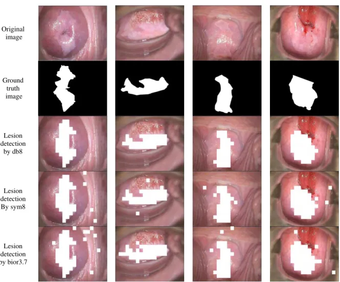
To evaluate the performance of the proposed system, many computer simulations and experiments are made on cervical images of normal and abnormal categories. The performance of the proposed method is tested on 200 normal colposcopy cervix images and 40 abnormal colposcopy cervix image obtained from Kasturibai Gandhi Hospital (KGH), Chennai, India. All the images are considered for the classification test. The wavelet decomposition level used in the proposed approach is 2. Fig 3 shows the detected lesion based on the proposed system for three wavelets.
In order to analyze the efficiency various wavelets, three wavelets bi-orthogonal (bior3.7), Daubechies-8(db8) and Symlet (sym8) are used. These wavelets are chosen, as they are used in many medical image classification tests such as mammogram classification systems. Table 1 shows the classification accuracy of the proposed system using the aforementioned wavelets.
Input image
Wavelet Transform Block Separation
Feature Calculation
Database
Unknown image
Wavelet Transform Block Separation
Feature Calculation
KNN
TABLE I
CLASSIFICATION ACCURACY OF THE PROPOSED SYSTEM USING THREE WAVELETS
Image Accuracy (%) Db7 Sym8 Bior3.7 Image 1 96.87 95.54 93.55 Image 2 97.01 96.36 96.06 Image 3 97.76 96.76 96.76 Image 4 97.25 96.25 95.91 Avg. accuracy (%) 97.22 96.22 95.57
It is observed from the table that the daubechies wavelet produces better performance of over approximately 1% than symlet and bi-orthogonal wavelets. The maximum average classification accuracy obtained by the proposed approach is 97.22% using daubechies family wavelet db8.
Original image
Ground truth image
Lesion detection
by db8
Lesion detection By sym8
Lesion detection by bior3.7
Fig. 3. The detected lesion based on the proposed system for three wavelets
5. CONCLUSION
In this paper, an efficient squamous cell carcinoma in cervical images based on DWT and KNN classifier is presented. The system scans the cancer detected region by a non overlapping region of size 32x32. The energy features are extracted for that region by decomposing the region using 2-level wavelet transform. The KNN classifier is used for lesion region classification into normal or cancer. The performance of the proposed system is evaluated by three wavelets namely bi-orthogonal (bior3.7), Daubechies-8(db8) and Symlet (sym8). Experimental results show the superiority of daubechies family wavelets in terms of classification accuracy.
REFERENCES
[3] Claude, I., P. Pouletaut, S. Huault and J.C. Boulanger (2001), “Integrated color and texture tools for colposcopic image segmentation” Proceedings of the IEEE International Conference on Image processing. pp: 311-314.
[4] Gordon, S. and H. Greenspan, 2007. Segmentation of non-convex regions within uterine cervix images. Proceedings of the IEEE 4th International Symposium on Biomedical Imaging, Apr. 12-15, IEEE Xplore Press, Arlington, VA, pp: 312-315.
[5] Artan, Y. and X. Huang, 2008. Combining multiple 2ν-svm classifiers for tissue segmentation. Proceedings of the 5th IEEE International Symposium on Biomedical Imaging, May 14-17, IEEE Xplore Press, Paris, pp: 488-491.
[6] Meslouhi, O.E., M. Kardouchi, H. Allali and T. Gadi, 2009. Semi-automatic cervical cancer segmentation using active contours without edges. Proceedings of the 5th International Conference on Signal Image Technology and Internet Based Systems, Nov. 29-Dec. 4, IEEE Xplore Press, Marrakesh, pp 54-58.
[7] Claude, I. Winzenrieth, “Contour features for colposcopic image classification by artificial neural networks”, IEEE International Conference on Pattern recognition, 2002, pp 771-774.
[8] Bhakti Tulpule, Shuyu Yang, “Segmentation and Classification of Cervix Lesions by Pattern and Texture Analysis”, IEEE International Conference on Fuzzy System, 2005, pp 173-176.
[9] Tulpule, B., S. Yang, Y. Srinivasan, S. Mitra and B. Nutter, 2005. Segmentation and classification of cervix lesions by pattern and texture analysis. Proceedings of the IEEE 14th International Conference on Fuzzy System, May 25-25, IEEE Xplore Press, Reno, NV, pp: 173-176.