Submitted 25 August 2015 Accepted 12 November 2015 Published1 December 2015 Corresponding author Mats Ellerstr¨om,
Academic editor Jenny Renaut
Additional Information and Declarations can be found on page 17
DOI10.7717/peerj.1469
Copyright 2015 Johansson et al.
Distributed under
Creative Commons CC-BY 4.0 OPEN ACCESS
A quick and robust method for
quantification of the hypersensitive
response in plants
Oskar N. Johansson∗
, Anders K. Nilsson∗
, Mikael B. Gustavsson, Thomas Backhaus, Mats X. Andersson and Mats Ellerstr¨om
Department of Biology and Environmental Sciences, University of Gothenburg, Gothenburg, Sweden
∗
These authors contributed equally to this work.
ABSTRACT
One of the most studied defense reactions of plants against microbial pathogens is the hypersensitive response (HR). The HR is a complex multicellular process that in-volves programmed cell death at the site of infection. A standard method to quantify plant defense and the HR is to measure the release of cellular electrolytes into water after infiltration with pathogenic bacteria. In this type of experiment, the bacteria are typically delivered into the plant tissue through syringe infiltration. Here we report the development of a vacuum infiltration protocol that allows multiple plant lines to be infiltrated simultaneously and assayed for defense responses. Vacuum infiltration did not induce more wounding response in Arabidopsis leaf tissue than syringe inoculation, whereas throughput and reproducibility were improved. The method was used to study HR-induced electrolyte loss after treatment with the bacterium
Pseudomonas syringaepv.tomatoDC3000 harboring the effector AvrRpm1, AvrRpt2 or AvrRps4. Specifically, the influence of bacterial titer on AvrRpm1-induced HR was investigated. Not only the amplitude, but also the timing of the maximum rate of the HR reaction was found to be dose-dependent. Finally, using vacuum infiltration, we were able quantify induction of phospholipase D activity after AvrRpm1 recognition in leaves labeled with33PO4.
Subjects Molecular Biology, Plant Science
Keywords Hypersensitive response (HR), Programmed cell death (PCD), Electrolyte leakage, Effector-triggered-immunity (ETI),Pseudomonas syringae, AvrRpm1
INTRODUCTION
in turn acquired immune receptors called resistance (R) proteins that recognize pathogenic effectors directly or via proteome perturbations and activate defense pathways (Spoel & Dong, 2012). The outcomes of effector-triggered-immunity (ETI) include an oxidative burst, production of anti-microbial compounds, expression of pathogenesis-related (PR) genes, and often a rapid programmed cell death (PCD) at the site of infection. The reaction as a whole is denoted hypersensitive response (HR) (Coll, Epple & Dangl, 2011;Hofius et al., 2011;Mur et al., 2008). All aspects of the HR are not prerequisites for resistance, as exemplified by severalArabidopsis thaliana(hereafter Arabidopsis) mutants showing re-duced pathogen triggered PCD but unaffected in their ability to restrict growth of avirulent bacteria (Johansson et al., 2014;Jurkowski et al., 2004;Yu, Parker & Bent, 1998). HR induced by avirulent bacteria depends on living cells of the pathogen and can be activated in the plant within 15 min after recognition of the intruder (Klement & Goodman, 1967).
In the post-genomic era, enormous progress has been made in the understanding of the underlying genetics of plant immunity. Large scale transcriptomic and proteomic studies have identified thousands of genes and gene products as directly or indirectly responding to effector elicitation (Katagiri, 2004;Zimaro et al., 2011). Candidate genes from such studies may later be tested for their role in plant immunity by the use reverse genetics. This has evoked the need for quick and effective screening methods for the identification of immune-depressed or hyperactive immune mutants.
Various read-outs and methods have been employed to quantify plant defense responses after pathogen elicitation. For example, the area of cell death (Alamillo & Garcia-Olmedo, 2001), K+
/H+
the required time period. For a normal effector response this is about four to eight hours of constant measuring. In this context, we felt that there was room for improvement of the quantification method of HR-PCD.
In here, we present a quick and simple vacuum-based infiltration method of leaf material suitable for quantification of HR-PCD and other defense associated responses in plants. The method was used to test how cultivation conditions of the bacterial pathogenPseudomonas syringaeinfluence its ability to elicit HR-PCD in Arabidopsis. To further evaluate the applicability of this method, we investigated the role of bacterial titer on the outcome of HR-PCD. We found that lower bacterial inocula did not only result in decreased cell death and electrolyte leakage in plants as previously reported, but also resulted in a delay of the full defense reaction. The presented vacuum infiltration method is time-saving over syringe inoculation, shows higher reproducibility, and allows instantaneous infiltration of multiple plant lines. The method may also be used on material which has been pretreated with toxic or harmful reagents, abolishing the need for manual handling of and minimizing the exposure to the experimenter.
MATERIALS AND METHODS
Plant material and growth conditionArabidopsis thalianawild type Columbia-0 (Col-0) andrpm1.3(Grant et al., 1995) were sown in soil, stratified at 4◦
C for 72 h and cultivated in growth chamber under short day conditions (8 h day/16 h night, 22◦
C/18◦
C) at 60% relative humidity. Seedlings were transplanted into individual pots two weeks after germination. Plants used in experiments were 5–6 weeks old.
Vacuum infiltration
Finally, fully infiltrated discs (sunken discs) without any visible damage from the coring were transferred using plastic bacterial inoculation loops to 6 well cell cultivation plates filled with 10 ml deionized water (four discs in each well in 6 replicates per treatment). Conductivity was measured by moving 5 ml of the bathing solution to a 15 ml centrifuge tube in which a conductivity meter (Orion, Thermo scientific, Waltham, MA, USA) was placed. The solution was thereafter returned to the plate well.
Pseudomonasculturing, cell staining and image acquisition
Pseudomonas syringaepv.tomatoDC3000 expressing the effector protein AvrRpm1, AvrRpt2 or AvrRps4, or a strain carrying an empty vector (DC3000) (Bisgrove et al., 1994;Grant et al., 1995) were propagated on solidPseudomonasagar F (King’s B medium, Biolife, Milano, Italy) containing kanamycin (50 mg l−1) and rifampicin (50 mg l−1)
in a drawer in darkness at room temperature unless stated otherwise.P. syringae hrcC
mutant (Rahme, Mindrinos & Panopoulos, 1991) were grown without kanamycin. Bacteria were re-plated 24 h prior to infiltration into plants. A loopfull of the overnight culture of bacteria was suspended in 30 mL of 10 mM MgCl2and optical density at 600 nm was
measured using a NanoDrop 2000c (Thermo Scientific, Waltham, MA, USA) in the cuvette mode. OD600=1.0 correspond to 3.55∗108CFU ml−1as determined by serial dilution
and plating. Trypan blue staining of discs was adapted from (Koch & Slusarenko, 1990). Four discs were placed in a micro centrifuge tube, covered with staining solution (0.025% trypan blue in equal volumes phenol, glycerol, lactic acid and water) and kept at 95◦
C in a heating block for 2.5 min. Samples were left in the staining solution for 30 min, then rinsed once in water and thereafter de-stained for two days in chloral hydrate:water (5:2 (w/v)) on an orbital shaker at 50 rpm. Stained discs were mounted in 60% (v/v) glycerol and images were acquired using a Zeiss Axioplan 2 Imaging microscope with a Plan-Neofluar 10x/0.30 objective connected to a Canon Powershot G6 camera. Images were subjected to Auto color filtering in Adobe Photoshop CS5 (Adobe Systems Incorporated, San Jose, CA, US).
Quantification of jasmonic acid and free and esterified OPDA In tissue quantification of glycerolipid-bound 12-Oxo-phytodienoic acid (OPDA) was performed as previously described (Nilsson et al., 2012). For the quantification of free OPDA and JA released by tissue, 6 leaf discs were placed in a glass tube with 2 ml dH2O
Quantification of phosphatidic acid
For labeling experiment, leaf discs were incubated with33PO4(New England Nuclear,
Perkin Elmer) over night as described (Andersson et al., 2006). Discs were then transferred to 50 ml centrifuge tubes and vacuum infiltrated withP. syringaeexpressing AvrRpm1 at OD600=0.1 as described above. Mock treatment was performed by inoculating plants
with 10 mM MgCl2. Lipids were extracted as previously described (Andersson et al., 2006).
Samples were loaded on activated thin layer chromatography plates and developed using the solvent system chloroform:methanol:acetic acid:water (80:20:10:3.5), and subjected to autoradiography. Developed films were scanned and the intensity of the phosphatidic acid bands was determined using the western blot tool in ImageJ (http://rsb.info.nih.gov/ ij/). Unlabeled PA was measured in a total lipid extract obtained from two leaf discs by LC-MS/MS. A total lipid extract was obtained as described (Andersson et al., 2006) and the lipids separated by reverse phase chromatography and detected by MS/MS on a triple quadrupole detector as described (Nilsson et al., 2014) using the neutral loss ofm/z115 (Li-Beisson et al., 2010;Schwudke et al., 2006) to detect PA species.
Statistical- and regression analysis
Data were handled in Excel (Microsoft, Redmond, WA, USA). Variances are expressed as standard deviation and significance was determined by Student’st-test. Modeling of the electrolyte leakage from Col-0 was performed in SAS version 9.1 (SAS Institute, Cary, NC, USA) using the five parameter Weibull regression model with a Box–Cox transformation of the concentration (Eq. (1)) (Scholze et al., 2001).
effect=θmin+(θmax−θmin)×
1−exp
−exp
θ1+θ2×
tθ3−1 θ3
, (1)
wheretrepresents time after vacuum infiltration,θminis the lower asymptote,θmax the upper asymptote, andθ1−3are additional parameters that describe the curve itself. Fixed
parameters for the regression analysis are presented inTable S2.
RESULTS AND DISCUSSION
Vacuum infiltration does not cause more wounding than syringe inoculation
Vacuum infiltration
Rinsing leaf disc Washing leaf discs Conductivity measurements Leaf tissue
preparation
Bacterial solution preparation and addition to leaf discs
Figure 1 Schematic representation of the vacuum infiltration method.Leaf discs are punched out and placed in plastic centrifuge tubes. Bacterial suspension is added to the discs and vacuum is applied. The discs are then poured into a tea strainer, washed with deionized water and transferred to Petri dishes filled with water. Fully infiltrated discs, distinguishable by their loss of buoyancy, are then transferred to 6 well cell cultivation plates and the conductivity of the bathing solution is monitored.
infiltration. Pressure was reduced until the bacterial suspension with the submerged leaf discs reached the boiling point at room temperature (approximately 80–100 kPa below ambient atmospheric pressure). The majority of the discs were infiltrated when they were quickly brought back to normal atmospheric pressure. An easy to follow step-by-step protocol of the procedure is available as aSupplemental Information 1.
In order to evaluate the use of vacuum for pathogen delivery into leaf discs, we first set out to determine and compare the wounding effect of syringe versus vacuum inoculation. To this end we measured the formation of jasmonic acid (JA) and both free and lipid-bound forms of its precursor 12-oxo-phytodienoic acid (OPDA), as these jasmonates are highly induced by mechanical wounding in Arabidopsis (Buseman et al., 2006;Glauser et al., 2009;Kourtchenko et al., 2007;Nilsson et al., 2012;Stelmach et al., 2001). Discs made from leaves previously syringe infiltrated with 10 mM MgCl2
were compared to discs that had been submerged in MgCl2and subjected to vacuum
1000 1500 2000 2500 0 10 20 30 40
0 0.5
Hours post infiltration
Esterified OPD
A (nmol gFW
-¹) 0 50 100 Vacuum Syringe Freeze-thaw F ree OPD
A (nmol gFW
-¹)
Hours post infiltration
4
0 0.5 4
A
B
J
A (nmol gFW
-¹) 0 1 2 3 4 5 6 7
Hours post infiltration
0 0.5 4
C
D
Uninfiltrated Vacuum infiltrated Syringe infiltrated
0.5 hpi 4 hpi Vacuum Syringe Freeze-thaw Vacuum Syringe Freeze-thaw
Figure 2 Wounding response after syringe and vacuum infiltration. Leaf discs from 5 weeks old Arabidopsis plants were either syringe or vacuum infiltrated with 10 mM MgCl2. Control samples
were frozen in liquid nitrogen and left to thaw at room temperature. Leaf discs were analyzed for glycerolipid-bound OPDA at indicated times (A). Free OPDA and JA released by plants are shown in (B) and (C), respectively. (D) Shows dead cells in leaf disc after trypan blue staining. Error bars represent standard deviation from replicate samples.
the borer during tissue preparation stained positive. It seems reasonable that these cells are the source of the low amounts of JA and OPDA detected in syringe- as well as vacuum inoculated samples. The presented experiments demonstrate that vacuum infiltration does not trigger a significant wounding response in Arabidopsis in terms of production of jasmonates. At the very least, the vacuum method does not cause a stronger wounding response than syringe infiltration. It should be noted that avoiding tissue damage during syringe infiltration takes a bit of practice, whereas vacuum infiltration in this respect can be considered as more or less foolproof and can be successfully performed by first time users.
Cultivation conditions influencesPseudomonasability to elicit HR-PCD
to recognize the effector and thus incapable of mounting a significant HR (Grant et al., 1995). This is an extensively studied host-pathogen model system (Katagiri, Thilmony & He, 2002) and was chosen to evaluate vacuum infiltration as a method to study ETI.
We wanted to test how the cultivation conditions of the bacteria used for the vacuum infiltration affect the defense reaction of the plant; this in order to standardize the procedure.Pseudomonasspecies are typically cultivated in King’s Medium B based on the formulation of King and colleagues (1954). The exact nutritional composition ofPseudomonasmedia does however differ between manufacturers. Using vacuum infiltration, we tested how different media recommended forPseudomonascultivation influenced the ability ofP. syringaepv.tomatoDC3000 expressing AvrRpm1 to elicit HR-PCD in Arabidopsis. Bacteria grown over night at room temperature on three different commercially available solid media (Table S1) were suspended in 10 mM MgCl2
(OD600 =0.1, 3.55∗107CFU ml−1) and infiltrated into leaf discs of wild-type Col-0
plants. Discs were washed in deionized water for 10 min, placed in 6 well culture plates filled with water and the conductivity of the bathing solution measured hourly for a total of eight hours (Fig. 3A). Bacteria cultivated onPseudomonasagar F (Biolife, Milano, Italy) elicited the strongest plant defense response in terms of electrolyte leakage and was selected for use in further experiments. Notably, bacteria cultivated onPseudomonasagar base (Biolife, Italy) elicited only approximately half the ion leakage and initiation of the response reaction appeared to be delayed.
It has been reported thatP. syringaepv.glycineasynthesizes substantially higher levels of the phytotoxin coronatine when grown at 18◦
C as compared to growth at 28◦
C (Palmer & Bender, 1993). Likewise, the AvrPto effector fromP. syringaepv.tomatois secreted when the bacteria are grown at 20◦
C but not at 30◦
C (Van Dijk et al., 1999). On the other hand, pre-inoculation temperature did not influenceP. syringaestrains’ ability to cause visual HR symptoms on tobacco (Budde & Ullrich, 2000). Next, we tested whether the growth temperature ofP. syringaeDC3000avrRpm1had any effect on the outcome of the HR on Arabidopsis (Fig. 3B). The HR-PCD was found to be significantly delayed when the bacteria had been cultivated at 28◦
C compared to when grown at 21◦
C (room temperature). The underlying mechanism to this thermosensitivity may be that the bacteria produce more toxins and have higher secretion of effectors at lower temperature as discussed above. Another possibility is that bacteria grown at the higher temperature will reach stationary phase at an earlier time point and that this influences their ability to induce HR. Taken together, these results demonstrate that differences in the cultivation of the pathogen can have profound impact on the outcome of the HR. This is something experimenters should consider and standardize as far as possible. Even rather subtle changes such as changes in batches of media or unstable growth temperature might affect the reproducibility substantially.
Vacuum infiltration shows high reproducibility
Figure 3 Media and temperature-dependent ability ofP. syringaeto elicit HR.P. syringaeDC3000 expressing the effector AvrRpm1 cultivated on three types of solid media (A) or at two different tem-peratures (B) were vacuum infiltrated into wild-type Col-0 leaf discs (OD600=0.1). Culture media used in B wasPseudomonasagar F (King’s B medium, Biolife, Milano, Italy). Mean and standard deviation wheren=6 (A) andn=6 (B) are shown. Experiments were replicated with similar results.
OD600 =0.1 (3.55∗107CFU ml−1) using syringe or vacuum were compared. The
Figure 4 Reproducibility in vacuum and syringe infiltration experiments.Data retrieved from 13 in-dependent experiments where plants were inoculated withP. syringaeDC3000 AvrRpm1 at OD600=0.1 using syringe or vacuum are presented in the chart. Dots show the deviation from average 6 h after infiltration of the pathogen in individual experiments. Boxes show the deviation from average in the 10–90% percentiles and the lines within boxes indicate median values.
Vacuum infiltration can be used to measure HR elicited by AvrRpt2 and AvrRps4
The AvrRpm1 effector induces a relatively fast HR in wild-type Col-0 plants (Debener et al., 1991;Grant et al., 2000). To extend the applicability of the vacuum method, electrolyte leakage was monitored after inoculation of twoP. syringaeDC3000 strains carrying effectors that are considerably slower in promoting HR than AvrRpm1, viz., AvrRpt2 and AvrRps4 (OD600=0.1, 3.55∗107CFU ml−1) (Fig. 5). Discs infiltrated withP. syringae
carrying the empty vector DC3000, MgCl2or uninfiltrated samples were also included
in the experiment. At the end of the measurements (24 h), no significant difference in conductivity was observed between untreated samples and samples mock inoculated with MgCl2. Again demonstrating that the vacuum procedure does not cause more cell death
Figure 5 Electrolyte leakage in leaf discs treated withP. syringaecarrying different effectors.Leaf discs from wild-type Col-0 plants were vacuum infiltrated withP. syringaeDC3000 carrying an empty vector (DC3000) or the effector AvrRpt2 or AvrRps4. Discs in control samples were infiltrated with 10 mM MgCl2 or left untreated. Mean and standard deviation wheren=6 are shown. The experiment was
replicated with similar results.
The results show that the vacuum infiltration method works well also with effectors which induce considerably slower HR responses than AvrRpm1.
The influence of bacterial titer on the outcome of the HR
The concentration of pathogenic bacteria used for electrolyte leakage experiments is typically several orders higher than the inocula that plants are exposed to in nature (Hirano & Upper, 2000). To be able to mimic natural plant-microbe interactions more closely in the lab, we sought to investigate the influence of the bacterial titer on the hypersensitive cell death response. Already some forty years ago,Turner & Novacky (1974)recognized that there exists a strong correlation between the inoculum of incompatible bacteria and the number of dead plant cells after the HR. However, the dynamic changes in cell death that follow effector-triggered HR at different bacterial titers have to the best of our knowledge not been thoroughly investigated. To address this, five concentrations of
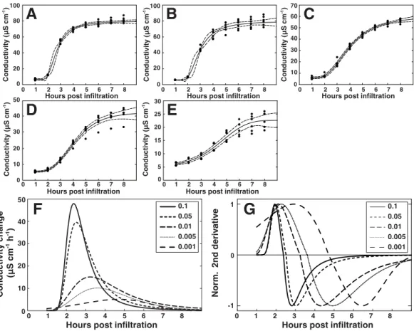
P. syringaeexpressing AvrRpm1, from the same overnight culture, were simultaneously vacuum infiltrated into leaf discs of wild-type Col-0 andrpm1.3plants (Figs. 6Aand6B). In addition, discs were trypan blue stained for visualization of cell death (Fig. 6C). As expected, initiation of the HR, and consequently a dramatic increase in conductivity, was observed 2–3 h after infiltration with the two highest bacterial titers (OD600=0.05 and
0.1, corresponding to 1.78 and 3.55∗107CFU ml−1, respectively). The rapid conductivity change was accompanied by a massive cell death where almost all of the mesophyll cells stained dark with trypan blue three hours after inoculation. A more moderate increase in electrolyte leakage was observed in Col-0 plants treated with lower bacterial titers (OD600=0.001–0.01, 3.55∗105–3.55∗106CFU ml−1). No statistical difference in the
0
10 20 30 40 50 60 70 80 90
0.1 0.05 0.01 0.005 0.001 0.1
0.05 0.01 0.005 0.001
Hours post infiltration
Conductivity (µS cm
-¹)
Conductivity (µS cm
-¹)
Hours post infiltration 0
10 20 30 40 50 60 70 80 90
0 1 2 3 4 5 6 7 8 0 1 2 3 4 5 6 7 8
A
B
C
Hours post infiltration
2 3 4 5 6
0.1
0.05
0.01
0.005
0.001 OD₆₀₀
0.1
rpm1.3
Col-0
Figure 6 Influence of bacterial titer on the outcome of HR.Leaf discs from wild-type Col-0 (A) or
rpm1.3(B) Arabidopsis were simultaneously vacuum infiltrated with five concentrations (OD600=
0.001–0.1) ofP. syringaeDC3000 expressing the effector AvrRpm1. Mean and standard deviation where
n=6 are shown. At indicated times, discs were stained with trypan blue for visualization of induced cell
were observed when wild-type was inoculated with bacterial titers as high or higher than OD600=0.05. In this context, it is tempting to speculate that mutants hyperactive in HR or
mutants that display kinetic differences in HR-PCD associated release of electrolytes may have been missed or overlooked in studies where high bacterial inoculum has been used. This since the number of bacteria necessary for activating HR-PCD is significantly lower than what is commonly used. Electrolyte release, as measured by conductivity, remained low at all concentrations throughout the experiment inrpm1.3samples, although a small dose-dependent increase in electrolyte leakage was noticed. This increase in electrolytes is likely the result of activation of the plant RPS2 protein and other R proteins by the AvrRpm1 effector (Kim et al., 2009) and responses evoked by bacterial PAMPs and other effectors delivered by the pathogen.
When tested, it took about 45 min for one person to prepare 5 plant lines with 6 replicates using syringe infiltration. Preparation of the same number of lines and replicates for the vacuum infiltration assay was found to be slightly faster. Although the two methods required approximately the same amount of time for experiment start-up, they differ in that the pathogen treatment is instantaneous in all samples using vacuum infiltration. When plants were syringe infiltrated, approximately 40 min passed from the start to the finishing of the inoculation procedure. With such a long time span between the samples, it is necessary to make conductivity measurements approximately every five minutes throughout the experiment. Using vacuum infiltration, on the other hand, all samples are simultaneously inoculated and only about ten minutes of measurements every hour is required. We conclude that the vacuum infiltration method allows synchronized treatment in multiple plant lines and replicates and shows higher reproducibility than pressure infiltration, thus making it suitable for detection of subtle differences in HR-mediated electrolyte leakage between samples. Furthermore, the vacuum method requires less training and is not as time consuming as syringe infiltration.
Modeling the kinetics of HR-induced cell death
Figure 7 Modeling of the hypersensitive response reveals bacterial dose dependence.A regression analysis was performed on the electrolyte leakage data from Col-0 presented inFig. 6A. (A)–(E) Shows functions fitted to the data from the five different titers ofP. syringaeDC3000 AvrRpm1 where (A) shows OD600=0.1, (B) OD600=0.05, (C) OD600=0.01, (D) OD600=0.005, and (E) OD600=0.001. The first derivative of the functions displayed in (A)–(E), which shows the rate of change in electrolyte leakage over time, is shown in (F). The second normalized derivative of the functions displayed in (A)–(E) is presented in (G). Dots in (A)–(E) are actual measured values and dashed lines indicate 95% confidence intervals. Modeling was performed on two different data sets with similar results.
inoculations. These results were confirmed in two additional independent experiments where plant material was inoculated withP. syringaeexpressing AvrRpm1 at three different concentrations (OD600=0.1, 0.01 and 0.001) and conductivity measured every 30 min
bacterial inocula. The appearance of the trypan blue stains for the two lowest bacterial titers lend some support to the latter explanation (Fig. 6C).
Vacuum infiltration as a tool to study PLD activity
Phosphatidic acid (PA) is a key signaling lipid involved in plant responses to both abiotic and biotic stresses (Arisz, Testerink & Munnik, 2009;Li, Hong & Wang, 2009). We have previously reported that PA is formed quickly in tissue after elicitation with AvrRpm1 and that PA by itself can induce HR-like cell death in Arabidopsis (Andersson et al., 2006). In the study we used a transgenic system within plantaexpression of the AvrRpm1 effector under the control of a dexamethasone inducible promoter (Mackey et al., 2003;Mackey et al., 2002). Prior to induction of the effector gene, lipids in leaf discs had been labeled with33PO4or14C-acetate. This experimental setup, with ectopic expression of AvrRpm1,
eliminated the need for manual handling of radioactive leaf material, something which would have been necessary if leaves were pressure infiltrated with a bacterial suspension after radiolabeling. Additionally, we have repeatedly failed to obtain conclusive results on phospholipase activation after syringe infiltration. We thus tested if the herein described vacuum infiltration procedure was suitable for determining if an increase in PA, as apparent using the transgenic system, could be observed after treatment with
P. syringaeexpressing AvrRpm1. Leaf discs labeled over night with33PO4were vacuum
infiltrated withP. syringaeDC3000avrRpm1(OD600=0.1, 3.55∗107CFU ml−1) or
mock treated with 10 mM MgCl2and placed in deionized water. Lipids were extracted
0.5 and 4 h after infiltration and subsequently subjected to thin layer chromatography and autoradiography. Films were scanned and the intensity of the radiolabeled PA was determined (Fig. 8A). A five-fold, significant (Student’st-test,p<0.05), increase in PA radiolabel could be observed after 4 h in discs treated with the bacterium. PA levels in mock samples remained low over the four hours of the experiment. To support this, leaf material from Col-0 andrpm1.3plants was vacuum infiltrated withP. syringaeDC3000avrRpm1
and analyzed for PA content with LC-MS/MS (Fig. 8B). A similar significant increase in PA as seen after radiolabeling was noted. The results show that radiolabeled PA accumulates quickly in plants also after elicitation with avirulent bacteria expressing AvrRpm1, and that vacuum infiltration provides a safe and reliable way of inoculating plants when syringe infiltration is not an option. The presented method allows a simple route for pre-treatment of leaf discs with for instance inhibitors or radiolabeled chemicals.
CONCLUSIONS
Figure 8 Phosphatidic acid accumulates in plants in response toP. syringaeexpressing AvrRpm1. Ara-bidopsis leaf tissue was analyzed for the lipid second messenger phosphatidic acid after treatment with the avirulentP. syringaeDC3000 AvrRpm1 at OD600=0.1. In (A), Arabidopsis leaf discs were overnight labeled with33PO4 and vacuum inoculated with P. syringaeor mock treated with 10 mM MgCl2.
Phospholipids were extracted, separated on thin layer chromatography and exposed on X-ray films. Phosphatidic acid was quantified from developed films. In (B), leaf discs from Col-0 and the RPM1 loss-of-function mutantrpm1.3were vacuum infiltrated with the bacteria and extracted lipids were analyzed by LC-MS. Samples were prepared in triplicates and error bars show standard deviation. Asterisk denotesp<0.05, statistically significant difference between mock/rpm1.3and pathogen treated wild-type at 4 hpi as determined by Student’st-test.
vacuum infiltration, we show that the kinetics of the HR-PCD correlates to the density of effector-expressingP. syringaeand we propose that this may be an important factor to consider when screening for mutants with perhaps weak or conditional phenotypes. The results also show that the status of the pathogen is important, as demonstrated by the effect of different media compositions and temperatures during pre-culture. However, we acknowledge the existence of additional factors, beyond our control, that affect both the kinetics and final amplitude of the conductivity curve.
presented here can be combined with several types of analytical methods and we have successfully used it in our lab to monitor early changes in metabolite content, gene expression and protein abundance after pathogen and chemical elicitation. The method is particularly useful when large quantities of inoculated leaf material are needed, for example when quantifying low abundant metabolites. Finally, we believe the described method will be an excellent tool to screen large populations of Arabidopsis mutants for aberrant defense response phenotypes.
ACKNOWLEDGEMENT
We thank Professor Murray Grant forPseudomonasstrains.
ADDITIONAL INFORMATION AND DECLARATIONS
Funding
The financial support of the Swedish Council for Environment, Agricultural Sciences and Spatial Planning to Mats Ellerstr¨om (project No. 2007-1051) and Mats Andersson (project No. 2007-1563 and 2009-888), the Olle Engkvist Byggm¨astare, Adlerbertska research foundation to Mats Andersson is gratefully acknowledged. The funders had no role in study design, data collection and analysis, decision to publish, or preparation of the manuscript.
Grant Disclosures
The following grant information was disclosed by the authors:
Swedish Council for Environment, Agricultural Sciences and Spatial Planning: 2007-1051, 2007-1563, 2009-888.
Olle Engkvist Byggm¨astare, Adlerbertska research foundation.
Competing Interests
Thomas Backhaus is an Academic Editor for PeerJ.
Author Contributions
• Oskar N. Johansson and Anders K. Nilsson conceived and designed the experiments,
performed the experiments, analyzed the data, wrote the paper, prepared figures and/or tables, reviewed drafts of the paper.
• Mikael B. Gustavsson conceived and designed the experiments, performed the
experiments, analyzed the data, wrote the paper, reviewed drafts of the paper.
• Thomas Backhaus conceived and designed the experiments, performed the
experi-ments, analyzed the data, contributed reagents/materials/analysis tools, wrote the paper, reviewed drafts of the paper.
• Mats X. Andersson analyzed the data, contributed reagents/materials/analysis tools,
wrote the paper, reviewed drafts of the paper.
• Mats Ellerstr¨om conceived and designed the experiments, analyzed the data,
Data Availability
The following information was supplied regarding data availability:
The research in this article did not generate any raw data apart from that in the included
Data S1.
Supplemental Information
Supplemental information for this article can be found online athttp://dx.doi.org/ 10.7717/peerj.1469#supplemental-information.
REFERENCES
Alamillo JM, Garcia-Olmedo F. 2001.Effects of urate, a natural inhibitor of
peroxynitrite-mediated toxicity, in the response ofArabidopsis thalianato the bacterial pathogenPseudomonas syringae.The Plant Journal25:529–540DOI 10.1046/j.1365-313x.2001.00984.x.
Andersson MX, Kourtchenko O, Dangl JL, Mackey D, Ellerstrom M. 2006.
Phospholipase-dependent signalling during the AvrRpm1- and AvrRpt2-induced disease resistance responses in Arabidopsis thaliana.The Plant Journal47:947–959DOI 10.1111/j.1365-313X.2006.02844.x. Andersson MX, Nilsson AK, Johansson ON, Boztas G, Adolfsson LE, Pinosa F, Petit CG,
Aronsson H, Mackey D, Tor M, Hamberg M, Ellerstrom M. 2015.Involvement of the
electrophilic isothiocyanate sulforaphane in Arabidopsis local defense responses.Plant Physiology167:251–261DOI 10.1104/pp.114.251892.
Arisz SA, Testerink C, Munnik T. 2009.Plant PA signaling via diacylglycerol kinase.Biochimica et
Biophysica ACTA/General Subjects1791:869–875DOI 10.1016/j.bbalip.2009.04.006.
Arnold DL, Jackson RW. 2011.Bacterial genomes: evolution of pathogenicity.Current Opinion in
Plant Biology14:385–391DOI 10.1016/j.pbi.2011.03.001.
Atkinson MM, Huang JS, Knopp JA. 1985.The hypersensitive reaction of tobacco toPseudomonas
syringaepv. pisi: activation of a plasmalemma K/H exchange mechanism.Plant Physiology 79:843–847DOI 10.1104/pp.79.3.843.
Baker CJ, O’Neill NR, Keppler LD, Orlandi EW. 1991.Early responses during plant-bacteria
interactions in tobacco cell suspensions.Phytopathology81:1504–1507 DOI 10.1094/Phyto-81-1504.
Bisgrove SR, Simonich MT, Smith NM, Sattler A, Innes RW. 1994.A disease resistance gene
in Arabidopsis with specificity for two different pathogen avirulence genes.The Plant Cell 6:927–933DOI 10.1105/tpc.6.7.927.
Brisset MN, Paulin JP. 1991.Relationships between electrolyte leakage from Pyrus communis
and virulence of Erwinia amylovora.Physiological and Molecular Plant Pathology38:443–453 DOI 10.1016/S0885-5765(05)80112-2.
Budde IP, Ullrich MS. 2000.Interactions ofPseudomonas syringaepv. glycinea with host and
nonhost plants in relation to temperature and phytotoxin synthesis.Molecular Plant-Microbe Interactions13:951–961DOI 10.1094/MPMI.2000.13.9.951.
Buell CR, Joardar V, Lindeberg M, Selengut J, Paulsen IT, Gwinn ML, Dodson RJ, Deboy RT, Durkin AS, Kolonay JF, Madupu R, Daugherty S, Brinkac L, Beanan MJ, Haft DH, Nelson WC, Davidsen T, Zafar N, Zhou L, Liu J, Yuan Q, Khouri H, Fedorova N, Tran B, Russell D, Berry K, Utterback T, Van Aken SE, Feldblyum TV, D’Ascenzo M, Deng WL, Ramos AR, Alfano JR, Cartinhour S, Chatterjee AK, Delaney TP, Lazarowitz SG, Martin GB,
genome sequence of the Arabidopsis andtomatopathogenPseudomonas syringaepv.tomato DC3000.Proceedings of the National Academy of Sciences of the United States of America 100:10181–10186DOI 10.1073/pnas.1731982100.
Buseman CM, Tamura P, Sparks AA, Baughman EJ, Maatta S, Zhao J, Roth MR, Esch SW,
Shah J, Williams TD, Welti R. 2006.Wounding stimulates the accumulation of glycerolipids
containing oxophytodienoic acid and dinor-oxophytodienoic acid in Arabidopsis leaves.Plant Physiology142:28–39DOI 10.1104/pp.106.082115.
Chen LQ, Hou BH, Lalonde S, Takanaga H, Hartung ML, Qu XQ, Guo WJ, Kim JG,
Underwood W, Chaudhuri B, Chermak D, Antony G, White FF, Somerville SC, Mudgett MB,
Frommer WB. 2010.Sugar transporters for intercellular exchange and nutrition of pathogens.
Nature468:527–532DOI 10.1038/nature09606.
Coll NS, Epple P, Dangl JL. 2011.Programmed cell death in the plant immune system.Cell Death
and Differentiation18:1247–1256DOI 10.1038/cdd.2011.37.
Coll NS, Vercammen D, Smidler A, Clover C, Van Breusegem F, Dangl JL, Epple P. 2010. Arabidopsis type I metacaspases control cell death.Science330:1393–1397
DOI 10.1126/science.1194980.
Debener T, Lehnackers H, Arnold M, Dangl JL. 1991.Identification and molecular mapping of
a single Arabidopsis thaliana locus determining resistance to a phytopathogenicPseudomonas syringaeisolate.The Plant Journal1:289–302DOI 10.1046/j.1365-313X.1991.t01-7-00999.x.
Dellagi A, Brisset MN, Paulin JP, Expert D. 1998.Dual role of desferrioxamine in Erwinia
amylovora pathogenicity.Molecular Plant-Microbe Interactions11:734–742 DOI 10.1094/MPMI.1998.11.8.734.
Dodds PN, Rathjen JP. 2010.Plant immunity: towards an integrated view of plant–pathogen
interactions.Nature Reviews Genetics11:539–548DOI 10.1038/nrg2812.
Glauser G, Dubugnon L, Mousavi SA, Rudaz S, Wolfender JL, Farmer EE. 2009.Velocity
estimates for signal propagation leading to systemic jasmonic acid accumulation in wounded Arabidopsis.Journal of Biological Chemistry284:34506–34513DOI 10.1074/jbc.M109.061432.
Grant M, Brown I, Adams S, Knight M, Ainslie A, Mansfield J. 2000.The RPM1 plant disease
resistance gene facilitates a rapid and sustained increase in cytosolic calcium that is necessary for the oxidative burst and hypersensitive cell death.The Plant Journal23:441–450 DOI 10.1046/j.1365-313x.2000.00804.x.
Grant MR, Godiard L, Straube E, Ashfield T, Lewald J, Sattler A, Innes RW, Dangl JL. 1995. Structure of the Arabidopsis RPM1 gene enabling dual specificity disease resistance.Science 269:843–846DOI 10.1126/science.7638602.
Hibberd AM, Stall RE, Bassett MJ. 1987.Different phenotypes associated with incompatible
races and resistance genes in bacterial spot disease of pepper.Plant Disease71:1075–1078 DOI 10.1094/PD-71-1075.
Hirano SS, Upper CD. 2000.Bacteria in the leaf ecosystem with emphasis onPseudomonas
syringae-a pathogen, ice nucleus, and epiphyte.Microbiology and Molecular Biology Reviews 64:624–653DOI 10.1128/MMBR.64.3.624-653.2000.
Hofius D, Munch D, BressendorffS, Mundy J, Petersen M. 2011.Role of autophagy in disease
resistance and hypersensitive response-associated cell death.Cell Death and Differentiation 18:1257–1262DOI 10.1038/cdd.2011.43.
Jurkowski GI, Smith RK, Yu IC, Ham JH, Sharma SB, Klessig DF, Fengler KA, Bent AF. 2004. Arabidopsis DND2, a second cyclic nucleotide-gated ion channel gene for which mutation causes the “defense, no death” phenotype.Molecular Plant-Microbe Interactions17:511–520 DOI 10.1094/MPMI.2004.17.5.511.
Katagiri F. 2004.A global view of defense gene expression regulation—a highly interconnected
signaling network.Current Opinion in Plant Biology7:506–511DOI 10.1016/j.pbi.2004.07.013.
Katagiri F, Thilmony R, He SY. 2002.The Arabidopsis thaliana–Pseudomonas syringaeinteraction.
Arabidopsis Book1:e0039DOI 10.1199/tab.0039.
Kim MG, Geng X, Lee SY, Mackey D. 2009.ThePseudomonas syringaetype III effector AvrRpm1
induces significant defenses by activating the Arabidopsis nucleotide-binding leucine-rich repeat protein RPS2.The Plant Journal57:645–653DOI 10.1111/j.1365-313X.2008.03716.x.
King EO, Ward MK, Raney DE. 1954.Two simple media for the demonstration of pyocyanin and
fluorescin.Journal of Laboratory and Clinical Medicine44:301–307.
Klement Z, Goodman RN. 1967.The role of the living bacterial cell and induction time in the
hypersensitive reaction of the tobacco plant.Phytopathology57:322–323.
Koch E, Slusarenko A. 1990.Arabidopsis is susceptible to infection by a downy mildew fungus.
The Plant Cell2:437–445DOI 10.1105/tpc.2.5.437.
Kourtchenko O, Andersson MX, Hamberg M, Brunnstrom A, Gobel C, McPhail KL,
Gerwick WH, Feussner I, Ellerstrom M. 2007.Oxo-phytodienoic acid-containing galactolipids
in Arabidopsis: jasmonate signaling dependence.Plant Physiology 145:1658–1669 DOI 10.1104/pp.107.104752.
Li M, Hong Y, Wang X. 2009.Phospholipase D- and phosphatidic acid-mediated signaling in
plants.Biochimica et Biophysica ACTA/General Subjects1791:927–935 DOI 10.1016/j.bbalip.2009.02.017.
Li-Beisson Y, Shorrosh B, Beisson F, Andersson MX, Arondel V, Bates PD, Baud S, Bird D, Debono A, Durrett TP, Franke RB, Graham IA, Katayama K, Kelly AA, Larson T,
Markham JE, Miquel M, Molina I, Nishida I, Rowland O, Samuels L, Schmid KM, Wada H,
Welti R, Xu C, Zallot R, Ohlrogge J. 2010.Acyl-lipid metabolism.Arabidopsis Book8:e0133
DOI 10.1199/tab.0133.
Mackey D, Belkhadir Y, Alonso JM, Ecker JR, Dangl JL. 2003.Arabidopsis RIN4 is a target of the
type III virulence effector AvrRpt2 and modulates RPS2-mediated resistance.Cell112:379–389 DOI 10.1016/S0092-8674(03)00040-0.
Mackey D, Holt 3rd BF, Wiig A, Dangl JL. 2002.RIN4 interacts withPseudomonas syringaetype
III effector molecules and is required for RPM1-mediated resistance in Arabidopsis.Cell 108:743–754DOI 10.1016/S0092-8674(02)00661-X.
Mur LAJ, Kenton P, Lloyd AJ, Ougham H, Prats E. 2008.The hypersensitive response; the
centenary is upon us but how much do we know?Journal of Experimental Botany59:501–520 DOI 10.1093/jxb/erm239.
Nilsson AK, Fahlberg P, Ellerstrom M, Andersson MX. 2012.Oxo-phytodienoic acid (OPDA) is
formed on fatty acids esterified to galactolipids after tissue disruption in Arabidopsis thaliana. FEBS Letters586:2483–2487DOI 10.1016/j.febslet.2012.06.010.
Nilsson AK, Johansson ON, Fahlberg P, Steinhart F, Gustavsson MB, Ellerstrom M,
Andersson MX. 2014.Formation of oxidized phosphatidylinositol and 12-oxo-phytodienoic
Palmer DA, Bender CL. 1993.Effects of environmental and nutritional factors on production of the polyketide phytotoxin coronatine byPseudomonas syringaepv. glycinea.Applied and Environmental Microbiology59:1619–1626.
Pan X, Welti R, Wang X. 2010.Quantitative analysis of major plant hormones in crude plant
extracts by high-performance liquid chromatography-mass spectrometry.Nature Protocols 5:986–992DOI 10.1038/nprot.2010.37.
Rahme LG, Mindrinos MN, Panopoulos NJ. 1991.Genetic and transcriptional organization of
the hrp cluster ofPseudomonas syringaepv. phaseolicola.Journal of Bacteriology173:575–586.
Scholze M, Boedeker W, Faust M, Backhaus T, Altenburger R, Grimme LH. 2001.A general
best-fit method for concentration–response curves and the estimation of low-effect concentrations.Environmental Toxicology and Chemistry20:448–457
DOI 10.1897/1551-5028(2001)020<0448:AGBFMF>2.0.CO;2.
Schwudke D, Oegema J, Burton L, Entchev E, Hannich JT, Ejsing CS, Kurzchalia T,
Shevchenko A. 2006.Lipid profiling by multiple precursor and neutral loss scanning driven
by the data-dependent acquisition.Analytical Chemistry78:585–595DOI 10.1021/ac051605m.
Sels J, Mathys J, De Coninck BMA, Cammue BPA, De Bolle MFC. 2008.Plant
pathogenesis-related (PR) proteins: a focus on PR peptides.Plant Physiology and Biochemistry46:941–950 DOI 10.1016/j.plaphy.2008.06.011.
Spoel SH, Dong X. 2012.How do plants achieve immunity? Defence without specialized immune
cells.Nature Reviews Immunology12:89–100DOI 10.1038/nri3141.
Stakman EC. 1915.Relation between Puccinia graminis and plants highly resistant to its attack.J
Agric Res4:193–200.
Stelmach BA, Muller A, Hennig P, Gebhardt S, Schubert-Zsilavecz M, Weiler EW. 2001.A novel
class of oxylipins, sn1-O-(12-oxophytodienoyl)-sn2-O-(hexadecatrienoyl)-monogalactosyl Diglyceride, from Arabidopsis thaliana.Journal of Biological Chemistry276:12832–12838 DOI 10.1074/jbc.M010743200.
Thordal-Christensen H, Zhang Z, Wei Y, Collinge DB. 1997.Subcellular localization of
H2O2 in plants. H2O2 accumulation in papillae and hypersensitive response during the barley—powdery mildew interaction.The Plant Journal11:1187–1194
DOI 10.1046/j.1365-313X.1997.11061187.x.
Turner J, Novacky A. 1974.The quantitative relation between plant and bacterial cells involved in
the hypersensitive reaction.Phytopathology64:885–890DOI 10.1094/Phyto-64-885.
Van Dijk K, Fouts DE, Rehm AH, Hill AR, Collmer A, Alfano JR. 1999.The Avr (effector)
proteins HrmA (HopPsyA) and AvrPto are secreted in culture fromPseudomonas syringae pathovars via the Hrp (type III) protein secretion system in a temperature- and pH-sensitive manner.Journal of Bacteriology181:4790–4797.
Venisse JS, Gullner G, Brisset MN. 2001.Evidence for the involvement of an oxidative stress
in the initiation of infection of pear by Erwinia amylovora.Plant Physiology125:2164–2172 DOI 10.1104/pp.125.4.2164.
Yu GL, Katagiri F, Ausubel FM. 1993.Arabidopsis mutations at the RPS2 locus result in loss of
resistance toPseudomonas syringaestrains expressing the avirulence gene avrRpt2.Molecular Plant-Microbe Interactions6:434–443DOI 10.1094/MPMI-6-434.
Yu IC, Parker J, Bent AF. 1998.Gene-for-gene disease resistance without the hypersensitive
response in Arabidopsis dnd1 mutant.Proceedings of the National Academy of Sciences of the United States of America95:7819–7824DOI 10.1073/pnas.95.13.7819.
Zimaro T, Gottig N, Garavaglia BS, Gehring C, Ottado J. 2011.Unraveling plant responses to