Submitted17 November 2015 Accepted 13 February 2016 Published3 March 2016
Corresponding author Cheng-Guang Liang, [email protected], [email protected] Academic editor Shao-Chen Sun
Additional Information and Declarations can be found on page 12
DOI10.7717/peerj.1761
Copyright 2016 Zhou et al.
Distributed under
Creative Commons CC-BY 4.0
OPEN ACCESS
The beneficial effects of cumulus cells
and oocyte-cumulus cell gap junctions
depends on oocyte maturation and
fertilization methods in mice
Cheng-Jie Zhou, Sha-Na Wu, Jiang-Peng Shen, Dong-Hui Wang, Xiang-Wei Kong, Angeleem Lu, Yan-Jiao Li, Hong-Xia Zhou, Yue-Fang Zhao and Cheng-Guang Liang
The Research Center for Laboratory Animal Science, College of Life Science, Inner Mongolia University, Hohhot, Inner Mongolia, China
ABSTRACT
Cumulus cells are a group of closely associated granulosa cells that surround and nourish oocytes. Previous studies have shown that cumulus cells contribute to oocyte maturation and fertilization through gap junction communication. However, it is not known how this gap junction signaling affectsin vivoversusin vitromaturation of oocytes, and their subsequent fertilization and embryonic development following insemination. Therefore, in our study, we performed mouse oocyte maturation and insemination using in vivo- or in vitro-matured oocyte-cumulus complexes (OCCs, which retain gap junctions between the cumulus cells and the oocytes), in vitro -matured, denuded oocytes co-cultured with cumulus cells (DCs, which lack gap junctions between the cumulus cells and the oocytes), andin vitro-matured, denuded oocytes without cumulus cells (DOs). Using these models, we were able to analyze the effects of gap junction signaling on oocyte maturation, fertilization, and early embryo development. We found that gap junctions were necessary for bothin vivoandin vitro
oocyte maturation. In addition, for oocytes maturedin vivo, the presence of cumulus cells during insemination improved fertilization and blastocyst formation, and this improvement was strengthened by gap junctions. Moreover, for oocytes maturedin vitro, the presence of cumulus cells during insemination improved fertilization, but not blastocyst formation, and this improvement was independent of gap junctions. Our results demonstrate, for the first time, that the beneficial effect of gap junction signaling from cumulus cells depends on oocyte maturation and fertilization methods.
SubjectsCell Biology, Zoology
Keywords Oocyte, Cumulus cells, Gap junction, Maturation, Fertilization, Early embryo
development
INTRODUCTION
proteins, and which allow for the exchange of small molecules between adjacent cells (Kidder & Mhawi, 2002).
Fully grown oocytes become competent to undergo two aspects of maturation: cytoplasmic and nuclear. Both of these processes are essential for the formation of an oocyte with the capacity to undergo fertilization and development to live offspring. Nuclear maturation encompasses the processes of reversing meiotic arrest at prophase I and driving the progression of meiosis to metaphase II. Cytoplasmic maturation refers to the processes that prepare the egg for activation and preimplantation development (Eppig, 1996).
Gap junctions between cumulus cells and oocytes are thought to be essential for oocyte maturation and fertilization. During oocyte nuclear maturation, gap junctions are the main connection between cumulus cells and oocytes (Feng et al., 2013;Furger et al., 1996; Santiquet et al., 2012), and they allow a rapid transfer of small metabolites and regulatory molecules from the cumulus cells into the oocyte (Van Soom et al., 2002). Duringin vitro
maturation (IVM), the gap junction protein Cx43 plays a functional role in gap junction breakdown in the oocyte-cumulus complexes (OCCs) (Sasseville et al., 2009). During fertilization, the cumulus cells attract (Eisenbach, 1999), trap (Bedford & Kim, 1993), and select spermatozoa (Carrell et al., 1993), and prevent the premature hardening of the zona pellucida (ZP) (Tanghe et al., 2002), all of which are necessary for successful fertilization. Furthermore, gap junctional communication between oocytes and cumulus cells has been shown to be an essential factor in supporting fertilization (Tanghe et al., 2003). To some extent, early embryo development depends on the successful coordination of processes that occur during oocyte cytoplasmic maturation, including molecular changes, organelle reorganization, and cytoskeletal changes (Damiani et al., 1996;Reyes & Ross, 2016;Salamone et al., 2001). Numerous studies have shown that the presence of cumulus cells can improve cytoplasmic maturation (Ikeda & Yamada, 2014;Tanghe et al., 2002). However, it is still not clear if cumulus cells and their gap junctions with oocytes are necessary for the successfulin vivoandin vitromaturation of oocytes. Furthermore, there is currently a lack of understanding of the contribution of cumulus cells and gap junctions to fertilization and early embryo development downstream of oocyte maturation.
Thus, in this study, we used four types of oocytes for our maturation and insemination procedures, includingin vivo- orin vitro-matured oocyte-cumulus complexes (OCCs, which possess cumulus cells and intact gap junctions),in vitro-matured, denuded oocytes co-cultured with cumulus cells (DCs, which have cumulus cells, but lack gap junctions), andin vitro-matured, denuded oocytes without cumulus cells (DOs). Using these culture models, we investigated oocyte maturation, fertilization, and early embryo development to evaluate contributions of cumulus cells and oocyte-cumulus cell gap junctions to each of these processes.
MATERIALS AND METHODS
Ethics statement and animal feeding regimens
Care and Use Committee at the Inner Mongolia University (Approval number: SYXK 2014-0002). Mice were maintained under the care of the Laboratory Animal Facility at Inner Mongolia University. Mice were kept at a constant temperature of 22±2 ◦C on a
12 h light/dark cycle and had unrestricted access to food and water.
Oocyte collection
Adult female (B6D2) F1 mice (4–8 weeks of age) were used for the oocyte collections. All chemicals and media were purchased from Sigma-Aldrich Company (St. Louis, MO, USA) unless stated otherwise. The germinal vesicle (GV) stage oocytes were collected by puncturing the follicles of ovaries at 48 h after pregnant mare serum gonadotropin (PMSG; SanSheng, Ningbo, China) injection. The cumulus cells were removed by gentle pipetting. Forin vivometaphase II (MII) stage oocyte collection, mice were superovulated by injection of 10 IU PMSG, followed by injection of 10 IU human chorionic gonadotropin (hCG; SanSheng, Ningbo, China) 48 h later. The cumulus cells were dispersed by 0.3 mg/mL hyaluronidase in HEPES-M2 medium.
Oocyte maturation
GV oocytes were cultured in MEM Alpha (α-MEM; Gibco, Pleasanton, CA, USA)
medium supplemented with 5% (v/v) fetal calf serum (FCS), 10 ng/mL epidermal growth factor (EGF), and 0.01 AU/mL follicle-stimulating hormone (FSH), under a humidified atmosphere of 5% CO2at 37 ◦C for 14–16 h. The number of oocytes with the first polar
body (PB1) was counted to determine the percentage of nuclear maturation.
IVF and embryo culture
Adult male (B6D2) F1 mice (12–14 weeks of age) were used for the sperm collections. The sperm suspension was capacitated for 2 h in 200µL T6 medium supplemented with 10
mg/mL bovine serum albumin (BSA). MII oocytes were incubated with spermatozoa for 6 h in 200µL T6 medium supplemented with 20 mg/mL BSA. The sperm concentration used for
fertilization was 1×10⁶/mL. The zygotes were collected and cultured in
Chatot-Ziomet-Bavister (CZB) medium containing 3 mg/ml BSA without glucose under a humidified atmosphere of 5% CO2at 37 ◦C for the first 2 days, and then transferred to CZB medium
supplemented with 5.5 mmol/L glucose when the embryos reached the 4-cell stage. The percentage of embryos that reached the 2-cell stage was used for fertilization evaluation. The embryos were checked at 48, 72, and 96 h after fertilization to calculate the percentage of 4-cell, morula, and blastocyst stages, respectively.
Experimental design
In our study, oocytes were randomly divided into four groups to mature: (1) OCCs from
collected from GV-stage OCCs by pipetting. The cumulus cells collected from each OCC were supplemented back to the corresponding oocyte. The combinations of different maturation procedures and different insemination procedures were tested to evaluate the roles of cumulus, resulting in a total of ten combinations (M-vivo-OCC +I-OCC,
M-vivo-OCC+I-DC, M-vivo-OCC+I-DO, M-vitro-OCC+I-OCC, M-vitro-OCC+
I-DC, M-vitro-OCC+I-DO, M-vitro-DC+I-DC, M-vitro-DC+I-DO, M-vitro-DO+
I-DC, and M-vitro-DO+I-DO), which are shown inFig. 1.
Statistical analysis
The data are presented as the means ±standard deviation (SD) from three replicate
experiments. Differences were evaluated using the Student’st test.P<0.05 was regarded
as statistically significant.
RESULTS
The role of cumulus cells and their gap junctions in oocyte maturation
To investigate the contribution of cumulus cells and their gap junctions during oocyte maturation, PB1 extrusion was calculated using the following maturation models, as shown inFig. 1: M-vivo-OCC, M-vitro-OCC, M-vitro-DC, and M-vitro-DO. Our results showed that the highest nuclear maturation percentage was obtained among the OCC groups, but there was no statistical difference between thein vivoandin vitroOCC models. However, if cumulus cells were removed from the oocytes, PB1 extrusion was significantly decreased (P<0.01), and this decrease was not reversed by co-culturing cumulus cells with oocytes
(P<0.01) (Fig. 2andTable S1), suggesting that intact gap junctions between the cumulus
cells and oocytes are necessary for efficient oocyte maturation.
The role of cumulus cells and their gap junctions in oocyte fertilization
Fertilization can be affected by both oocyte maturation and insemination procedures. Thus, to assess the effects of cumulus cells and gap junctions during maturation and insemination, we normalized the fertilization percentage by quantifying the number of 2-cell embryos relative to the number of total GV-stage oocytes or total MII-stage oocytes in each group.
Fertilization based on the number of GV oocytes
Using the same maturation method, we observed significant increases in the percentage of 2-cell-stage embryos when comparing oocytes cultured with, to those cultured without, cumulus cells during insemination (P<0.01) (Fig. 3AandTable S2), suggesting that
the presence of cumulus cells improves fertilization. For the oocytes matured in vivo
(M-vivo-OCC), detachment of the cumulus cells from the oocytes before insemination, which disrupted the gap junctions between the cells, reduced the fertilization percentage (P<0.01). However, under the same conditions, fertilization was not affected for the
Figure 1 Experimental design.In vivoMII stage oocyte-cumulus complexes (OCC) were collected from oviduct in mice superovulated by PMSG, followed by hCG. GV stage OCCs were collected from ovaries of the mice 48 h after the administration of PMSG. Cumulus cells were removed by gentle pipetting. Oocytes were divided into four groups for maturation:in vivoOCC,in vitroOCC,in vitrodenuded oocyte (DO)+cumulus cells, andin vitroDO. For OCCs maturedin vivoorin vitro, oocytes were divided into three groups for insemination: OCC, DO+cumulus, and DO. Forin vitromatured DOs with or without
Figure 2 Percent of PB1 under different maturation methods.Oocytes were divided into four groups for maturation: M-vivo-OCC, M-vitro-OCC, M-vitro-DC, and M-vitro-DO. The percentages of the PB1 were calculated. Percentages without a common letter are statistical significant different (P<0.05).
Next, we analyzed the data to investigate the influence of maturation methods on fertilization outcomes. Using the same insemination method, successful fertilization was dependent on the maturation method of the oocytes. For example, the oocytes matured
in vivo(M-vivo-OCC) showed the highest fertilization percentage, the OCCs maturedin vitro(M-vitro-OCC) showed a median fertilization percentage, and the oocytes maturedin vitrowith dispersed cumulus cells (M-vitro-DC) or without cumulus cells (M-vitro-DO) showed the lowest fertilization percentages (P<0.01) (Fig. 3AandTable S2).
Fertilization based on the number of MII oocytes
To evaluate the contribution of cumulus cells and gap junctions during insemination, we quantified the percentage of 2-cell-stage embryos among the total MII oocytes. Using the same maturation method, we found that the presence of cumulus cells in the insemination medium improved the fertilization percentages (P<0.01) (Fig. 3BandTable S3). For the
M-vivo-OCC oocytes, the removal of cumulus cells from oocytes during insemination (I-DO) reduced the fertilization percentages; however, this reduction could be partially reversed by the addition of cumulus cells into the insemination medium (I-DC) (P<0.01)
(Fig. 3BandTable S3). Using the same insemination method, the oocytes maturedin vivohad higher fertilization percentages than those maturedin vitro(P<0.01), but the
presence of cumulus cells during IVM did not affect fertilization (P>0.05) (Fig. 3Band
Table S3).
The role of cumulus cells and their gap junctions in embryo development
Figure 3 Fertilization of oocytes under different maturation and insemination combinations.(A) Per-cent of fertilization based on the number of GV oocytes. (B) PerPer-cent of fertilization based on the number of MII oocytes. Percentages without a common letter are statistical significant different (P<0.05).
oocytes (i.e., the progression from GV stage to MII stage), the procedure of insemination (i.e., the progression from MII stage to 2-cell stage), and the development starting from the 2-cell stage (i.e., the progression after 2-cell stage). To assess the effects of cumulus cells and gap junctions during maturation, insemination, and early embryo development, we quantified each embryonic stage as a percentage of the total number of GV-stage oocytes, MII-stage oocytes, and 2-cell-stage embryos, respectively.
Embryonic development based on the number of GV oocytes
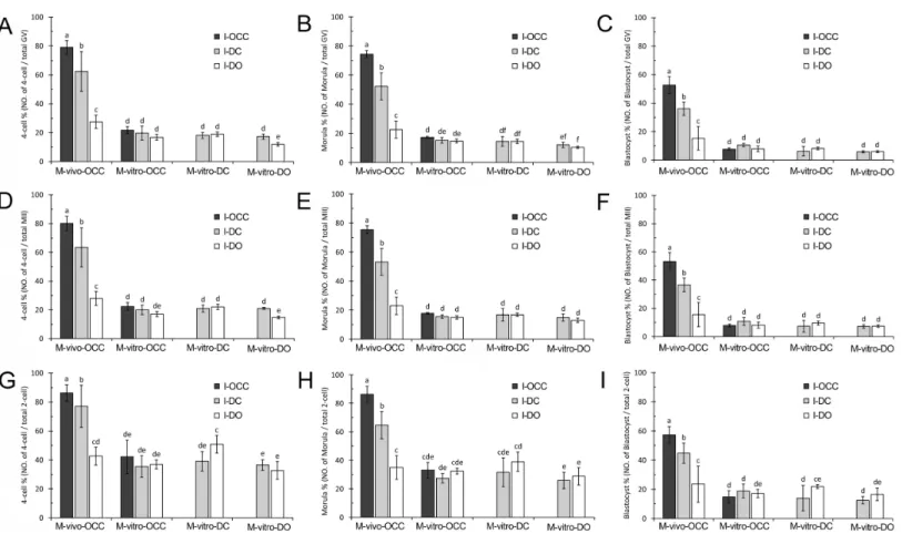
Figure 4 Development of embryos generated from combinations of different maturation and insemination methods.(A–C), Embryonic devel-opment based on the number of GV oocytes. (D–F), Embryonic develdevel-opment based on the number of MII oocytes. (G–I), Embryonic develdevel-opment based on number of 2-cell embryos. Percentages without a common letter are statistical significant different (P<0.05).
embryo development percentage, those inseminated in the presence of cumulus cells but without gap junctions (I-DC) showed a median embryo development percentage, and those inseminated in the absence of cumulus cells (I-DO) had the lowest embryo development percentage (4-cell stage:P<0.01,Fig. 4AandTable S4; morula stage:P<0.01,Fig. 4B
andTable S4; and blastocyst stage:P<0.01,Fig. 4CandTable S4).
For the oocytes maturedin vitrowith cumulus cells (M-vitro-OCC and M-vitro-DC), the loss of oocyte-cumulus cell gap junctions (I-DC) or the absence of cumulus cells (I-DO) during insemination did not affect early embryo development (4-cell stage: P>0.05,
Fig. 4AandTable S4; morula stage:P>0.05,Fig. 4BandTable S4; and blastocyst stage:
P>0.05,Fig. 4CandTable S4).
Interestingly, for the oocytes maturedin vitrowithout cumulus cells (M-vitro-DO), the absence of cumulus cells during insemination (I-DO) reduced the percentage of 4-cell embryos (P<0.05) (Fig. 4AandTable S4). However, this absence did not affect formation
of the morula (P>0.05) (Fig. 4BandTable S4) or blastocyst stages (P>0.05) (Fig. 4C
andTable S4).
Notably, the oocytes maturedin vivo(M-vivo-OCC), regardless of the presence or absence of cumulus cells during insemination (I-OCC, I-DC, and I-DO), had higher embryo development percentages than those matured in vitro(OCC, M-vitro-DC, and M-vitro-DO) (4-cell stage:P<0.01,Fig. 4AandTable S4; morula stage:P<0.01,
Fig. 4BandTable S4; and blastocyst stage:P<0.01,Fig. 4CandTable S4).
Embryonic development based on the number of MII oocytes
To evaluate the contribution of cumulus cells and gap junctions during insemination, we quantified the percentage of embryos from each stage relative to the total number of MII oocytes. For thein vivo-matured oocytes (M-vivo-OCC), we observed a greater percentage of embryo development when cumulus cells were present during insemination. More specifically, if cumulus cells retained gap junctions with oocytes (I-OCC), the highest embryo development percentage was obtained. If cumulus cells were separated from oocytes, but were still present in the insemination medium (I-DC), a median embryo development percentage was obtained. If cumulus cells were removed from the insemination medium (I-DO), the percentage of embryo development was much lower (4-cell stage:P<0.01,Fig. 4DandTable S5; morula stage:P<0.01,Fig. 4EandTable S5;
and blastocyst stage:P<0.01,Fig. 4FandTable S5).
For the oocytes maturedin vitrowith cumulus cells (M-vitro-OCC and M-vitro-DC), the absence of cumulus cells during insemination (I-DO) did not affect early embryo development (4-cell stage:P>0.05,Fig. 4DandTable S5; morula stage:P>0.05,Fig. 4E
andTable S5; and blastocyst stage:P>0.05,Fig. 4FandTable S5).
For the oocytes matured in vitrowithout cumulus cells (M-vitro-DO), the absence of cumulus cells during insemination (I-DO) reduced the percentage of 4-cell embryos (P<0.05) (Fig. 4DandTable S5), but did not affect the percentages of morula or blastocyst
embryos (morula stage:P>0.05,Fig. 4EandTable S5; blastocyst stage:P>0.05,Fig. 4F
andTable S5).
For the oocytes maturedin vitro(M-vitro-OCC, M-vitro-DC, and M-vitro-DO), the absence of cumulus cells during IVM and insemination (I-DO) did not affect blastocyst formation (P>0.05) (Fig. 4FandTable S5).
The oocytes maturedin vivo(M-vivo-OCC), regardless of the presence or absence of cumulus cells during insemination (I-OCC, I-DC, and I-DO), had a greater percentage of embryo development than those matured in vitro(M-vitro-OCC, M-vitro-DC and M-vitro-DO) (4-cell stage:P<0.01,Fig. 4DandTable S5; morula stage:P<0.01,Fig. 4E
andTable S5; and blastocyst stage:P<0.01,Fig. 4FandTable S5).
Embryonic development based on number of 2-cell embryos
oocytes, but were still present in the insemination medium (I-DC), a median embryo development percentage was obtained. If cumulus cells were removed from the oocytes during insemination (I-DO), the lowest embryo development percentage was obtained (4-cell stage: P<0.01, Fig. 4G and Table S6; morula stage: P<0.01, Fig. 4H and
Table S6; and blastocyst stage:P<0.01,Fig. 4IandTable S6).
For the OCCs maturedin vitro(M-vitro-OCC), the absence of cumulus cells during insemination (I-DO) did not affect embryo development (4-cell stage:P>0.05,Fig. 4G
andTable S6; morula stage:P>0.05,Fig. 4HandTable S6; and blastocyst stage:P>0.05,
Fig. 4IandTable S6). Similar results were obtained with DOs maturedin vitro (M-vitro-DO) (4-cell stage:P>0.05,Fig. 4GandTable S6; morula:P>0.05,Fig. 4HandTable S6;
and blastocyst:P>0.05,Fig. 4IandTable S6).
However, for the oocytes maturedin vitrowith cumulus cells (M-vitro-DC), the absence of cumulus cells during insemination increased the percentage of 4-cell-stage and blastocyst, but not morula embryos (4-cell stage:P<0.05,Fig. 4GandTable S6; and morula stage:
Fig. 4HandTable S6; and blastocyst stage:P<0.05,Fig. 4IandTable S6).
Compared to the oocytes maturedin vitro(OCC, DC, and M-vitro-DO), the presence of cumulus cells during insemination (I-OCC and I-DC) increased the percentage of embryo development fromin vivo-matured oocytes (M-vivo-OCC) (4-cell stage:P<0.01,Fig. 4GandTable S6; morula stage:P<0.01,Fig. 4HandTable S6; and
blastocyst stage:P<0.01,Fig. 4IandTable S6).
DISCUSSION
Cumulus cells and oocyte-cumulus cell gap junctions contribute to oocyte maturation
In this study, the contribution of cumulus cells and oocyte-cumulus cell gap junctions to oocyte maturation was analyzed under different culture conditions, includingin vivoor
in vitromaturation, with or without cumulus cells or gap junctions. This comprehensive experimental design allowed us to better understand the functions of cumulus cells and oocyte-cumulus cell gap junctions during maturation. Previous studies have shown that gap junctions between cumulus cells and oocytes are the main channels allowing the exchange of ions and small molecules, such as purines (Downs & Eppig, 1986; Eppig, Ward-Bailey & Coleman, 1985) and cyclic adenosine monophosphate (cAMP) (Kumar & Gilula, 1996;Yoshimura et al., 1992), to inhibit resumption of premature oocyte meiotic progression. Our study demonstrates that these gap junctions are also essential for oocytes to achieve higher nuclear maturation rates. We showed that disruption of these junctions impaired PB1 extrusion, and that this impairment could not be rescued by culturing dispersed cumulus cells with oocytes, which is consistent with previous studies showing that co-culturing with dispersed cumulus cells does not improve the nuclear maturation of oocytes (Downs, 2001;Luciano et al., 2005;Tao et al., 2008).
is similar to what we observed. For oocyte growth and developmentin vitro, the gap junctions between cumulus cells and oocytes must be maintained (Eppig, 1979;Hashimoto et al., 1998;Tanghe et al., 2002). Once gap junctions are disrupted by removing the cumulus cells, the passage of necessary signaling molecules is interrupted. Consistent with previous data, we demonstrated that gap junction signaling between oocytes and cumulus cells was critical for oocyte maturation.
Cumulus cells and oocyte-cumulus cell gap junctions contribute to oocyte fertilization
Although cumulus cell function during oocyte maturation has been widely studied, less is known about cumulus cell contribution during insemination. To the best of our knowledge, this study is the first to demonstrate that cumulus cells affected fertilization by influencing
in vitro oocyte nuclear maturation. Using the same insemination method, successful fertilization depended on the method of oocyte nuclear maturation, within vivo-matured oocytes showing the highest fertilization percentages. Using the same IVM method, the presence of cumulus cells during insemination resulted in higher cleavage rates. Therefore, our data suggest thatin vivoOCC maturation followed by OCC insemination is the optimal combination for obtaining the highest fertilization percentage.
A new finding in our study was that whenin vitro-matured MII oocytes were used for fertilization, the presence of cumulus cells during insemination improved the cleavage percentages, even when the gap junctions were destroyed. It has been proposed that gap junctional communication between the oocyte and corona cells is needed for supporting fertilization (Tanghe et al., 2003). We hypothesize that the improved fertilization percentage obtained when the DOs were inseminated in the presence of cumulus cells, but in the absence of gap junctions, was due to secreted factors from the cumulus cells (Guidobaldi et al., 2008;Sun et al., 2005). This hypothesis is quite plausible, as cumulus cells have been shown to secrete chemotactic factors that guide the spermatozoon to the oocyte (Ito, Smith & Yanagimachi, 1991;Sun et al., 2005), which increases the chance of fertilization.
Another new finding of our study was thatin vivo-matured oocytes achieved higher cleavage percentages than those from IVM, even in the absence of cumulus cells during insemination. To explain these data, we hypothesize that thein vivo-matured oocytes had undergone more complete cytoplasmic maturation than the in vitro-matured oocytes. Moreover, for the oocytes maturedin vivo, removal of cumulus cells during insemination (I-DO) reduced the fertilization percentage, but this reduction was partially reversed by the addition of cumulus cells to the insemination medium. Interestingly, for the in vitro-matured OCCs, this reduction was reversed completely by adding cumulus cells to the insemination medium. These data suggest that the gap junctions of thein vitro-matured OCCs are defective, leading to the reduced fertilization percentages during IVF compared with thein vivo-matured OCCs.
Cumulus cells and oocyte-cumulus cell gap junctions contribute to early embryo development
been suggested that co-culturing DOs embedded in cumulus cell clumps can improve cytoplasmic maturation (Feng et al., 2013), and denuding mouse oocytes of cumulus cells impairsin vitrocytoplasmic maturation (Ge et al., 2008). However, our data support the concept that cumulus cells do not contribute to cytoplasmic maturation during IVM. These different outcomes may be due to the employment of different species, different standards for evaluating cytoplasmic maturation, or different maturation conditions used in each study.
Our study showed that oocytes matured in vivowere the most suitable for embryo development, indicating thatin vivo-matured oocytes possess more complete cytoplasmic maturation thanin vitro-matured oocytes. Previously, pronuclear (PN) transfer between
in vivo-matured andin vitro-matured oocytes was used to examine the nuclear-ooplasm effects on resultant embryo development. The results showed that as long as the ooplasm was derived fromin vivosamples, reconstructed embryos had a higher developmental ability regardless of PN origin (Chang et al., 2005). These data showed that the ooplasm is the decisive factor for embryo development, which is consistent with our hypothesis.
In conclusion, our study systematically evaluated the contribution of cumulus cells and oocyte-cumulus cell gap junctions to oocyte maturation, fertilization, and embryo development, using various oocyte maturation and insemination methods. Our results demonstrate that the beneficial effects of cumulus cells and oocyte-cumulus cell gap junctions depend on oocyte maturation and insemination methods.
ADDITIONAL INFORMATION AND DECLARATIONS
Funding
This work was supported by the National Natural Science Foundation of China (31160243, 31371454), the Natural Science Foundation of Inner Mongolia Autonomous Region of China (2015JQ02), Program of Science and Technology Supporting for Homecoming People in Inner Mongolia to CL and Program of Graduate Student Education and Innovation Plan in Inner Mongolia (B20151012615) to CZ. The funders had no role in study design, data collection and analysis, decision to publish, or preparation of the manuscript.
Grant Disclosures
The following grant information was disclosed by the authors: National Natural Science Foundation of China: 31160243, 31371454.
Natural Science Foundation of Inner Mongolia Autonomous Region of China: 2015JQ02. Program of Science and Technology.
Program of Graduate Student Education and Innovation Plan in Inner Mongolia: B20151012615.
Competing Interests
Author Contributions
• Cheng-Jie Zhou conceived and designed the experiments, performed the experiments,
analyzed the data, contributed reagents/materials/analysis tools, wrote the paper, prepared figures and/or tables, reviewed drafts of the paper.
• Sha-Na Wu and Jiang-Peng Shen performed the experiments, contributed
reagents/materials/analysis tools.
• Dong-Hui Wang, Xiang-Wei Kong, Angeleem Lu, Yan-Jiao Li and Yue-Fang Zhao
performed the experiments.
• Hong-Xia Zhou conceived and designed the experiments, performed the experiments. • Cheng-Guang Liang conceived and designed the experiments, analyzed the data, wrote
the paper, prepared figures and/or tables, reviewed drafts of the paper.
Animal Ethics
The following information was supplied relating to ethical approvals (i.e., approving body and any reference numbers):
All studies adhered to procedures consistent with the National Research Council Guide for the Care and Use of Laboratory Animals and were approved by the Institutional Animal Care and Use Committee at the Inner Mongolia University. Mice were maintained under the care of the Laboratory Animal Facility, Inner Mongolia University.
The approval number is SYXK 2014-0002.
Data Availability
The following information was supplied regarding data availability: The raw data was supplied asData S1.
Supplemental Information
Supplemental information for this article can be found online athttp://dx.doi.org/10.7717/ peerj.1761#supplemental-information.
REFERENCES
Bedford JM, Kim HH. 1993.Cumulus oophorus as a sperm sequestering device,in vivo.
Journal of Experimental Zoology265:321–328 DOI 10.1002/jez.1402650314.
Carrell DT, Middleton RG, Peterson CM, Jones KP, Urry RL. 1993.Role of the cumulus in the selection of morphologically normal sperm and induction of the acrosome reaction during humanin vitrofertilization.Archices of Andrology31:133–137
DOI 10.3109/01485019308988391.
Chang HC, Liu H, Zhang J, Grifo J, Krey LC. 2005.Developmental incompetency of denuded mouse oocytes undergoing maturationin vitrois ooplasmic in nature and is associated with aberrant Oct-4 expression.Human Reproduction20:1958–1968
DOI 10.1093/humrep/dei003.
Damiani P, Fissore RA, Cibelli JB, Long CR, Balise JJ, Robl JM, Duby RT. 1996. Evaluation of developmental competence, nuclear and ooplasmic matura-tion of calf oocytes.Molecular Reproduction and Development 45:521–534
Downs SM. 2001.A gap-junction-mediated signal, rather than an external paracrine factor, predominates during meiotic induction in isolated mouse oocytes.Zygote 9:71–82.
Downs SM, Eppig JJ. 1986.The role of purines in the maintenance of meiotic arrest in mouse oocytes.Tokai Journal of Experimental and Clinical Medicine11:463–469. Eisenbach M. 1999.Sperm chemotaxis.Reviews of Reproduction4:56–66
DOI 10.1530/ror.0.0040056.
Eppig JJ. 1979.A comparison between oocyte growth in coculture with granulosa cells and oocytes with granulosa cell-oocyte junctional contact maintainedin vitro.
Journal of Experimental Zoology209:345–353 DOI 10.1002/jez.1402090216. Eppig JJ. 1996.Coordination of nuclear and cytoplasmic oocyte maturation in
eutherian mammals.Reproduction, Fertility, and Development 8:485–489
DOI 10.1071/RD9960485.
Eppig JJ, Ward-Bailey PF, Coleman DL. 1985.Hypoxanthine and adenosine in murine ovarian follicular fluid: concentrations and activity in maintaining oocyte meiotic arrest.Biology of Reproduction33:1041–1049DOI 10.1095/biolreprod33.5.1041. Feng G, Shi D, Yang S, Wang X. 2013.Co-culture embedded in cumulus clumps
promotes maturation of denuded oocytes and reconstructs gap junctions between oocytes and cumulus cells.Zygote21:231–237DOI 10.1017/S0967199412000305. Furger C, Cronier L, Poirot C, Pouchelet M. 1996.Human granulosa cells in culture
ex-hibit functional cyclic AMP-regulated gap junctions.Molecular Human Reproduction 2:541–548DOI 10.1093/molehr/2.8.541.
Ge L, Sui HS, Lan GC, Liu N, Wang JZ, Tan JH. 2008.Coculture with cumulus cells improves maturation of mouse oocytes denuded of the cumulus oophorus: ob-servations of nuclear and cytoplasmic events.Fertility and Sterility90:2376–2388
DOI 10.1016/j.fertnstert.2007.10.054.
Guidobaldi HA, Teves ME, Unates DR, Anastasia A, Giojalas LC. 2008.Progesterone from the cumulus cells is the sperm chemoattractant secreted by the rabbit oocyte cumulus complex.PLoS ONE 3:e3040DOI 10.1371/journal.pone.0003040. Hashimoto S, Saeki K, Nagao Y, Minami N, Yamada M, Utsumi K. 1998.Effects of
cumulus cell density duringin vitromaturation of the developmental competence of bovine oocytes.Theriogenology49:1451–1463DOI 10.1016/S0093-691X(98)00091-0. Ikeda S, Yamada M. 2014.Midkine and cytoplasmic maturation of mammalian oocytes
in the context of ovarian follicle physiology.British Journal of Pharmacology 171:827–836DOI 10.1111/bph.12311.
Ito M, Smith TT, Yanagimachi R. 1991.Effect of ovulation on sperm transport in the hamster oviduct.Journal of Reproduction and Fertility93:157–163
DOI 10.1530/jrf.0.0930157.
Kidder GM, Mhawi AA. 2002.Gap junctions and ovarian folliculogenesis.Reproduction 123:613–620DOI 10.1530/rep.0.1230613.
Lorenzo PL, Rebollar PG, Illera MJ, Illera JC, Illera M, Alvarino JM. 1996.Stimulatory effect of insulin-like growth factor I and epidermal growth factor on the maturation of rabbit oocytesin vitro.Journal of Reproduction and Fertility107:109–117
DOI 10.1530/jrf.0.1070109.
Luciano AM, Lodde V, Beretta MS, Colleoni S, Lauria A, Modina S. 2005. Develop-mental capability of denuded bovine oocyte in a co-culture system with intact cumulus-oocyte complexes: role of cumulus cells, cyclic adenosine 3′
,5′
-monophosphate, and glutathione.Molecular Reproduction and Development 71:389–397DOI 10.1002/mrd.20304.
Reyes JM, Ross PJ. 2016.Cytoplasmic polyadenylation in mammalian oocyte matura-tion.Wiley Interdisciplinary Reviews: RNA7:71–89.
Salamone DF, Damiani P, Fissore RA, Robl JM, Duby RT. 2001.Biochemical and devel-opmental evidence that ooplasmic maturation of prepubertal bovine oocytes is com-promised.Biology of Reproduction64:1761–1768DOI 10.1095/biolreprod64.6.1761. Santiquet NW, Develle Y, Laroche A, Robert C, Richard FJ. 2012.Regulation of
gap-junctional communication between cumulus cells duringin vitro mat-uration in swine, a gap-FRAP study.Biology of Reproduction87(2):46, 1–8
DOI 10.1095/biolreprod.112.099754.
Sasseville M, Gagnon MC, Guillemette C, Sullivan R, Gilchrist RB, Richard FJ. 2009. Regulation of gap junctions in porcine cumulus-oocyte complexes: contributions of granulosa cell contact, gonadotropins, and lipid rafts.Molecular Endocrinology 23:700–710DOI 10.1210/me.2008-0320.
Sun F, Bahat A, Gakamsky A, Girsh E, Katz N, Giojalas LC, Tur-Kaspa I, Eisenbach M. 2005.Human sperm chemotaxis: both the oocyte and its surrounding cumulus cells secrete sperm chemoattractants.Human Reproduction20:761–767
DOI 10.1093/humrep/deh657.
Tanghe S, Van Soom A, Mehrzad J, Maes D, Duchateau L, De Kruif A. 2003.Cumulus contributions during bovine fertilizationin vitro.Theriogenology60:135–149
DOI 10.1016/S0093-691X(02)01360-2.
Tanghe S, Van Soom A, Nauwynck H, Coryn M, De Kruif A. 2002.Minireview: func-tions of the cumulus oophorus during oocyte maturation, ovulation, and fertiliza-tion.Molecular Reproduction and Development61:414–424 DOI 10.1002/mrd.10102. Tao Y, Cao C, Zhang M, Fang F, Liu Y, Zhang Y, Ding J, Zhang X. 2008.Effects of
cumulus cells on rabbit oocytein vitromaturation.Journal of Animal Physiology and Animal Nutrition92:438–447 DOI 10.1111/j.1439-0396.2007.00729.x.
Van Soom A, Tanghe S, De Pauw I, Maes D, De Kruif A. 2002.Function of the cumulus oophorus before and during mammalian fertilization.Reproduction in Domestic Animals37:144–151DOI 10.1046/j.1439-0531.2002.00345.x.