6. Jahrgang 2009 // Nummer 2 // ISSN 1810-2107
Journal für
Reproduktionsmedizin
und Endokrinologie
No.2
2009
– Journal of Reproductive Medicine and Endocrinology
–
www.kup.at/repromedizin
Online-Datenbank mit Autoren- und Stichwortsuche
Krause & Pachernegg GmbH, Verlag für Medizin und Wirtschaft, A-3003 Gablitz
Offizielles Organ: AGRBM, BRZ, DIR, DVR, DGA, DGGEF, DGRM, EFA, OEGRM, SRBM/DGE
Indexed in EMBASE/Excerpta Medica
Andrologie
•Embryologie & Biologie
•Endokrinologie
•Ethik & Recht
•Genetik
Gynäkologie
•Kontrazeption
•Psychosomatik
•Reproduktionsmedizin
•Urologie
Member of the
Interactions between Oocyte and Surrounding Cumulus
Cells Influence the Results of Assisted Reproduction
Fritzsche H, Michelmann HW, Siebzehnrübl E
Schmedemann RKA
Finden Sie in der Rubrik „Tipps und Tricks im Gyn-Ultraschall“ aktuelle
Fallbeispiele von Univ.Prof. Dr. Christoph Brezinka, Innsbruck.
Mitteilungen aus der Redaktion:
Die meistgelesenen Artikel
Journal für Urologie und
Urogynäkologie
Journal für
Reproduktions-medizin und Endokrinologie
Speculum
P
P
P
Journal für Gynäkologische
Endokrinologie
373
J. REPRODUKTIONSMED. ENDOKRINOL. 6/2006
Received: March 24, 2006; accepted after revision: October 6, 2006 From the 1Zentrum für Reproduktionsmedizin, Jena, 2
Universitäts-Frauenklinik Göttingen, Arbeitsgruppe Reproduktionsmedizin,
3Zentrum für Reproduktionsmedizin, Frankfurt and 4FERRING
Arznei-mittel GmbH, Kiel
Correspondence: PD Dr. med. habil. Heidi Fritzsche, Zentrum für Reproduktionsmedizin, D-07743 Jena, Markt 4, Germany; e-mail: [email protected]
Interactions between Oocyte and Surrounding
Cumulus Cells Influence the Results of
Assisted Reproduction
H. Fritzsche
1, H. W. Michelmann
2, E. Siebzehnrübl
3, R. K. A. Schmedemann
4The interactions between oocyte and surrounding cumulus cells, as well as between cumulus oophorus and theca cells, were investigated in IVF/ ICSI cycles. Gap junctions connect cumulus cells with the oocyte, thereby enabling a bi-directional exchange of products essential for optimal oocyte development. GnRH, FSH, LH and E2 play a major role during oocyte maturation. In general, FSH and LH are prerequisites for folliculogenesis,
as well as oogenesis, but it is the quantitative threshold value of both that seems to determine oocyte quality and pregnancy rate. It remains to be determined how apoptosis and the anti-Muellerian hormone (AMH) can be used as predictive factors regarding the success of ART. In a retrospec-tive sub-analysis of compararetrospec-tive stimulation regimens, using either LH + FSH (hMG-HP) or FSH (rFSH) alone in GnRH-antagonist down-regulated cycles, it was possible to demonstrate that stimulation with LH and FSH results in a significantly higher pregnancy rate in IVF-patients compared to a stimulation with only FSH. Further prospective and randomised trials are needed to answer the question whether, in relation to pregnancy, IVF-patients benefit significantly from hMG stimulation when compared to FSH stimulation. J Reproduktionsmed Endokrinol 2006; 3 (6) 373–8.
Key words: oocyte-cumulus-complex (OCC), predictive factors, apoptosis, anti-muellerian-hormone (AMH), GnRH-agonist, gonadotrophin, FSH, LH, IVF
T
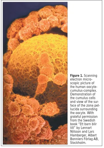
he interaction between oocyte and follicular cells, in particular cumulus oophorus (Fig. 1) and theca in-terna cells, has been investigated intensively in the last 25 years. Nevertheless, there are still many unanswered questions, especially in relation to humans. This paper highlights, in quite a novel way, the importance of cumu-lus cells for oocyte quality in the course of in vitro ferti-lisation (IVF) and intracytoplasmatic sperm injection (ICSI). Here, we present a comprehensive account of results from basic research and preliminary clinical find-ings.We focus our attention on the following points:
1. Cumulus cells and their interaction with oocytes and spermatozoa.
2. Predictive factors resulting from this interaction. 3. The influence of gonadotrophins on the
oocyte-cumu-lus-complex (OCC).
4. Clinical findings related to the interaction of oocyte and surrounding cumulus cells.
Interaction of Cumulus Cells with Oocytes
and Spermatozoa
At the primordial stage, oocytes are surrounded by a sin-gle layer of cells. These cells are the original source which later contribute to the zona pellucida and granu-losa cells. During the transition from the primary to the tertiary follicle, the development of multi-layered granu-losa cells and basal membranes takes place. The forma-tion of an antrum inside the granulosa cells leads to the formation of an antral follicle. The antrum is lined by the follicular epithelium. Finally, the whole follicle is shaped by the surrounding connective tissue. The layer of granu-losa cells does not contain blood vessels. It is separated
Figure 1. Scanning electron micro-scopic picture of the human oocyte-cumulus-complex. Demonstration of the cumulus cells and view of the sur-face of the zona pel-lucida surrounding the oocyte. With grateful permission from the Swedish book “Ett barn blir till” by Lennart Nilsson and Lars Hamberger, Albert Bonniers Förlag AB, Stockholm.
from the theca interna by the basal membrane. In con-trast to the theca interna, the theca externa contains few blood vessels.
The theca interna is a particular endocrine tissue and a very important site for the production of estrogens. Dur-ing all phases of folliculogenesis, those granulosa cells surrounding the oocyte form the cumulus oophorus. The cell layer that directly covers the oocyte is called the co-rona radiata. Its cells are connected with the oolemma by cytoplasmic filaments penetrating the zona pellucida.
374
J. REPRODUKTIONSMED. ENDOKRINOL. 6/2006ing the oocyte spread away from the zona and arrange themselves in a circular, radial form (sunburst phenom-enon). Both processes generate numerous intercellular spaces which play a decisive role during fertilisation, by guiding spermatozoa to the zona pellucida.
Hunter [6] realised that molecular factors released by the cumulus cells are jointly responsible for the passage of spermatozoa through the zona pellucida. The OCC is probably also involved in the prevention of polyspermia. But, this only remains an assumption. According to van Soom et al [7], the total volume of the OCC is important for the orientation of spermatozoa towards the oocyte. Spermatozoa are able to find the oocyte by virtue of chemotactic stimuli released by cumulus cells. Those cells, located closest to the oocyte, generate the most sig-nificant chemotactic gradient.
It is not clear, whether the capacitation of spermatozoa is promoted by the OCC. However, there are some stages of capacitation involving the cumulus oophorus in different ways. In contrast to capacitation, acrosomal reaction is solely controlled by the zona pellucida. Hyaluronidase, which is secreted by cumulus cells, binds the plasma membrane factor of the spermatozoon, thus elevating the basal calcium which, in turn, improves the acrosomal re-action.
A variety of models have been proposed for the interac-tion of cumulus cells and spermatozoa [7]:
– The arrangement of cumulus cells, on the one hand, determines the orientation of progressive motile sper-matozoa to the oocyte and, on the other hand, pre-vents an interaction of abnormal motile spermatozoa with the zona pellucida [8].
– Cumulus cells create a milieu which favours capacita-tion and penetracapacita-tion of spermatozoa [9].
– Cumulus cells impede morphological changes in the oocyte [10].
– Toxic products of sperm metabolism are kept away by cumulus cells.
Predictive Factors in Relation to
Reproductive Therapies
In particular, mechanisms leading to the selection of the dominant follicle and vice versa to the apoptosis of the remaining ones are not entirely clear. During folliculo-genesis, apoptosis, programmed cell death, plays an im-portant role. It occurs not only with oocytes, but also with theca and cumulus cells. Apoptosis in cumulus cells is associated with follicular atresia. A very low rate of apo-ptosis in OCCs is correlated not only with oocyte quality, but also with its fertilising capacity [11]. This means that the rate of apoptosis in cumulus cells acts as an indicator of oocyte quality. This was verified by Alisch [12], who analysed the rate of apoptosis in cumulus cells with a comet assay, using the DNA status of the cell as an indi-cator for oocyte maturity. She confirmed that a low rate of apoptosis in cumulus cells is correlated with high quality embryos.
Al Hasani et al [13] related the rate of apoptosis in cumulus cells and spermatozoa to fertilisation and em-bryo score after ICSI. He, too, found a positive correla-tion. In contrast, Clavero et al [14] were unable to dem-onstrate any correlation between the apoptosis index and Gap junctions enable a bi-directional exchange of
mo-lecular messages between cumulus cells and oocyte, to ensure the development of the oocyte (Fig. 2).
Consequently, granulosa cells may contribute signifi-cantly to the maturation of the oocyte, though this proc-ess is mainly controlled by the oocyte. It is also the oocyte which co-ordinates the development and differ-entiation of granulosa cells inside the cumulus complex. Therefore, this kind of bidirectional regulation is neces-sary, not only for the normal development of the follicle, but also for the oocyte [1].
The removal of cumulus cells from an immature oocyte leads to zona hardening in mice and to a premature cor-tical reaction in pigs. This means that the cumulus com-plex is an important factor which, among others, prevents spontaneous hardening of the zona pellucida.
The precise process of folliculogenesis, including its bio-logical control, is not fully understood. However, it is well known that FSH and EGF stimulate oocyte matura-tion in vitro and LH promotes the meiotic maturation of the oocyte in vivo [2]. If there is no simultaneous, synchronised maturation of the oocyte nucleus and the cytoplasm, the oocyte will loose its fertilising capacity [3–5].
After ovulation, it is important that spermatozoa and the oocyte will meet in due time, i. e. within hours, inside the ampulla of the oviduct. On their way to the oocyte, movement of the spermatozoa is facilitated by contrac-tions of the inner genital tract.
These contractions do not play any role during IVF or ICSI, but the presence of the cumulus oophorus plays a decisive role in the fertilisation process. Animal research has established that the cumulus not only attracts sper-matozoa, but it also has a supporting effect on sperm motility.
375
J. REPRODUKTIONSMED. ENDOKRINOL. 6/2006
oocyte maturity or fertilisation rate. Mikkelsen et al [15] analysed the rate of apoptosis in granulosa cells of imma-ture follicles compared to maimma-ture follicles. They investi-gated follicles after FSH stimulation, follicles from PCO patients, and follicles from non-stimulated cycles. In non-stimulated cycles, more granulosa cells with apopto-sis could be found compared to granulosa cells from FSH cycles. There was no difference in relation to the rate of apoptosis in granulosa cells from PCO and non-stimulated patients. In non-stimulated cycles, granulosa cells origi-nating from dominant follicles revealed a lower rate of apoptosis compared to cells from non-dominant follicles. These studies support the statement that the rate of apo-ptosis might be used as a predictive factor during ART. The most important variable factor influencing the out-come of ART is maternal age. As many women seeking help with ART are older than 35, their ovarian reserves play an important role. Numerous investigations have shown that the number of follicles depends, in a nearly linear manner, on the age of the women. Despite enor-mous interindividual variations, there is a drastic reduc-tion in follicle numbers and, therefore, in female fertility of women aged 40 or older. For this reason, any sterility treatment should only be given as an exception to women of this age. However, the circumstances of each individual patient, especially biological age, should be considered. The question is whether there are prognostic factors for the ovarian response already before the beginning of hor-monal stimulation with gonadotrophins.
So far, the following predictive factors have been used: – FSH value during the early follicular phase
– The amount of inhibin B on the third day of the cycle – Ovarian morphology
– Number of follicles at the beginning of stimulation – Vascular resistance inside the uterine and
intra-ovar-ian arteries.
The anti-Muellerian hormone (AMH) must be regarded as a new marker for assessing the ability of the ovary to re-spond. AMH belongs to the family of growth hormones, is expressed by granulosa cells of primary and secondary follicles, and can be measured in the serum. Te Velde et al [16] define AMH as a qualitative and quantitative marker for ovarian reserve.
Van Rooij et al [17] evaluated this statement with a large group of unselected women. He analysed FSH, inhibin, estradiol and AMH in women with immature follicles, in order to control whether AMH is able to provide detailed information about ovarian reserve; i. e. about the number of primordial follicles and oocyte quality. According to his findings, the AMH value correlates positively with the number of granulosa cells inside the growing follicle. As AMH also increases when small, immature follicles are abundant, as in PCO patients, it has no predictive value for oocyte quality, but it does for ovarian reserve.
In contrast to non-stimulated cycles, the AMH value is a significant prognostic factor for the patient’s response in stimulated cycles. This was proven in IVF cycles by Cohen-Bacrie and Hazout [18] who were able to recog-nise the “old” ovary with its diminishing ovarian reserve. This low reserve manifests itself by a reduced ovarian response to any hormonal stimulation, by a reduction in oocyte quality, a decline in the fertilisation rate, and a
lower pregnancy rate after IVF or ICSI. This is why AMH measurement could be a prognostic tool in scheduling hormonal stimulation regimes in elderly women. AMH measurement outmatches analysis of FSH or inhibin B, as it is independent of the ovarian cycle. However, more clinical studies are needed.
The Influence of Gonadotrophins on the OCC
The regulation of the ovarian cycle is a multifactorial pro-cess influenced by numerous mediators, such as GnRH, FSH, LH and estradiol. The development of mature folli-cles and oocytes is possible only if a certain FSH plasma level has been exceeded.So far, the relevance of LH receptors for early follicular development, at the time of theca cell formation, is un-clear. In addition, it has been a subject of research even today, whether LH is, at all, necessary for the develop-ment of the leading follicle to reach the stage of ovula-tion. Earlier, it was believed that LH has no importance for the maturation of the oocyte. But now, a basic LH value is considered to be absolutely necessary for optimal maturation of the oocyte. In an in vitro assay, Wu et al [19] demonstrated that follicles with a size between 85µm and 110µm were only able to grow, if a minimal
dose of LH was present in the culture medium.
In vitro OCCs display one of the following modes of de-velopment in different culture media:
1. In culture media only containing LH, follicles and their oocytes and granulosa cells are able to grow, but without the development of an antrum.
2. If there is only FSH in the culture media, the develop-ment of an antrum takes place but the oocyte, as well as cumulus cells, appear degenerated.
3. If both LH and FSH are present in the culture medium, the growth of normal oocytes with surrounding granu-losa cells inside an antrum can be observed.
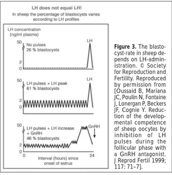
Investigations in sheep also demonstrated that a normal, pulsatile LH pattern during the follicular phase is neces-sary for the optimal development of the oocyte (Fig. 3).
376
J. REPRODUKTIONSMED. ENDOKRINOL. 6/2006In addition, LH stimulates the production of IL-1β inside
the granulosa cells. This leads to inhibition of E2
produc-tion of about 20 % [20]. The combinaproduc-tion of FSH and hCG has an additive effect on the secretion of a meiosis-activating substance which is produced inside the cumu-lus cells of the OCC [21]. The substitution of hCG by LH also stimulates the meiotic maturation of oocytes [22]. In patients who did not react sufficiently to FSH stimulation during the early follicular phase, it was found that mini-mal dosages of LH in the late follicular phase resulted in the development of mature oocytes and the attainment of good fertilisation rates [23]. FSH alone promotes follicu-lar development; however, estradiol synthesis is raised significantly by the combination of FSH and LH [24]. Also, in cycles down-regulated by GnRh agonists or an-tagonists, estradiol synthesis and the development of the endometrium can be enhanced by supplementing LH during the late follicular phase [25].
The LH peak at mid cycle is essential for the withdrawal of corona radiata cells from the zona pellucida. Further-more, this peak induces the final stages of oocyte matura-tion and leads to the transformamatura-tion of granulosa cells into luteinising cells. Women suffering from a hypogo-nadotrophic hypogonadism need some support with LH to avoid an increased rate of miscarriages. Westergaard et al [26] were able to demonstrate that down regulation with GnRH agonists or antagonists is able to create such a hypogonadotrophic situation. Therefore, a physiological amount of LH is of importance, not only for optimal maturation of follicles and oocytes, but also for the luteal phase.
In conclusion, FSH as well as LH are important for folli-culogenesis and oogenesis. There must be a quantitative LH threshold for optimal oocyte development. Below a plasma concentration of < 1 IU/l LH, there is a decline in oocyte quality and pregnancy rate (Fig. 4).
During follicular puncture of stimulated ovaries, imma-ture oocytes may also be obtained [27]. In vitro matura-tion (IVM) of these oocytes, however, is only possible if these oocytes originate from antral follicles and if ovula-tion is not induced by hCG. IVM of oocytes from pre-an-tral follicles has only been accomplished so far in mice. Nevertheless, in view of the treatment of young female cancer patients, cryopreservation and IVM of all stages of follicular development is becoming increasingly impor-tant. Canipari [28] showed that many factors in culture media, such as EGF, bFGF, TGFβ, IGF, GnRH, GRF, GH,
PACAP, VIP and forskolin, are important for achieving successful IVM.
During IVM, LH is the physiological signal which initiates meiosis and activates all processes leading to a fertilis-able oocyte. Vanderhyden et al [29] demonstrated that steroid production in granulosa cells is controlled, to a great degree, by the oocyte. It produces a progesterone inhibitor which prevents the production or storage of pro-gesterone by granulosa cells. This means that the devel-opment of follicles does not only depend on their initial maturity, but also on the hormonal milieu and their vas-cularisation.
Clinical Impact of the Dialogue
between Oocyte and Cumulus Cells
In an open, randomised, multi-national clinical study, two different stimulation regimes (hMG-HP versus RecFSH) were compared in relation to pregnancy rate in 727 patients [30] (Fig. 5). The study concluded that there is no significant difference with reference to pregnancy rate. Other studies have also shown that there are no dif-ferences in pregnancy rate after using one of the two stimulation regimes (Fig. 6, Tab. 1).A split evaluation of IVF and ICSI patients independently showed no differences in the ICSI group. IVF patients, however, who were stimulated by hMG, had significantly higher pregnancy rates [31]. This discrepancy could not be explained by the different hormone profiles of the pa-tients (LH, hCG, and E2) (Tab. 2).
This subgroup analysis of the MFK study emphasises the following results [31]:
1. After ICSI, there were no significant differences in re-lation to pregnancy rate (10th week of pregnancy) in
patients treated either with FSH or hMG.
Figure 4. The importance of various parameters in the ovulation cas-cade. Reprinted with permission from [Chappel SC et al. Reevaluation of the roles of luteinizing hormone and follicle-stimulating hormone in
377
J. REPRODUKTIONSMED. ENDOKRINOL. 6/2006
2. In IVF patients stimulated with hMG, a higher preg-nancy rate (10th week of pregnancy) could be achieved
compared to patients stimulated with FSH.
3. Compared to the FSH group, hMG-stimulated patients showed higher elevated hCG values on the 6th day of
stimulation.
4. These results raise the question of whether the LH ac-tivity of hMG plays a fundamental role in relation to pregnancy rate and baby take-home rate.
The fact that cumulus cells are in close contact with the oocyte by virtue of gap junctions could be evidence of
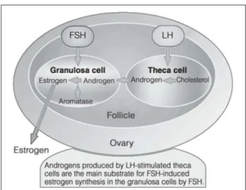
their importance in the course of oocyte maturation [32, 33]. In addition, the two-cell theory of theca granulosa cells has much to say about the relevance of LH for oocyte development (Fig. 7).
In theca interna cells, LH supports androgen production. After diffusion into granulosa cells, androgens are aroma-tised into estradiol by FSH. This production of steroid hor-mones and their metabolising under the influence of LH and FSH plays an important role during oocyte matura-tion. The extent to which LH supplementation during hor-monal stimulation will produce better pregnancy rates is something which has to be proven in future studies using a larger patient sample. Furthermore, it has to be taken into account that two different modes of fertilisation are being compared. This raises the question of whether, dur-ing IVF, cumulus cells have an identical impact on the oocyte as with in vivo fertilisation. Quite big differences between in vivo and in vitro have already been detected in a number of mammals [34]. The presumption that, af-ter in vitro fertilisation, cumulus cells succeed in setting up a microenvironment to assist embryonic development was strengthened by the work of Talbot et al [10]. It has been demonstrated that the removal of these cells leads to a reduction in or a total loss of sperm penetration due to zona hardening [35].
In summary, it should be concluded that the interaction between the oocyte and its surrounding cumulus cells is of biological importance. Therefore, a change in our way of thinking about the processes occurring during the ferti-lisation cascade is obviously useful and necessary for our daily routine in assisted reproduction technology.
References:
1. Eppig JJ. Oocyte control of ovarian follicular development and function in mammals. Reproduction 2001; 122: 829–38. 2. Webb RJ, Bains H, Cruttwell C, Carroll J. Gap-junctional
com-munication in mouse cumulus-oocyte complexes: implications for the mechanism of meiotic maturation. Reproduction 2002; 123: 41–52.
3. Hegele-Hartung C, Kuhnke J, Lessl M, Gröndahl C, Ottesen JL, Beier HM, Eisner S, Eichenlaub-Ritter U. Nuclear and cytoplasmic maturation of mouse oocytes after treatment with synthetic meio-sis-activating sterol in vitor. Biol Reprod 1999; 61: 1362–72. 4. Hegele-Hartung C, Grützner M, Lessl M, Gröndahl C, Ottesen JL,
Brännström M. Activation of meiotic maturation in rat oocytes after treatment with follicular fluid meiosis-activating sterol in vitro and ex vivo. Biol Reprod 2001; 64: 418–24.
Table 2. Pregnancy rates after IVF- or ICSI-treatment via hmg_HP or rFSH-stimulation. Reprinted from [31].
IVF ICSI
HP-hMG rFSH valuep HP-hMG rFSH valuep
Number of
patients 121 112 –– 237 221 –– Clinical
pregnancy 42 (35 %) 22 (20 %) 0.009 56 (24 %) 55 (25 %) n.s. Ongoing
pregnancy 38 (31 %) 22 (20 %) 0.037 49 (21 %) 50 (23 %) n.s. Implantation
rate 21 % 15 % 0.054 12 % 12 % n.s.
Table 1. Comparison of efficacy of hMG vs. rFSH referring to clinical pregnancy rate after ART-treatment. © Cochrane Collaboration, repro-duced with permission from [Van Wely M, Westergaard LG, Bossuyt PMM, Van der Veen F. Human menopausal gonadotropin versus re-combinant follicle stimulation hormone for ovarian stimulation in assisted reproductive cycles. Cochrane Database of Systematic Reviews 2003, Issue 1. Art. No.: CD003973. DOI: 10.1002/14651858.CD003973. Date of Most Recent Substantive Amendment: 30 August 2002]
Clinical pregnancy rates
n % ICSI hMG rFSH
Gordon 2001 68 0 38 % 28 % Westergaard 2001 379 25 40 % 34 % EISG 2002 727 64 26 % 22 % Ng 2001 40 100 25 % 20 %
Figure 7: Two-cell-theory. Importance of FSH and LH for granulosa-and theca cells. (Source: Ferring Pharmaceuticals)
378
J. REPRODUKTIONSMED. ENDOKRINOL. 6/20065. Herrler A, Beier HM. Neue molekulare und funktionelle Aspekte der Zona pellucida während der frühembryonalen Entwicklung. Reproduktionsmedizin 1999; 15: 268–75.
6. Hunter RH. Sperm:egg ratios and putative molecular signals to modulate gamete interactions in polytocous mammals. Mol Reprod Dev 1993; 35: 324–7.
7. van Soom A, Tanghe S, De Pauw I, Maes D, de Kruif A. Function of the cumulus oophorus before and during mammalian fertilization. Reprod Domest Anim 2002; 37: 144–51.
8. Bedford JM, Mori T, Oda S. The unusual state of the cumulus oophorus and of sperm behaviour within it, in the musk shrew, Suncus murinus. J Reprod Fertil 1997; 110: 127–34.
9. Ito M, Smith TT, Yanagimachi R. Effect of ovulation on sperm trans-port in the hamster oviduct. J Reprod Fertil 1991; 93: 157–63. 10. Talbot P, Shur BD, Myles DG. Cell adhesion and fertilization: steps
in oocyte transport, zona pellucida interactions, and sperm-egg fusion. Biol Reprod 2003; 68: 1–9.
11. Host E, Mikkelsen AL, Lindenberg S, Smidt-Jensen S. Apoptosis in hu-man cumulus cells in relation to maturation stage and cleavage of the corresponding oocyte. Acta Obstet Gynecol Scand 2000; 79: 936–40. 12. Alisch A, Ruping K, Koster F, Schopper B, Baum M, Finas D, Felber-baum R, Dor S, Al-Hasani S, Diedrich K. Cumulus cell apoptosis as a predictor for oocyte quality in artificial reproduction technique. Zentralbl Gynakol 2003; 125: 452–7.
13. Al Hasani S, Abu-Hassan D, Köster F, Schöppe B. Comet assay of cumulus cells and spermatozoa DNA status and relationship to oocyte fertilization and embryo quality following ICSI. 19th ESHRE
Annual Meeting 29.6.–02.07.2003, Madrid, Spain.
14. Clavero A, Castilla JA, Nunez AI, Garcia-Pena ML, Maldonado V, Fontes J, Mendoza N, Martinez L. Apoptosis in human granulosa cells after induction of ovulation in women participating in an in-tracytoplasmic sperm injection program. Eur J Obstet Gynecol Reprod Biol 2003; 110: 181–5.
15. Mikkelsen AL, Host E, Lindenberg S. Incidence of apoptosis in granulosa cells from immature human follicles. Reproduction 2001; 122: 481–6.
16. Te Velde ER, Pearson PL. The variability of female reproductive age-ing. Hum Reprod Update 2002; 8: 141–54.
17. van Rooij IA, Broekmans FJ, te Velde ER, Fauser BC, Bancsi LF, de Jong FH, Themmen AP. Serum anti-Mullerian hormone levels: a novel measure of ovarian reserve. Hum Reprod 2002; 17: 3065–71. 18. Cohen-Bacrie P, Hazout A. Follicle stimulating hormone and
in-hibin B in the woman. Gynecol Obstet Fertil 2001; 29: 320–3. 19. Wu J, Nayudu PL, Michelmann HW. Luteinizing hormone has a
stage-limited effect on preantral follicle development in vitro. Biol Reprod 2000; 63: 320–7.
20. Chen HF, Shew JY, Chao KH, Chang LJ, Ho HN, Yang YS. Luteiniz-ing hormone up-regulates the expression of interleukin-1 beta mRNA in human granulosa-luteal cells. Am J Reprod Immunol 2000; 43: 125–33.
21. Byskov AG, Yding Andersen C, Hossaini A, Guoliang X. Cumulus cells of oocyte-cumulus complexes secrete a meiosis-activating
substance when stimulated with FSH. Mol Reprod Dev 1997; 46: 296–305.
22. Zelinski-Wooten MB, Hutchison JS, Chandrasekher YA, Wolf DP, Stouffler RL. Administration of human luteinizing hormone (hLH) to macaques after follicular development: further titration of LH surge requirements for ovulatory changes in primate follicles. J Clin Endocrinol Metab 1992; 75: 502–7.
23. Anderiesz C, Ferraretti A, Magli C, Fiorentino A, Fortini D, Gia-naroli L, Jones GM, Trounson AO. Effect of recombinant human gonadotrophins on human, bovine and murine oocyte meiosis, fer-tilization and embryonic development in vitro. Hum Reprod 2000; 15: 1140–8.
24. Baird DT, McNeilly AS. Gonadotrophic control of follicular devel-opment and function during the oestrous cycle of the ewe. J Reprod Fertil Suppl 1981; 30: 119–33.
25. Shoham Z. Treatment of female infertility with recombinant human luteinizing hormone: is there a benefit over other available drugs? Expert Opin Pharmacother 2003; 4: 1985–94.
26. Westergaard LG, Erb K, Laursen SB, Rex S, Rasmussen PE. Human menopausal gonadotropin versus recombinant follicle-stimulating hormone in normogonadotropic women down-regulated with a gonadotropin-releasing hormone agonist who were undergoing in vitro fertilization and intracytoplasmic sperm injection: a prospec-tive randomized study. Fertil Steril 2001; 76: 543–9.
27. Smitz J, Cortvrindt RG. The earliest stages of folliculogenesis in vitro. Reproduction 2002; 123: 185–202.
28. Canipari R. Oocyte-granulosa cell interactions. Hum Reprod Up-date 2000; 6: 279–89.
29. Vanderhyden BC, Telfer EE, Eppig JJ. Mouse oocytes promote pro-liferation of granulosa cells from preantral and antral follicles in vitro. Biol Reprod 1992, 46: 1196–204.
30. The European and Israeli Study Group on Highly Purified Meno-tropin versus Recombinant Follicle-Stimulating Hormone. Efficacy and safety of highly purified menotropin versus recombinant fol-licle-stimulating hormone in in vitro fertilization/intracytoplasmic sperm injection cycles: a randomized; comparative trial. Fertil Steril 2002; 78: 520–8.
31. Platteau P, Smitz J, Albano C, Sorensen P, Arce JC, Devroey P. Exo-genous luteinizing hormone activity may influence the treatment outcome in in vitro fertilization but not in intracytoplasmic sperm injection cycles. Fertil Steril 2004; 81: 1401–4.
32. Kidder GM, Mhawi AA. Gap junctions and ovarian folliculo-genesis. Reproduction 2002; 123: 613–20.
33. Matzuk MM, Burns KH, Viveiros MM, Eppig JJ. Intercellular com-munication in the mammalian ovary: oocytes carry the conversa-tion. Science 2002; 296: 2178–80.
34. Catt JW, Rhodes SL. Comparative intracytoplasmic sperm injection (ICSI) in human and domestic species. Reprod Fertil Dev 1995; 7: 161–6.
Besuchen Sie unsere Rubrik
聺
Medizintechnik-Produkte
C200 und C60 CO2-Inkubatoren
Labotect GmbH
CTE2200-Einfriersystem MTG Medical Technology Vertriebs-GmbH
OCTAX Ferti Proof-Konzept MTG Medical Technology Vertriebs-GmbH
Hot Plate 062 und Hot Plate A3 Labotect GmbH