Identification and Characterization of
Bifunctional Proline Racemase/
Hydroxyproline Epimerase from Archaea:
Discrimination of Substrates and Molecular
Evolution
Seiya Watanabe*, Yoshiaki Tanimoto, Hisashi Nishiwaki, Yasuo Watanabe
Faculty of Agriculture, Ehime University, Matsuyama, Ehime, 790–8566, Japan
Abstract
Proline racemase (ProR) is a member of the pyridoxal 5’-phosphate-independent racemase family, and is involved in the Stickland reaction (fermentation) in certain clostridia as well as the mechanisms underlying the escape of parasites from host immunity in eukaryotic Trypa-nosoma. Hydroxyproline epimerase (HypE), which is in the same protein family as ProR, cat-alyzes the first step of thetrans-4-hydroxy-L-proline metabolism of bacteria. Their substrate specificities were previously considered to be very strict, in spite of similarities in their struc-tures and catalytic mechanisms, and no racemase/epimerase from the ProR superfamily has been found in archaea. We here characterized the ProR-like protein (OCC_00372) from the hyperthermophilic archaeon,Thermococcus litoralis(TlProR). This protein could reversibly catalyze not only the racemization of proline, but also the epimerization of 4-hydroxyproline and 3-hydroxyproline with similar kinetic constants. Among the four (putative) ligand binding sites, one amino acid substitution was detected between TlProR (tryptophan at the position of 241) and natural ProR (phenylalanine). The W241F mutant showed a significant preference for proline over hydroxyproline, suggesting that this (hydrophobic and bulky) tryptophan resi-due played an importance role in the recognition of hydroxyproline (more hydrophilic and bulky than proline), and substrate specificity for hydroxyproline was evolutionarily acquired separately between natural HypE and ProR. A phylogenetic analysis indicated that such unique broad substrate specificity was derived from an ancestral enzyme of this superfamily.
Introduction
L-Proline can serve as a complete source of carbon and energy or of nitrogen for organisms. The metabolism of L-proline is generally initiated through its oxidation by L-proline dehydro-genase (EC 1.5.99.8; L-ProDH) to formΔ1-pyrroline-5-carboxylate (Pyr5C) [1] (Fig. 1A). OPEN ACCESS
Citation:Watanabe S, Tanimoto Y, Nishiwaki H, Watanabe Y (2015) Identification and
Characterization of Bifunctional Proline Racemase/ Hydroxyproline Epimerase from Archaea:
Discrimination of Substrates and Molecular Evolution. PLoS ONE 10(3): e0120349. doi:10.1371/journal. pone.0120349
Academic Editor:Paul Taylor, University of Edinburgh, UNITED KINGDOM
Received:October 31, 2014
Accepted:January 21, 2015
Published:March 18, 2015
Copyright:© 2015 Watanabe et al. This is an open access article distributed under the terms of the
Creative Commons Attribution License, which permits unrestricted use, distribution, and reproduction in any medium, provided the original author and source are credited.
Data Availability Statement:All relevant data are within the paper and its Supporting Information files.
Funding:The authors have no support or funding to report.
Following its spontaneous hydrolysis, the L-glutamateγ-semialdehyde produced is then oxi-dized to L-glutamate by Pyr5C dehydrogenase (EC 1.2.1.88). L-Proline is alternatively metabo-lized to 5-aminopentanoate through two enzymatic steps. L-Proline racemase (EC 5.1.1.4; ProR) first catalyzes the racemization of L-proline to D-proline, followed by reductive cleavage to yield 5-aminopentanoate by D-proline reductase (EC 1.21.4.1). This pathway is only opera-tive in certain clostridia includingClostridium sticklandii[2] andClostridium difficile[3], clini-cally significant nosocomial pathogens, andTrypanosomaspecies includingTrypanosoma cruzi[4,5] andTrypanosoma vivax[6], the causative agents of Chagas disease and animal try-panosomiasis, respectively. In the former, the pathway is involved in the so-called“Stickland reaction (fermentation)”, whereas ProR in the latter has been implicated in the mechanisms underlying the escape of parasites from host immunity as a B-cell mitogen.
Hydroxyproline has been detected in certain proteins, especially collagen, as well as some peptide antibiotics. In mammalian systems [1], the L-proline residue in procollagen is post-translationally hydroxylated totrans-4-hydroxy-L-proline (T4LHyp) ortrans -3-hydroxy-L-proline (T3LHyp). Among the several stereoisomers of hydroxy-3-hydroxy-L-proline including T4LHyp and T3LHyp (Fig. 2), T4LHyp is the most common in nature. In contrast to mammalians [1], some bacteria have been shown to metabolize T4LHyp toα-ketoglutarate via three intermediates through four enzymatic steps [7,8]. Of these, hydroxyproline 2-epimerase (HypE; EC 5.1.1.8) first catalyzes the isomerization of T4LHyp tocis-4-hydroxy-D-proline (C4DHyp), which is then converted toΔ1-pyrroline-4-hydroxy-2-carboxylate (Pyr4H2C) by C4DHyp dehydroge-nase (D-HypDH). In the metabolism of T3LHyp (Fig. 1A), T3LHyp dehydratase (EC 4.2.1.77; T3LHypD) initially catalyzes the dehydration of T3LHyp toΔ1-pyrroline-2-carboxylate (Pyr2C) via a putativeΔ2-pyrroline-2-carboxylate intermediate [9]. The Pyr2C is then con-verted by NAD(P)H-dependent Pyr2C reductase (EC 1.5.1.1) to yield L-proline, which is me-tabolized through the general degradation of L-proline described above. This pathway is most commonly found in mammals [9], bacteria [10] and archaea [11].
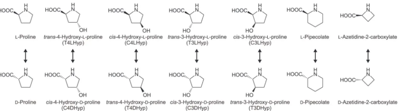
Racemase enzymes catalyze the deprotonation/reprotonation of the chiral carbon (Cα) of both amino acid enantiomers, resulting in the stereoinversion of chiral centers. ProR is a mem-ber of the pyridoxal 5’-phosphate (PLP)-independent racemase family along with HypE and T3LHypD (although the reaction involving the latter is not epimerization) [9,12,13]. These enzymes have been classified into three types based on the two specific residues at the active sites [9]: the Cys-Cys type (ProR and HypE), Cys-Thr type (T3LHypD), and Cys or Ser-Thr type (function unknown). In the cases of ProR and HypE, catalysis is based on the same 1,1-proton transfer mechanism using two general acidic/basic cysteine residues located on Fig 2. Library of proline derivatives.TlProR can enable the reversible racemization and epimerization of all proline derivatives (arrows).
opposite faces of the active site [12]. On the other hand, their substrate specificities were previ-ously considered to be very strict: proline for ProR [13] and T4LHyp andcis -4-hydroxy-L-pro-line (C4LHyp) for HypE [13,14]. Although the catalytic mechanism of T3LHypD currently remains unclear, dehydration may start by the abstraction of a proton from the Cαof the sub-strate by an active site cysteine residue as a general basic catalyst [9]. This enzyme can also only utilize T3LHyp as a substrate.
As described above, no racemase/epimerase from the ProR superfamily had been found in archaea. We here identified and characterized a ProR-like protein with a pair of Cys-Cys as (putative) active sites from the hyperthermophilic marine archaeon,Thermococcus litoralis
[15], which differed from the previously isolated T3LHypD enzyme [11]. This protein could re-versibly catalyze not only the racemization of proline, but also the epimerization of 4-hydroxy-proline and 3-hydroxy4-hydroxy-proline with similar kinetic constants. Site-directed mutagenesis revealed several important amino acid residue(s) responsible for discriminating between pro-line and hydroxypropro-line. A phylogenetic analysis indicated that such unique broad substrate specificity was derived from an ancestral enzyme of this superfamily. To the best of our knowl-edge, this is the first study on the bifunctional enzyme of ProR and HypE from archaea.
Materials and Methods
Materials
T3LHyp was purchased from Kanto Chemical (Tokyo, Japan). C4LHyp, L-pipecolate, and D-pipecolate were obtained from Tokyo Chemical Industry (Tokyo, Japan). T4DHyp and C3LHyp were from Sigma Aldrich (USA). T4LHyp, C4DHyp, L-proline, and D-proline were from Wako Pure Chemical Industries (Osaka, Japan).
General procedures
Basic recombinant DNA techniques were performed as described by Sambrooket al. [16]. Ar-chaeal genomic DNA was prepared using a DNeasy Tissue Kit (Qiagen). PCR was carried out using a GeneAmp PCR System 2700 (Applied Biosystems) for 30 cycles in 50μl of reaction
mixture containing 1 U of KOD FX DNA polymerase (TOYOBO), appropriate primers (15 pmol), and template DNA under the following conditions: denaturation at 98°C for 10 s, an-nealing at 50°C for 30 s, and extension at 68°C for time periods calculated at an extension rate of 1 kbp∙min−1. DNA sequencing was carried out using the BigDye Cycle Sequencing Kit ver.3.1 (Applied Biosystems) and appropriate primers with the Genetic Analyzer 3130 (Ap-plied Biosystems). High-pressure liquid chromatography (HPLC) was performed using an Agi-lent 1120 Compact LC system (TOSOH). Protein concentrations were determined by the method of Lowry et al. [17] with bovine serum albumin as the standard. SDS-PAGE was per-formed as previously described by Laemmli [18]. Western blot analysis was carried out using an ECL Western Blotting Analysis System (GE Healthcare) and RGS∙His HRP antibody (horse-radish peroxidase-fused mouse monoclonal antibody against Arg-Gly-Ser-His6in the
N-termi-nal additioN-termi-nal peptide of the expressed recombinant proteins (Qiagen))
Plasmid construction for expression of recombinant proteins
The primer sequences used in this study are shown inS1 Table. In this study, the prefixes Tl (T.litoralis), Fa (Ferroplasma acidiphilum), Hj (Haloarcula japonica), Cd (C.difficile), and Ab (Azospirillum brasilense) were added to gene symbols or protein designations where necessary for clarity. TheTlProR(GenBank accession number OCC_00372),FaProR
(BAN78527) were amplified by PCR using primers containing appropriate restriction enzyme sites at the 5’- and 3’-ends and the genomic DNA ofT.litoralisDSM 5473,F.acidiphilumDSM 12658,H.japonicaDSM 6131,C.difficile630, orA.brasilenseATCC 29145 as a template, re-spectively. Each amplified DNA fragment was introduced into BamHI-SalI sites (for the
TlProR,FaProR, andCdProRgenes), BamHI-PstI (for theHjProRgene), or BamHI-HindIII (for theAbHypEgene) in pETDuet-1 (Novagen), a plasmid vector used to confer an N-termi-nal His6tag on expressed proteins, to obtain pET/TlProRWT, pET/FaProRWT, pET/HjProR,
pET/CdProR, and pET/AbHypEWT, respectively.
In the present study, we usedF.acidiphilumDSM 12658 instead ofFerroplasma acidarma-nusfer1 as a target microorganism, for which there was 99% identity in the 16S rRNA se-quence. We successfully amplified theFaProRgene by genomic PCR using oligonucleotide primers designed from theF.acidarmanusfer1 genome sequence. Unless otherwise noted, Fer-roplasmasp. hereafter indicates strain DSM 12658.
Expression and purification of His
6-tagged recombinant proteins
Escherichia coliBL21-CodonPlus(DE3)-RIL (Novagen) harboring the constructed pETDuet-1 plasmid was grown at 37°C to a turbidity of 0.6 at 600 nm in Super broth medium (pH 7.0, 12 g tryptone, 24 g yeast extract, 5 ml glycerol, 3.81 g KH2PO4, and 12.5 g K2HPO4per liter)
con-taining 50 mg/liter ampicillin. After the addition of 1 mM isopropyl-β -D-thiogalactopyrano-side (IPTG), the culture was grown for a further 6 h to induce the expression of the His6
-tagged protein. The grown cells were harvested by centrifugation at 30,000 ×gfor 20 min, sus-pended in Buffer A (50 mM sodium phosphate buffer (pH 8.0) containing 300 mM NaCl and 10 mM imidazole), disrupted by sonication for 20 min at appropriate intervals on ice using Ultra Sonic Disruptor UD-211 (TOMY SEIKO Co., Ltd, Tokyo, Japan), and then centrifuged at 108,000 ×gfor 20 min at 4°C. The supernatant was loaded onto a Ni-NTA Superflow umn (Qiagen) equilibrated with Buffer A linked to the BioAssist eZ system (TOSOH). The col-umn was washed with Buffer B (50 mM sodium phosphate buffer (pH 8.0) containing 300 mM NaCl, 10% (v/v) glycerol, and 50 mM imidazole). The enzymes were then eluted with Buffer C (pH 8.0, Buffer B containing 250 mM imidazole instead of 50 mM imidazole), concentrated by ultrafiltration with Centriplus YM-30 (Millipore), dialyzed against 50 mM Tris-HCl buffer (pH 8.0) containing 50% (v/v) glycerol, and stored at−35°C until use.
The native molecular mass of recombinant proteins was estimated by gel filtration, which was carried out using a HPLC system at a flow rate of 1 ml/min. The purified enzyme was load-ed onto a TSKgel G3000SWXL column (TOSOH) equilibratload-ed with 50 mM Tris-HCl buffer (pH 8.0). A high molecular weight gel filtration calibration kit (GE Healthcare) was used for molecular markers.
Enzyme assay by HPLC
Purified protein was added to 50 mM Tris-HCl buffer (pH 8.0) (1 ml) containing 10 mM sub-strate, and incubated at 50°C (for TlProR and HjProR) or 30°C (FaProR, CdProR, and AbHypE). After varying the incubation times, the enzyme reaction was stopped by rapidly incubating at−80°C. To this solution, 25μl each of 10 mM 4-fluoro-7-nitrobenzofurazan
(NBD-F) in ethanol and 100 mM borate buffer (pH 8.0) was then added and incubated at 60°C for 1 min. A total of 1.15 ml of 5 mM HCl was finally added to the reaction mixture, followed by filtration through a 0.22-μm filter (Millipore). HPLC analysis was carried out using an
conversion of L$D isomers was observed even after being incubated for 12 h, and the
differ-ence in background turnover between 30°C and 50°C was negligible. This method was used to estimate the effects of substrate specificity and inhibition by pyrrole-2-carboxylate (PYC) on activity.
Spectrophotometric enzyme assay
Racemase/epimerase activity for proline or hydroxyproline(s) was spectrophotometrically as-sayed at 50°C by monitoring the reduction rate of an artificial electron acceptor in the coupling system with dehydrogenase for proline or hydroxyproline, using a Shimadzu UV-1800 spectro-photometer (Shimadzu GLC Ltd., Tokyo, Japan).
Activity toward L-proline and T3LHyp. The standard reaction mixture contained 0.05 mM 2,6-dichloroindophenol (Cl2Ind) and purified D-ProDH fromPyrobaculum islandicum(20μg)
[19] in 50 mM Tris-HCl buffer (pH 8.0).
Activity toward D-proline. The standard reaction mixture contained 0.05 mM Cl2Ind and purified L-ProDH fromAeropyrum pernix(20μg) [20] in 50 mM Tris-HCl buffer (pH 8.0).
Activity toward T4LHyp. The standard reaction mixture contained 0.25 mMp -iodonitrote-trazolium violet (INT), 0.06 mM phenazine methosulfate (PMS), and purified D-HypDH from
A.brasilense(20μg) [21] in 50 mM Tris-HCl buffer (pH 8.0).
All reactions were initiated by the addition of 100 mM substrate (100μl) with a final
reac-tion volume of 1 ml. The millimolar absorpreac-tion coefficients (ε) for Cl2Ind and INT were 19.1 mM−1
∙cm−1at 600 nm and 15.0 mM−1∙cm−1at 490 nm, respectively. The detectable limit of specific activity was*0.001 unit/mg protein, due to non-enzymatic absorbance change by
Cl2Ind and PMS/INT. This method was used to determine the kinetic parameters,Kmandkcat,
which were calculated by a Lineweaver-Burk plot.
Identification of reaction products
To remove glycerol in the stored buffer, purified TlProR (10μg) was dialyzed at 4°C overnight in
50 mM potassium phosphate buffer (pH 7.0), lyophilized, and solved in D2O (600μl) containing
20 mM T4LHyp or T3LHyp. NMR spectra were recorded at 25°C on a JEOL JNM-EC400 NMR spectrometer (JEOL Ltd., Tokyo, Japan) operating at 400 MHz. 2,2-Dimethyl-2-silapentane-5-sulfonate was used as an internal standard.1H NMR spectra of T4LHyp and T3LHyp (400 MHz, D2O) were as follows: T4LHyp, 4.41 (1H, t,J= 4 Hz), 4.33 (1H, dd,J1= 10 Hz,J2= 8 Hz), 3.47
(1H, dd,J1= 12 Hz,J2= 4 Hz), 3.35 (1H, ddd,J1= 12 Hz,J2=J3= 2 Hz), 2.42 (1H, dddd,J1= 14 Hz,J2= 8 Hz,J3=J4= 2 Hz), and 2.15 (1H, ddd,J1= 14 Hz,J2= 10 Hz,J3= 4 Hz); C4DHyp, 4.57 (1H, m), 4.21 (1H, dd,J1= 11 Hz,J2= 4 Hz), 3.36 (1H, dd,J1= 12 Hz,J2= 4 Hz), 2.50 (1H, ddd,J1= 14 Hz,J2= 10 Hz,J3= 4 Hz), and 2.25 (1H, m); T3LHyp, 4.68 (1H, m), 4.07 (1H, s), 3.60 (1H, m), 3.50 (1H, m), and 2.04 (2H, m); C3DHyp, 4.71 (1H, m), 4.14 (1H, d,J= 4 Hz), 3.56 (1H, m), 3.48 (1H, m), and 2.18 (2H, m).
Site-directed mutagenesis
A mutation was introduced into theTlProR,FaProR, orAbHypEgenes by sequential steps of PCR [22] using sense and antisense primers (S1 Table) and pET/TlProRWT(for TlProRF62S
(CTG!AGG), TlProRF62V(CTG!GTT), TlProRL221H(CTG!GAG), TlProRF62S/L221H,
TlProRW241F(GTG!GAC), TlProRW241C(CTG!TGT), and TlProRW241Ymutants
(CTG!TAT)), pET/FaProRWT(for FaProRF240Wmutant (GCC!AAG)), or
pET/AbHy-pEWT(for AbHypEC226Fmutant (GTG!TTC)) as a template (the codons used for each
subsequent sequencing in both directions. Mutant proteins were expressed and purified by the same procedures as those for the wild-type enzyme.
Amino acid sequence alignment and phylogenetic analysis
Protein sequences were analyzed using the Protein-BLAST and Clustal W program distributed by DDBJ (DNA Data Bank of Japan) (www.ddbj.nig.ac.jp). The phylogenetic tree was pro-duced using the TreeView 1.6.1. program.
Results
Putative ProR or HypE, but not the T3LHypD gene from archaea
We previously reported that OCC_00387 and OCC_00362(7) fromT.litoralisencoded T3LHypD and Pyr2C reductase are involved in (putative) T3LHyp metabolism, respectively [11] (Fig. 1A). These genes were clustered on the genome together with several other hypothet-ical genes (Fig. 1B), among which OCC_00357 (α-subunit) and OCC_00352 (β) (genes) were homologous toα4β4-type L-ProDH fromPyrococcus horikishii(PH1363 and PH1364 with 69 and 80% identities, respectively) [23], while OCC_00347 (α), OCC_00342 (γ), OCC_00337 (δ), and OCC_00332 (β) (genes) were similar toαβγδ-type L-ProDH fromThermococcus profun-dus(pdhA, pdhF, pdhX, and pdhB with 81, 72, 72, and 64% identities, respectively) [24]. Since T3Lhyp is metabolized to L-proline via a Pyr2C intermediate [9–11], the gene cluster may be related to both the (general) metabolism of L-proline and T3LHyp. Another ProR-like gene (OCC_00372) was also detected in this gene cluster, and the putative amino acid sequence con-tained two cysteine residues at the active sites (Cys88-Cys251) specific for ProR or HypE, but not T3LHypD (see below). Furthermore, by a homology search using the Protein-BLAST pro-gram, some archaea includingF.acidarmanus(F.acidiphilum) andH.japonicawere found to possess the homologous gene (see below in detail). Therefore, in the present study, we selected three ProR-like genes fromT.litoralis,F.acidiphilum, andH.japonicaas target genes (referred to as TlProR, FaProR, and HjProR genes, respectively). Furthermore, ProR fromC.difficile
[13] and HypE fromA.brasilense[21] were used as controls (referred to as CdProR and AbHypE, respectively).
Preparation of recombinant His
6-tag proteins
After cloning all target genes into the vector pETDuet-1, the recombinant enzymes were suc-cessfully expressed inE.colicells as His6-tagged enzymes, and purified to homogeneity using a
nickel-chelating affinity column (Fig. 1C). Additional amino acid residues including the His6
-tag at the N-terminal was confirmed by western blot analysis with an anti-His6-tag antibody
(Fig. 1D). The apparent molecular masses of TlProR, FaProR, HjProR, CdProR, and AbHypE, as estimated by SDS-PAGE, were 40 (38.1), 40 (38.5), 42 (35.8), 40 (37.7) and 37 (34.1) kDa (values in parentheses indicate the calculated molecular mass of the enzyme with His6-tag),
while those estimated by analytic gel filtration were*80 kDa, respectively (data not shown).
Therefore, all these enzymes appeared to be dimeric.
Substrate specificity of TlProR
(putative) C3DHyp, 8.5; C3LHyp, 9.8; (putative) T3DHyp, 7.3; L-pipecolate, 15.4; D-pipeco-late, 14.7; L-azetidine-2-carboxyD-pipeco-late, 9.1; (putative) D-azetidine-2-carboxyD-pipeco-late, 8.6.
TlProR could catalyze the conversion of not only L-proline, but also 4-hydroxy-L-proline (T4LHyp (63) and C4LHyp (69)) and 3-hydroxy-L-proline (T3LHyp (73) and C3LHyp (47)) (values in parentheses indicate specific activity as a percentage of L-proline) (Fig. 3). The reac-tion was completely bi-direcreac-tional, and the reverse reacreac-tions were also similar: D-proline (130), C4DHyp (160), and T4DHyp (97) (values in parentheses indicate specific activity as a
Fig 3. Substrate specificities of TlProR, FaProR, HjProR, CdProR, and AbHypE.The reaction mixture (1 ml) consisted of 10 mM of substrate and each purified enzyme as follows in Tris-HCl buffer (pH 8.0): TlProR, 1.3μg; FaProR, 100μg; HjProR, 570μg; CdProR, 3.6μg; AbHypE, 4.9μg. A2C is L-azetidine-2-carboxylate. Samples at the indicated times were analyzed by HPLC (means±S.D.,n= 3). Values on the bars of TlProR indicate specific activity (unit/mg protein), which are similar to those in Tables1and2.
percentage of L-enantiomer). Furthermore, using large amounts of the purified enzyme, we could also estimate specific activities for L-pipecolate (0.7), D-pipecolate (3.6), and L-azeti-dine-2-carboxylate (0.16), which are the six- and four-membered ring analogs of
proline, respectively.
Another previously reported difference in the properties of ProR and HypE is the profile of inhibition by pyrrole-2-carboxylate (PYC), a transition state analogue of proline [6,13,25]: ProR enzymes from trypanosomatids and clostridia were inhibited at even 0.01 and 1 mM, re-spectively, whereas HypE reactions were only affected by high amounts of PYC (*10 mM), as
described in CdProR and AbHypE (Fig. 4). In spite of the significant HypE activity, the IC50
value of TlProR (0.217 mM) was*28-fold higher than that of AbHypE (6.00 mM), confirming
dual activity toward proline and 4-hydroxyproline.
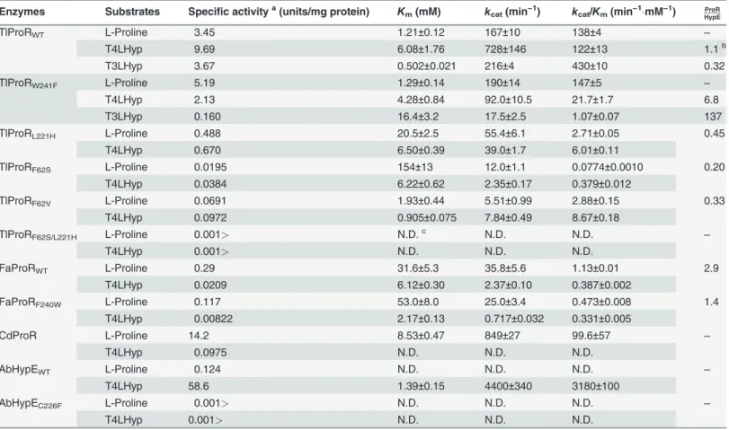
Kinetic analysis of TlProR
A more sensitive spectrophotometric assay method using proline or hydroxyproline dehydro-genase as a coupling enzyme was developed to estimate kinetic properties. The kinetic parame-ters of proline and hydroxyproline determined from the Lineweaver-Burk plot are shown in Tables1(L!D) and2(D!L). The catalytic efficiency (kcat/Km) values for L-proline and
T4LHyp were similar, whereas a preference for D-proline over C4DHyp (45-fold) was identi-fied and was caused by a 75-fold lowerKmfor D-proline. This may have been partially due to differences in theKmvalues of L-ProDH (0.28 mM) [20] and D-ProDH (4.2 mM) [19] as the coupling enzyme used. TlProR was assayed at 50°C, which was far from the optimum tempera-ture (90*100°C; data not shown). Although thekcat/Kmvalue for T4LHyp of TlProR was
27-fold lower than that for AbHypE, the specific activity of the former at 100°C, estimated by HPLC analysis, was similar to that of the latter: 29.1 and 58.6 unit/mg protein, respectively.
Zhao et al. [26] recently characterized 51 ProR-like proteins from bacteria. Of these, the
kcat/Kmvalue for T4LHyp of HypE fromPseudomonas putidawas 192 and 3430-fold higher than those for T3LHyp and L-proline, respectively, and was caused by a markedly higherkcat
value. Similar results were also obtained for other (putative) HypE proteins. In contrast, the ki-netic constant for T3LHyp of TlProR was similar to those of L-proline and T4LHyp. Thus, to Fig 4. Unique inhibition of archaeal ProR-like enzymes by pyrrole-2-carboxylate (PYC; inset).Reactions were performed for 30 min with the same conditions as those inFig. 3, except for the presence of several concentration of PYC. L-Proline (for TlProR, FaProR, HjProR, and CdProR) or T4LHyp (for AbHypE) was used as a substrate. Relative specific activity values were expressed as percentages of the values obtained in the absence of PYC (means± S.D.,n= 3). Data for the ProR ofT.cruzi(TcProR) are from Berneman et al. [25]. IC50values were calculated by curve fitting using ImageJ software (http:// rsb.info.nih.gov/ij/).
the best of our knowledge, this is the first example of a ProR-like protein being capable of uti-lizing not only proline, but also 4-hydroxyproline and 3-hydroxyproline as a substrate.
Identification of reaction products by TlProR
T4LHyp was incubated with TlProR in D2O at various incubation times.1H NMR spectra
showed the progressive loss of H1peaks, and additionally contained resonances associated with
C4DHyp, a potential product: an exchange of theα-proton with solvent deuterium (Fig. 5A). Similar phenomena were also observed when T3LHyp was used as a substrate instead of T4LHyp (Fig. 5B). These results were expected for a 1,1-proton transfer reaction that equili-brates the configurations at Cαof not only 4-hydroxyproline, but also 3-hydroxyproline (novel activity), as proposed previously [12].
Phylogenetic analysis of TlProR
As expected from the preliminary annotation, TlProR belongs to the ProR superfamily, which consists of four subfamilies: the archetype ProR (blue inFig. 6), HypE (light green), T3LHypD (orange), and function unknown protein (yellow). Only two putative proteins from the
Table 1. Kinetic parameters for L-proline, T4LHyp, and T3LHyp.
Enzymes Substrates Specific activitya(units/mg protein) K
m(mM) kcat(min−1) kcat/Km(min−1mM−1) HypEProR
TlProRWT L-Proline 3.45 1.21±0.12 167±10 138±4 ‒
T4LHyp 9.69 6.08±1.76 728±146 122±13 1.1b
T3LHyp 3.67 0.502±0.021 216±4 430±10 0.32
TlProRW241F L-Proline 5.19 1.29±0.14 190±14 147±5
‒
T4LHyp 2.13 4.28±0.84 92.0±10.5 21.7±1.7 6.8
T3LHyp 0.160 16.4±3.2 17.5±2.5 1.07±0.07 137
TlProRL221H L-Proline 0.488 20.5±2.5 55.4±6.1 2.71±0.05 0.45
T4LHyp 0.670 6.50±0.39 39.0±1.7 6.01±0.11
TlProRF62S L-Proline 0.0195 154±13 12.0±1.1 0.0774±0.0010 0.20
T4LHyp 0.0384 6.22±0.62 2.35±0.17 0.379±0.012
TlProRF62V L-Proline 0.0691 1.93±0.44 5.51±0.99 2.88±0.15 0.33
T4LHyp 0.0972 0.905±0.075 7.84±0.49 8.67±0.18
TlProRF62S/L221H L-Proline 0.001> N.D.c N.D. N.D. ‒
T4LHyp 0.001> N.D. N.D. N.D.
FaProRWT L-Proline 0.29 31.6±5.3 35.8±5.6 1.13±0.01 2.9
T4LHyp 0.0209 6.12±0.30 2.37±0.10 0.387±0.002
FaProRF240W L-Proline 0.117 53.0±8.0 25.0±3.4 0.473±0.008 1.4
T4LHyp 0.00822 2.17±0.13 0.717±0.032 0.331±0.005
CdProR L-Proline 14.2 8.53±0.47 849±27 99.6±57 ‒
T4LHyp 0.0975 N.D. N.D. N.D.
AbHypEWT L-Proline 0.124 N.D. N.D. N.D. ‒
T4LHyp 58.6 1.39±0.15 4400±340 3180±100
AbHypEC226F L-Proline 0.001> N.D. N.D. N.D. ‒
T4LHyp 0.001> N.D. N.D. N.D.
aUnder standard assay conditions in the“Materials and Methods”. b
Ratio of L-proline to T4LHyp or T3LHyp inkcat/Km. c
Not determined due to trace activity.
hyperthermophilic archaeonThermococcus sibiricus(TSIB_0633) andThermococcussp. ES1 (TES1_1056) showed>85% sequential identity (referred to as TlProR subfamily; red), and the corresponding genes formed a similar gene cluster toT.litoralis, together with (putative) L-ProDH and Pyr2C reductase genes (Fig. 1B). Although TlProR (subfamily) is not strongly re-lated to any of the subclasses of the other members (30*40% sequence identity), a
Protein-BLAST analysis revealed that TlProR was a close, but distinct subfamily of the ProR subfamily; the (putative) ligand binding sites were similar to those of ProR, but not HypE or T3LHypD, as described below. Furthermore, we found that two putative proteins (Cys-Cys type) from the hyperacidophilic archaeonF.acidarmanus(FaProR) andFerroplasmasp. type II were located between the TlProR and ProR subfamilies (purple inFig. 6). Several halophilic archaea includ-ingH.japonica(HjProR) (pink) also possessed similar types of proteins (genes), but formed a distinct subfamily to any other subfamily, and were often clustered with putative bacterial type (but not archaeal) L-ProDH (PutA) (Fig. 1B).
Characterization of FaProR and HjProR
To further estimate the evolutionary insight of archaeal ProR-like protein(s), FaProR and HjProR were characterized enzymatically using purified recombinant proteins (Fig. 1C). The racemization activity of proline was found to be significant in FaProR, although specific activity was*10-fold lower than that of TlProR (Fig. 3). On the other hand, thekcat/Kmvalue for
T4LHyp was*3-fold lower than that for L-proline, which was attributed to a 17-fold lower
kcatvalue, and we could not determine the kinetic parameters of the epimerization of C4DHyp Table 2. Kinetic parameters for D-proline and C4DHyp.
Enzymes Substrates Specific activitya(units/mg protein) K
m(mM) kcat(min−1) kcat/Km(min−1mM−1) ProRHypE
TlProRWT D-Proline 3.45 0.0919±0.006 133±2 1570±90 45.1b
C4DHyp 3.48 6.65±0.69 230±12 34.8±1.8
TlProRW241F D-Proline 4.87 0.194±0.020 191±5 990±82 158
C4DHyp 0.681 6.70±0.68 41.8±2.7 6.25±0.24
TlProRL221H D-Proline 0.299 2.54±0.46 17.3±2.7 6.84±0.19 4.3
C4DHyp 0.111 4.18±0.81 6.66±1.31 1.59±0.00
TlProRF62S D-Proline 0.0283 4.00±0.17 1.55±0.09 0.387±0.008 1.4
C4DHyp 0.0186 3.14±0.15 0.859±0.012 0.274±0.010
TlProRF62V D-Proline 0.0309 0.156±0.004 1.41±0.03 9.04±0.23 ‒
C4DHyp 0.00661 N.D. N.D. N.D.
TlProRF62S/L221H D-Proline >0.001 N.D. N.D. N.D. ‒
C4DHyp >0.001 N.D. N.D. N.D.
FaProRWT D-Proline 0.164 1.17±0.09 7.09±0.12 6.09±0.35 ‒
C4DHyp 0.00234 N.D.c N.D. N.D.
FaProRF240W D-Proline 0.141 3.12±0.08 7.21±0.06 2.31±0.04 ‒
C4DHyp 0.00230 N.D. N.D. N.D.
CdProR D-Proline 3.92 0.612±0.158 149±34 245±8 ‒
C4DHyp >0.001 N.D. N.D. N.D.
aUnder standard assay conditions in the
“Materials and Methods”. bRatio of D-proline to C4DHyp inkcat/Km.
cNot determined due to trace activity.
to T4DHyp due to this low activity (Tables1and2). Although similar results were obtained from the HPLC analysis (Fig. 3), L- or D-hydroxyproline(s) was significantly converted to an-other enantiomer(s) after being incubated for 12 h. These results suggested that the substrate specificity of FaProR was more similar to the known ProR enzymes than that of TlProR, con-firming a phylogenetic relationship, as described above.
Although HjProR could also catalyze proline racemization, (low) specific activity was un-measurable possibly due to the partial proteolysis (Fig. 1CD) and/or the enzyme from extreme halophilus: activation by salts is frequency necessary for full activity recovery [27]. Therefore, substrate specificity was preliminarily estimated by HPLC analysis using samples that had been incubated for 12 h (Fig. 3), and the enzyme could utilize both proline and hydroxyproline(s) as a substrate, similar to TlProR.
Fig 5. Representative1H NMR spectra for epimerase activity toward T4LHyp (A) and T3LHyp (B) by
TlProR.Asterisks are peaks derived from an internal standard. Left panels show the assignments of protons in D2O. The dashed line box indicates the progressive loss of H1peaks.
Identification of amino acid residue(s) for discriminating between proline
and hydroxyproline
In the crystal structure of the enzyme-T4LHyp complex of HypE fromPseudomonas protegens
(PDB ID 4J9X) (33% identity with TlProR) [26], seven amino acid residues were detected close to the ligands, except for two catalytic cysteine residues (referred to as sites 1*7) (Fig. 7).
Among them, sites 3, 6, and 7 were previously reported to be contained in two conserved mo-tifs for the ProR superfamily (Met87-Cys-Gly-His90and Asp247 -Arg-Ser-Pro-Cys-Gly-Thr-Gly254; the numbers for TlProR), and formed hydrogen bonds with the carboxyl group or pyr-rolidine nitrogen atom (shaded in green) [13].
TlProR possessed a tryptophan residue (Trp241) at site 5 (shaded in pink), which was substi-tuted to phenylalanine or cysteine/tyrosine in all known ProR (and also FaProR and HjProR) and HypE enzymes, respectively. Therefore, we constructed TlProRW241F, TlProRW241C,
Fig 6. Phylogenetic tree of the ProR superfamily.The number on each branch indicates the bootstrap value. The circles, squares, and triangles at the end of each branch are enzymes from bacteria, archaea, and eukaryotes, respectively. Proteins with asterisks were used forFig. 7. Proteins in circles were functionally characterized.
TlProRW241Y, FaProRF241W, and AbHypEC226Fmutants at an equivalent position. Of these,
only the TlProRW241F, FaProRF240W, and AbHypEC226Fmutants successfully expressed inE. colicells, as well as the wild-type (WT) enzyme (Fig. 1C). TlProRW241Fshowed 5.3, 430, and
5.8-fold lowerkcat/Kmvalues for T4LHyp, T3LHyp, and C4DHyp respectively, mainly due to a
marked decrease inkcatvalues, whereas no significant effects were found in the kinetic con-stants of proline (Tables1and2). FaProRF241Wshowed similar kinetic constants for proline
and hydroxyproline to the WT enzyme. The AbHypEC226Fmutant could utilize neither proline
nor hydroxyproline(s) as a substrate. These results clearly indicated that tryptophan in the TlProR residue played important role(s) in both substrate specificity and structural folding, and favorably interacted with both the 4 and 3-hydroxy groups of hydroxyproline.
Phenylalanine at site 1 for ligand binding may only exist in ProR(-like) enzymes including TlProR, while histidine at site 4 could be specific for HypE. The three constructed TlProR mu-tants, F62S, F62V, and L221H, which mimic the natural HypE enzyme, moderately shifted sub-strate specificity toward 4-hydroxyproline (in particular, direction of L!D), and this was
mainly attributed to a marked decrease inkcatvalues; however, the prominent loss of both ProR and HypE catalysis was also observed, and there was no synergistic effect in a double mu-tant, F62S/L221H (Tables1and2).
Discussion
The most interesting finding of the present study was the manner by which TlProR recognized both proline and 4-hydroxyproline (and 3-hydroxyproline) as substrates. Goytia et al. [13] pro-posed that several amino acid residues were responsible for this discrimination, in which two aromatic phenylalanine residues of ProR (at sites 1 and 5 inFig. 7) may play the most impor-tant role(s) through hydrophobic interaction(s) between the enzyme and pyrrolidine ring of proline. If this hypothesis is correct, a mutation at the corresponding sites in ProR or HypE could significantly change each substrate specificity. However, when these amino acid residues are exchanged between ProR and HypE, the resultant mutant(s) can generally utilize neither proline nor hydroxyproline as a substrate: V60F for HypE fromPseudomonas aeruginosa[13], F62S and F62V for TlProR (this study), and C226F for AbHypE (this study). On the other hand, there is no doubt that TlProR (directly) evolved from an ancestor with phenylalanine at site 5 such as FaProR (Fig. 6). In other words, the W241F mutation (but not W241C and W241Y) mimicked the natural evolutionary process, through which no radical loss of activity occurred.
Fig 7. Partial multiple sequence alignment of deduced amino acid sequences of TlProR.A, B, and E are enzymes from archaea, bacteria, and eukaryotes, respectively. Consensus segments of the ProR superfamily are shown as a line on the sequence. Catalytic cysteine and/or threonine residues are shaded in red. Gray-shaded letters indicate highly conserved amino acid residues. Seven substrate binding sites are shaded in yellow and green, and the former interact with the carboxyl group or pyrrolidine nitrogen atom. The tryptophan residue, which is important for the discrimination of substrates (Typ241in TlProR), is shaded in pink. Gray-shaded letters indicate highly conserved amino acid residues. The secondary structures of ProR fromT.cruzi(PDB ID 1W61),α-helix (rectangles) andβ-sheets (arrows), are shown under the sequence.
FaProR possessed the same ligand binding sites as natural ProR (Fig. 7). Therefore, it is un-expected that the (hydrophobic and bulky) tryptophan at site 5 of TlProR played an important role in the activity of hydroxyproline, which is more hydrophilic and bulkier than proline. No equivalent tryptophan was detected in the other members of the ProR superfamily, and many HypE enzymes includingSinorhizobium meliloti[28] possessed (the same aromatic and hydro-philic) tyrosine residue at that position, strongly suggesting that substrate specificity had been convergently acquired between TlProR and (natural) HypE. On the other hand, the environ-ment in the active site pocket of TlProR seems to be suitable for recognizing hydroxyproline as a substrate independently of the amino acid residue at site 5, because FaProR, which possesses phenylalanine, but not tryptophan, at this position, as well as TlProR, shows similar IC50value
for inhibition by PYC to TlProR (Fig. 4). Several bacterial ProR-like proteins of the Ser-Thr type at the (putative) active sites (see in“Introduction”) have recently been functionally anno-tated as T3LHypD (Type II inFig. 6) [26]. The ligand binding sites (1, 2, 4, and 5) were completely different from those of known T3LHypD (Type I) (Fig. 7). These results indicate that convergent evolution frequently occurred in the ProR superfamily, and also that there was more than one pattern for favorable binding of the same substrate. A previous study proposed that the last universal common ancestor (well denoted“LUCA”or“LCA”) was a hyperthermo-philic organism [29] such asT.litoralis(98°C of maximum temperature for the growth) [15], and also that they possessed enzymes with significant broad substrate specificity [30]; there-fore, the unique properties of TlProR may be (partially) derived from an ancient protein of the ProR superfamily.
The physiological role of TlProR has not yet been established. SinceT.litoralispossesses no homologous gene to D-proline reductase, the physiological function of TlProR may be to epi-merize hydroxyproline, but not racemize proline, confirming the evolutionary scenario pro-posed above.T.litoralissignificantly accumulated T4LHyp as an intercellular organic solute, while otherThermococcusspecies includingT.celer,T.stetteri, andT.zilligiidid not [31]. Fur-thermore, archaeal D-ProDH was previously shown to be capable of converting C4DHyp to Pyr4H2C [19]; therefore, the homologous pathway involved in the metabolism of T4LHyp by bacteria may also be operative inT.litoralis[7,8]. In another hypothesis, Pyr5C was suggested to be spontaneously taumerized to L-glutamateγ-semialdehyde via a“5-hydroxy-L-proline”
intermediate (Fig. 1A). If TlProR is also able to epimerize 5-hydroxy-L-proline to the D-enan-tiomer due to broad substrate specificity, D-glutamate is finally produced via D-glutamateγ -semialdehyde. In bacteria, D-glutamate was reported to be produced from L-glutamate by glu-tamate racemase (EC 5.1.1.3), and used as a building block of cell-wall peptidoglycan [32]. Al-though D-glutamate has also been detected in several archaea, the homolog from archaea functions as an aspartate racemase (EC 5.1.1.13) [33]. Therefore, TlProR may be involved in an alternative pathway of D-glutamate biosynthesis. The development of the gene disruption of
T.litoraliswill be useful for understanding the physiological role(s) of theTlProRgene in more detail.
Supporting Information
S1 Table. Primers used in this study.aLower case letters indicate additional bases for intro-ducing the digestion sites of restriction enzymes in parentheses.bOnly sense primers are shown. Underlining indicates mutated regions.
Acknowledgments
We thank Dr. Takenori Satomura (University of Fukui, Japan) and Dr. Hiroshi Sekiya (Matsu-yama University, Japan) for the gift of L-ProDH fromA.pernixand D-ProDH fromP. islandi-cum, and genomic DNA ofC.difficile630, respectively. Our thanks extend especially to Mr. Masashi Yoshida (Shinwa Chemical Industries, Kyoto, Japan) for his help in the HPLC analysis of chiral proline derivatives.
Author Contributions
Conceived and designed the experiments: SW. Performed the experiments: SW YT. Analyzed the data: SW HN. Contributed reagents/materials/analysis tools: SW YW. Wrote the paper: SW.
References
1. Wu G, Bazer FW, Burghardt RC, Johnson GA, Kim SW, Knabe DA, et al. Proline and hydroxyproline metabolism: implications for animal and human nutrition. Amino Acids. 2011; 40: 1053–1063. doi:10. 1007/s00726-010-0715-zPMID:20697752
2. Bouillaut L, Self WT, Sonenshein AL. Proline-dependent regulation ofClostridium difficileStickland metabolism. J Bacteriol. 2013; 195: 844–854. doi:10.1128/JB.01492-12PMID:23222730
3. Jackson S, Calos M, Myers A, Self WT. Analysis of proline reduction in the nosocomial pathogen
Clos-tridium difficile. J Bacteriol. 2006; 188: 8487–8495. PMID:17041035
4. Chamond N, Grégoire C, Coatnoan N, Rougeot C, Freitas-Junior LH, da Silveira JF, et al. Biochemical characterization of proline racemases from the human protozoan parasiteTrypanosoma cruziand defi-nition of putative protein signatures. J Biol Chem. 2003; 278: 15484–15494. PMID:12735293
5. Reina-San-Martín B, Degrave W, Rougeot C, Cosson A, Chamond N, Cordeiro-Da-Silva A, et al. A B-cell mitogen from a pathogenic trypanosome is a eukaryotic proline racemase. Nat Med. 2000; 6: 890– 897. PMID:10932226
6. Chamond N, Cosson A, Coatnoan N, Minoprio P. Proline racemases are conserved mitogens: charac-terization of aTrypanosoma vivaxproline racemase. 2009;Mol Biochem Parasitol. 165: 170–179. doi: 10.1016/j.molbiopara.2009.02.002PMID:19428664
7. Watanabe S, Yamada M, Ohtsu I, Makino K.α-Ketoglutaric semialdehyde dehydrogenase isozymes in-volved in metabolic pathways of D-glucarate, D-galactarate and hydroxy-L-proline: molecular and met-abolic convergent evolution. J Biol Chem. 2007; 282: 6685–6695. PMID:17202142
8. Watanabe S, Morimoto D, Fukumori F, Shinomiya H, Nishiwaki H, Kawano-Kawada M, et al. Identifica-tion and characterizaIdentifica-tion of D-hydroxyproline dehydrogenase andΔ1 -pyrroline-4-hydroxy-2-carboxyl-ate deaminase involved in novel L-hydroxyproline metabolism of bacteria: metabolic convergent evolution. J Biol Chem. 2012; 287: 32674–32688. PMID:22833679
9. Visser WF, Verhoeven-Duif NM, de Koning TJ. Identification of a humantrans-3-hydroxy-L-proline dehydratase, the first characterized member of a novel family of proline racemase-like enzymes. J Biol Chem. 2012; 287: 21654–21662. doi:10.1074/jbc.M112.363218PMID:22528483
10. Watanabe S, Tanimoto Y, Yamauchi S, Tozawa Y, Sawayama S, Watanabe Y. Identification and char-acterization oftrans-3-hydroxy-L-proline dehydratase andΔ1-pyrroline-2-carboxylate reductase in-volved intrans-3-hydroxy-L-proline metabolism of bacteria. FEBS Open Bio. 2014; 4: 240–250. doi:10. 1016/j.fob.2014.02.010PMID:24649405
11. Watanabe S, Tozawa Y, Watanabe Y. Ornithine cyclodeaminase/μ-crystallin homolog from hyperther-mophilic archaeonThermococcus litoralisfunctions as a novelΔ1-pyrroline-2-carboxylate reductase in-volved in putativetrans-3-hydroxy-L-proline metabolism. FEBS Open Bio. 2014; 4: 617–626. doi:10. 1016/j.fob.2014.07.005PMID:25161870
12. Buschiazzo A, Goytia M, Schaeffer F, Degrave W, Shepard W, Grégoire C, et al. Crystal structure, cata-lytic mechanism, and mitogenic properties ofTrypanosoma cruziproline racemase. Proc Natl Acad Sci U S A. 2006; 103: 1705–1710. PMID:16446443
13. Goytia M, Chamond N, Cosson A, Coatnoan N, Hermant D, Berneman A, et al. Molecular and structural discrimination of proline racemase and hydroxyproline-2-epimerase from nosocomial and bacterial pathogens. PLoS One. 2007; 2: e885. PMID:17849014
15. Neuner A, Jannasch HW, Belkin S, Stetter KO.Thermococcus litoralissp. nov.: a new species of ex-tremely thermophilic marine archaebacteria Arch Microbiol. 1990; 153: 205–207.
16. Sambrook J, Fritsch EF, Maniatis T. Molecular Cloning: a Laboratory Manual, 3rd ed. Cold Spring Har-bor, NY: Cold Spring Harbor Laboratory; 2001.
17. Lowry OH, Rosebrough NJ, Farr AL, Randall RJ. Protein measurement with the folin phenol reagent. J Biol Chem. 1951; 193: 265–275. PMID:14907713
18. Laemmli UK. Cleavage of structural proteins during the assembly of the head of bacteriophage T4. Na-ture. 1970; 227: 680–685. PMID:5432063
19. Satomura T, Kawakami R, Sakuraba H, Ohshima T. Dye-linked D-proline dehydrogenase from hyper-thermophilic archaeonPyrobaculum islandicumis a novel FAD-dependent amino acid dehydrogenase. J Biol Chem. 2002; 277: 12861–12867. PMID:11823469
20. Sakuraba H, Satomura T, Kawakami R, Kim K, Hara Y, Yoneda K, et al. Crystal structure of novel dye-linked L-proline dehydrogenase from hyperthermophilic archaeonAeropyrum pernix. J Biol Chem. 2012; 287: 20070–20080. doi:10.1074/jbc.M111.319038PMID:22511758
21. Watanabe S, Hiraoka Y, Endo S, Tanimoto Y, Tozawa Y, Watanabe Y. An enzymatic method to esti-mate the content of L-hydroxyproline. J Biotechnol. 2015 doi:10.1016/j.jbiotec.2015.01.026
22. Penning TM, Jez JM. Enzyme redesign. Chem Rev. 2001; 101: 3027–3046. PMID:11710061
23. Kawakami R, Sakuraba H, Tsuge H, Goda S, Katunuma N, Ohshima T. A second novel dye-linked L-proline dehydrogenase complex is present in the hyperthermophilic archaeonPyrococcus horikoshii
OT-3. FEBS J. 2005; 272: 4044–4054. PMID:16098188
24. Kawakami R, Sakuraba H, Ohshima T. Gene and primary structures of dye-linked L-proline dehydroge-nase from the hyperthermophilic archaeonThermococcus profundusshow the presence of a novel het-erotetrameric amino acid dehydrogenase complex. Extremophiles. 2004; 8: 99–108. PMID:15064976
25. Berneman A, Alves-Ferreira M, Coatnoan N, Chamond N, Minoprio P. Medium/high throughput D-amino acid oxidase colorimetric method for determination of D-D-amino acids. application for D-amino acid racemases. J Microbial Biochem Technol. 2010; 2: 139–146.
26. Zhao S, Sakai A, Zhang X, Vetting MW, Kumar R, Hillerich B, et al. Prediction and characterization of enzymatic activities guided by sequence similarity and genome neighborhood networks. Elife. 2014; 3: e03275.
27. Madern D, Ebel C, Zaccai G. Halophilic adaptation of enzymes. Extremophiles. 2000; 4: 91–98. PMID: 10805563
28. White CE, Gavina JM, Morton R, Britz-McKibbin P, Finan TM. Control of hydroxyproline catabolism in
Sinorhizobium meliloti. Mol Microbiol. 2012; 85: 1133–1147. doi:10.1111/j.1365-2958.2012.08164.x
PMID:22804907
29. Akanuma S, Nakajima Y, Yokobori S, Kimura M, Nemoto N, Mase T, et al. Experimental evidence for the thermophilicity of ancestral life. Proc Natl Acad Sci U S A. 2013; 110: 11067–11072. doi:10.1073/ pnas.1308215110PMID:23776221
30. Jensen RA. Enzyme recruitment in evolution of new function. Annu Rev Microbiol. 1976; 30: 409–425. PMID:791073
31. Lamosa P, Martins LO, Da Costa MS, Santos H. Effects of temperature, salinity, and medium composi-tion on compatible solute accumulacomposi-tion byThermococcusspp. Appl Environ Microbiol. 1998; 64: 3591–3598. PMID:9758772
32. Fisher SL. Glutamate racemase as a target for drug discovery. Microb Biotechnol. 2008; 1: 345–360. doi:10.1111/j.1751-7915.2008.00031.xPMID:21261855
33. Long Z, Lee JA, Okamoto T, Sekine M, Nimura N, Imai K, et al. Occurrence of D-amino acids and a pyri-doxal 5’-phosphate-dependent aspartate racemase in the acidothermophilic archaeon,Thermoplasma