345 345 345 345 345 Mem Inst Oswaldo Cruz, Rio de Janeiro, Vol. 95(3): 345-352, May/Jun. 2000
SHORT COMMUNICATION
Plasmodium yoelii
: Identification of a Gene Encoding a
Putative ADP-ribosylation Factor-1 GTPase-activating
Protein, PyAG1
Rémi Hienne
+, Alain Rico, Daniel Parzy, Jean-Claude Doury
Unité de Parasitologie, Institut de Médecine Tropicale du Service de Santé des Armées, Parc du Pharo, BP 46, 13998 Marseille Armées, France
The PyAG1 gene, identified by the screening of a Plasmodium yoelii genomic DNA library with a rhoptry-specific Mab, encodes a protein with a zinc finger structure immediately followed by the con-sensus sequence of the Arf GAP catalytic site. The serum of mice immunized with the recombinant protein recognized specifically the rhoptries of the late infected erythrocytic stages. Blast analysis using the Genbank database gave the highest scores with four proteins presenting an Arf1 GAP activity. If presenting also this activity, the PyAG1 protein could be involved in the regulation of the secreted protein vesicular transport and, consequently, in the rhoptry biogenesis.
Key words: Plasmodium yoelii - gene PyAG1 - immature rhoptries - vesicular transport
Rhoptries are located at the apical end of the invasive stages of all Apicomplexan parasites and have morphological characteristics of secretion organelles. Their contents are secreted into the host cell during invasion and play a critical role in the invasion process (Sam-Yellowe 1996). It has been proposed that rhoptries are formed de novo at the end of the asexual erythrocytic cycle by budding of secretory vesicles from the Golgi apparatus (GA), a process analogous to the secretory gran-ules of mammalian cells (Porchet & Torpier 1977). Therefore, the rhoptry components must be syn-thesized every cycle and transported to the or-ganelles via a secretory pathway involving the GA. The fact that this transport (Ogun & Holder 1994, Howard & Schmidt 1995) as well as the rhoptry maturation (Ward et al. 1997) can be blocked by brefeldin A (BFA) might indicate the intervention of coated vesicles the formation of which is regu-lated by the GTP-binding ADP-ribosylation fac-tor (Arf) cycle (Becker & Melkonian 1996).
In-deed, this fungal toxin maintains the Arf protein in an inactive form (Arf-GDP) by preventing the GDP/GTP exchange. This data and the microscopic observation of coated vesicles during apical or-ganelles maturation (Bannister & Mitchell 1995) could imply that the GTP-Arf cycle plays an im-portant role in the rhoptry biogenesis.
This short communication reports the cloning of a novel Plasmodium yoelii gene encoding a pu-tative Arf1 GAP (GTPase-activating protein) which seems to be associated with the immature roptries of the 4-8 nucleus schizonts.
An EcoRI library of P. yoelii genomic DNA, in
λExCell EcoRI/CIP (Amersham Pharmacia Biotech), was screened with a monoclonal antibody (Mab), named C5-10. This Mab belongs to a Mab library which specifically reacts, by immunofluo-rescence assay (IFA), with the P. yoelii rhoptries (Fig. 1A); an immunoelectron microscopy analy-sis confirmed this localization (Hienne et al. 1998). By immunoblot under reducing conditions (IB/R), the C5-10 Mab recognizes a major protein of 68 kDa and a minor doublet of 31/34 kDa (Fig. 2A). From the genomic DNA library, a recombi-nant lambda phage, λAT711, containing a 1011-bp insert, was isolated. This DNA insert pre-sented an open reading frame of 885-bp but no initiation codon. To obtain the full-length se-quence at the 5' end of the gene, we performed inverse PCR with three combinations of six oli-gonucleotides (C1, C2, C3, D1, D2, D3), using
P. yoelii genomic DNA HindIII digests (Fig. 3). This work is supported by grants from GDR
“CNRS-DGA/DSP”.
+Corresponding author. Fax: +33-4-9159.4477. E-mail: [email protected]
346 346 346 346
346 Arfl GAP Activity and Malaria Rhoptry Rémi Hienne et al.
A
B
Fig. 1: immunofluorescence on air-dried erythrocytes parasitized by late asexual blood stage of Plasmodium yoelii. A: with the C5-10 Mab; B: with the serum of a female BALB/c mouse (Charles River) immunized against the GST-PyAG1 recombinant protein. Bar = 10 µm.
31 kDa 46 kDa 68 kDa
-a
71 kDa
41,8 kDa
-30,6 kDa -a
68 kDa
45 kDa
31 kDa
-1 2
b
Fig. 2A, B: immunoblots of whole schizont extracts of Plasmodium yoelii, prepared under reducing conditions and incubated with (A) the C5-10 Mab or (B) the serum of a female BALB/c mouse (Charles River) immunized against the GST-PyAG1 recombinant protein; C: immunoblot of the GST-PyAG1 recombinant protein (lane 1) or the native GST protein (lane 2), prepared in reducing conditions and incubated with the C5-10 Mab. The development is realized with horseradish peroxidase-conjugated goat anti-mouse IgG (H+L) (Jakson) and the ECL™ Western blotting detection reagents (Amersham). The interesting bands positions are marked by arrows - α: PyAG1 (MWapp≅ 34 kDa); β: GST-PyAG1 recombinant protein (MWapp≅ 58 kDa).
AT711
ATG D4
C1 C2C3 C4
D3 D2 D1 TAA
E E H
100bp
Fig. 3: schematic presentation of the gene encoding putative zinc finger protein AT711. The open reading frame is represented by the shaded open box. The primers position is indicated by arrows: C165-43 (5'-CAT TTA TTA TTA CTT TCA TCG TC-3'); C2
161-141 (5'-ACC CCC AAA CTT CTA TGA ACC-3'); C3192-168 (5'-CAT TTT TAT ACT TCT TAC TAC ACT T-3'); C4843-819
(5'-TGG TTT TGA CTC GTT GTT ATT GTT C-3'); D1933-957 (5'-ACC TCG GAA TGC AAA TAT AA-3'); D2868-890 (5'-GGA AAT GGT ACA AAT GAA GCA TA-3'); D3821-843 (5'-ACA ATA ACA ACG AGT CAA AAC CA-3'); D427-51 (5'-TAC CAA ATT AAA AAA AGA CGA TGA A-3'). The restriction sites used in the cloning strategy and in the restricted PCR are marked: E, EcoR I; H, Hind III.
347 347347 347347 Mem Inst Oswaldo Cruz, Rio de Janeiro, Vol. 95(3), May/Jun. 2000
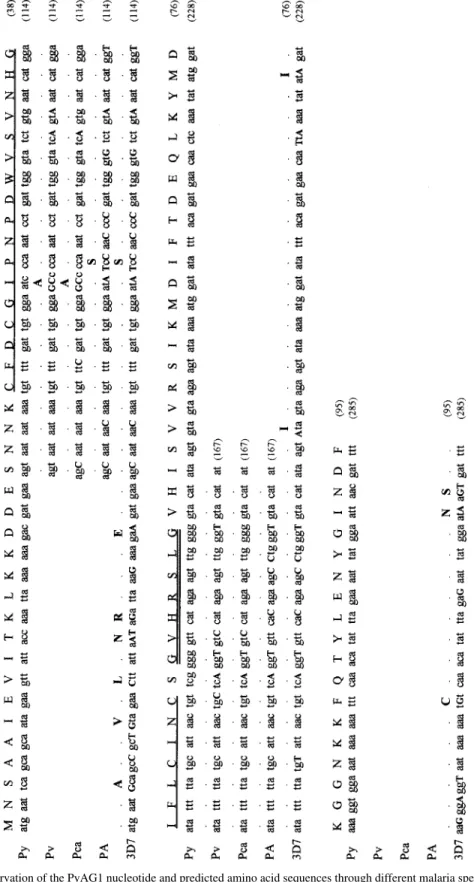
Two nested amplifications were carried out with the oligonucleotide pairs C3/D3 and C2/D2. The sequence, obtained with the oligonucleotide C1, contained a methionine codon as well as up-stream stop codons in frame with the putative initiation codon. The complete nucleotide and derived amino acid sequences (Fig. 4) of this novel gene, named PyAG1, are available in the GenBank data base under the accession num-ber AF055920. To confirm the synthesis of this putative protein during Plasmodium erythro-cytic cycle, we isolated poly(A)+RNA from late asexual stages of P. yoelii with Dynabeads kit (Dynal), after DNase treatment of the total RNA solution, and carried out RT PCR. The PCR-amplification and sequencing of cDNA, using the two oligonucleotides C4 and D4 (Fig. 3), demonstrated that PyAG1 gene is transcribed (data not shown).
This gene has an open reading frame of 888-bp in length which encodes a hydrophilic protein of 296 amino acids (33 kDa). This protein presents, at its N-terminus, two interesting motifs: a zinc fin-ger element (spanning residues 22-45) of the form [C-(X)2-C-(X)16-C-(X)2-C] (with C, cysteine; X, any amino acid) immediately followed by the con-sensus sequence of the Arf GAP catalytic site (Scheffzek et al. 1998) (spanning residues 47-53) of the form [s-h-H-R-x-h-x] (with s, glycine or ala-nine; h, hydrophobic amino acid; H, histidine; R, arginine; x, any amino acid).
A phylogenetic analysis by sequencing with the D4 and C3 oligonucleotides, using genomic DNA of rodent (P. yoelii nigeriensis, P. berghei, P. chabaudiadami, P. vinckei petteri) and human (P. falciparum Palo Alto and 3D7) plasmodial species, revealed an important preservation of this inter-esting region (Fig. 5, Table IA). This observation was confirmed by the sequencing of the 285 first nucleotides of the P. falciparum homologous gene (Fig. 5 and Table IB).
The PyAG1 gene product, expressed as glu-tathione S-transferase fusion protein (GST-PyAG1) in Escherichia coli (pGEX-3X plasmid/ GST Gene Fusion System, Amersham Pharmacia Biotech), was recognized by the Acm C5-10, us-ing IB/R (Fig. 2C). By IFA, the serum of female BALB/c mice (Charles River, France), immunized with the recombinant protein, recognized specifi-cally red blood cells infected by P. yoelii young schizonts (4-8 nuclei), with a rhoptry-like label-ling pattern (Fig. 1B). An immunoelectron micros-copy study will be required to confirm this ultra-structural localization. By IB/R, this polyclonal antibody confirmed the presence of the PyAG1 gene product in a reduced antigenic extract of P.
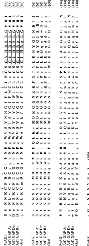
yoelii mature erythrocytic stages (Fig. 2B). Blast analysis using the Genbank™ database gave the highest homology scores with four pro-teins presenting the same two interesting motifs in a similar position and an Arf1 GAP activity: Arf1 GAP of Arabidopsis thaliana (Genbank accession number AC004684), Drosophila melanogaster
(Genbank accession number AF011427), Rattus norvegicus (Cukierman et al. 1995), and Gcs1 of
Saccharomyces cerevisiae (Ireland et al. 1994, Poon et al. 1996) (Fig. 6, Table II). The structural homology with these proteins and the presence of the consensus sequence of the Arf GAPs catalytic site allowed us to hypothetize that the PyAG1 gene product may possess an Arf1 GAP activity. This activity steps in the Arf-GTP cycle by catalysing the GTP hydrolysis and, consequently, the trans-port vesicle uncoating, indispensable step for the membrane fusion between the vesicles and the tar-get membrane.
The specific labelling of the immature rhoptries with polyclonal anti-PyAG1 serum corroborates this putative activity. Indeed, at first schizont stages, the parasites present immature rhoptries with low density (1.12 g.ml-1) on sucrose gradient, even though the rhoptries have a significantly greater density in sucrose (1.16 g.ml-1) at the mature sch-izonts, consequence of the accumulation of rhoptry proteins probably transported through coated vesicles (Jaikaria et al. 1993).
Therefore, the PyAG1 protein may interfere with the regulation of the secreted proteins vesicu-lar transport and, consequently, with the biogen-esis of the secreting organelles like rhoptries. The identification of such an activity supports the pres-ence of a classical eukaryotic transport pathway involving coated vesicules in malarial parasite which has been suggested by BFA-inhibition ex-periences (Crary & Haldar 1992, Benting et al. 1994, Das et al. 1994, Hinterberg et al. 1994, Ogun & Holder1995, Howard & Schmidt 1995) and P. falciparum Arf or Arl (ADP-ribosylation factor-like) characterization (Stafford et al. 1996, Lee et al. 1997, Truong et al. 1997).
Through this preliminary study, we have iden-tified a new element of the intracellular protein transport between plasmodial organelles. Due to its putative regulator activity on the secreting or-ganelles biogenesis, this protein could become a new target with a view to inhibit the parasite de-velopment.
ACKNOWLEDGEMENTS
348 348 348 348
348 Arfl GAP Activity and Malaria Rhoptry Rémi Hienne et al.
Fig. 4: nucleotide and predicted amino acid sequences of the gene encoding the putative zinc finger protein. Inserts obtained from
349 349349 349349 Mem Inst Oswaldo Cruz, Rio de Janeiro, Vol. 95(3), May/Jun. 2000
350 350 350 350
350 Arfl GAP Activity and Malaria Rhoptry Rémi Hienne et al.
351 351351 351351 Mem Inst Oswaldo Cruz, Rio de Janeiro, Vol. 95(3), May/Jun. 2000
REFERENCES
Bannister LH, Mitchell GH 1995. The role of the cytosqueleton in Plasmodium falciparum merozo-ite biology: an electronic-microscopic view. Ann TropMed Parasitol89: 105-111.
Becker B, Melkonian M 1996. The secretory pathway of protists: spatial and functional organization and evolution. Microbiol Rev60: 697-721.
Benting J, Mattei D, Lingelbach K 1994. Brefeldin A inhibits transport of the glycophorin-binding protein from Plasmodium falciparum into the host erythro-cyte. Biochem J300: 821-826.
Crary JL, Haldar K 1992. Brefeldin A inhibits protein secretion and parasite maturation in the ring stages of Plasmodium falciparum. Mol BiochemParasitol 53: 185-192.
Cukierman E, Hubert I, Rotman M, Cassel D 1995. The ARF1 GTPase-activating protein: zinc finger motif and golgi complex localization. Science270: 1999-2002.
Das A, Elmendorf HG, Li WL, Haldar K 1994.
Biosyn-thesis, export and processing of a 45 kDa protein de-tected in membrane clefts of erythrocytes infected with
Plasmodium falciparum. Biochem J302: 487-496. Hienne R, Ricard G, Fusaï T, Fujioka H, Pradines B,
Aikawa M, Doury J-C 1998. Plasmodium yoelii: identification of rhoptry proteins using monoclonal antibodies. Exp Parasitol90: 230-235.
Hinterberg K, Scherf A, Gysin J, Toyoshima T, Aikawa M, Mazie J-C, Pereira Da Silva L, Mattei D 1994.
Plasmodium falciparum: the Pf332 antigen is se-creted from the parasite by a brefeldine A-depen-dant pathway and is translocated to the erythrocyte membrane via the Maurer’s cleft. Exp Parasitol79: 279-291.
Howard RF, Schmidt CM 1995. The secretory pathway of Plasmodium falciparum regulates transport of p82/ RAP-1 to the rhoptries. Mol Biochem Parasitol74: 43-54.
Ireland LS, Johnston GC, Drebot MA, Dhillon N, DeMaggio AJ, Hoekstra MF, Singer RA 1994. A member of a novel family of yeast ‘Zn-finger’ pro-teins mediates the transition from stationary phase TABLE I
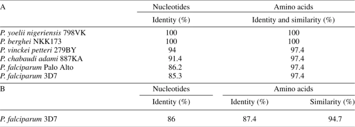
Preservation of the PyAG1 nucleotide and predicted amino acid sequences through different malaria species. A: variability of the nucleotide sequence [52-167] amplified with the oligonucleotide pair D4/C3; B: variability of the 285 first nucleotides of the Plasmodium falciparum 3D7 homologous gene. The following groups of amino
acids were designated as similar: [K, R], [M, V, L, I, F], [F, Y, W], [S, T], [E, D], [N, S]
A Nucleotides Amino acids
Identity (%) Identity and similarity (%)
P. yoelii nigeriensis 798VK 100 100
P. berghei NKK173 100 100
P. vinckei petteri 279BY 94 97.4
P. chabaudi adami 887KA 91.4 97.4
P. falciparum Palo Alto 86.2 97.4
P. falciparum 3D7 85.3 97.4
B Nucleotides Amino acids
Identity (%) Identity (%) Similarity (%)
P. falciparum 3D7 86 87.4 94.7
TABLE II
Homology between the deduced amino acid sequences of PyAG1 gene product and the Arf1 GAP of Arabidopsis thaliana (At), Drosophila melanogaster (Dm), Rattus norvegicus (Rn) and Saccharomyces cerevisiae (Gcs1)
Amino acids 17-121 Amino acids 22-53
(minimal sequence presenting an Arf (sequence containing the zinc finger GAP activity in R. norvegicus Arf1 GAP) element and the Arf GAP consensus
(Cukierman et al. 1995) sequence)
Identity (%) Similarity (%) Identity (%) Similarity (%)
Arf1 GAP At 45.7 63.8 56.3 75
Arf1 GAP Dm 38.1 56.2 65.6 81.3
Arf1 GAP Rn 38.1 57.1 62.5 78.1
352 352 352 352
352 Arfl GAP Activity and Malaria Rhoptry Rémi Hienne et al.
to cell proliferation. EMBO 13: 3812-3821. Jaikaria NS, Rozario C, Ridley RG, Perkins ME 1993.
Biogenesis of rhoptry organelles in Plasmodium falciparum. Mol Biochem Parasitol57: 269-280. Lee F-JS, Patton WA, Lin CY, Moss J, Vaughan M,
Goldman ND, Syin C 1997. Identification and char-acterization of an ADP-ribosylation factor in Plas-modium falciparum. Mol Biochem Parasitol87: 217-223.
Ogun SA, Holder AA 1994. Plasmodium yoelii: Brefeldin A-sensitive processing of proteins targeted to the rhoptries. Exp Parasitol79: 270-278. Poon PP, Wang X, Rotman M, Hubert I, Cukierman E,
Cassel D, Singer RA, Johnston GC 1996. Saccharo-myces cerevisiae Gcs1 is an ADP-ribosylation fac-tor GTPase-activating protein. P Natl Acad Sci USA 93: 10074-10077.
Porchet E, Torpier G 1977. Etude du germe infectieux de Sarcocystis tenella et Toxoplasma gondii par la technique de cryodécapage. Zeitschrift für
Parasitendunke54: 101-124.
Sam-Yellowe TY 1996. Rhoptry organelles of the apicomplexa: their role in host cell invasion and in-tracellular survival. Parasitol Today12: 308-316. Scheffzek K, Ahmadian MR, Wittinghofer A 1998.
GTPase-activating proteins: helping hands to complement an active site. Trends Biochem Sci23: 257-262.
Stafford WHL, Stockley RW, Ludbrook SB, Holder AA 1996. Isolation, expression and characterization of the gene for an ADP-ribosylation factor from the human malaria parasite, Plasmodium falciparum.
Eur J Biochem242: 104-113.
Truong RM, Francis SE, Chakrabarti D, Goldberg DE 1997. Cloning and characterization of Plasmodium falciparum ADP-ribosylation factor and factor-like genes. Mol Biochem Parasitol84: 247-253. Ward GE, Tilney LG, Langsley G 1997. Rab GTPases
and the unusual secretory pathway of Plasmodium.