A Novel Class of Mitochondria-Targeted Soft
Electrophiles Modifies Mitochondrial Proteins
and Inhibits Mitochondrial Metabolism in
Breast Cancer Cells through Redox
Mechanisms
Praveen K. Vayalil1,2, Joo-Yeun Oh1,2, Fen Zhou1,2, Anne R. Diers1,2¤, M. Ryan Smith1,2,
Hafez Golzarian4, Patsy G. Oliver3, Robin A. J. Smith5, Michael P. Murphy6, Sadanandan E. Velu4, Aimee Landar1,2*
1Department of Pathology, University of Alabama at Birmingham, Birmingham, Alabama, United States of America,2Center for Free Radical Biology, University of Alabama at Birmingham, Birmingham, Alabama, United States of America,3Department of Radiation Oncology, University of Alabama at Birmingham, Birmingham, Alabama, United States of America,4Department of Chemistry, University of Alabama at Birmingham, Birmingham, Alabama, United States of America,5Department of Chemistry, University of Otago, Dunedin, New Zealand,6MRC Mitochondrial Biology Unit, Cambridge, United Kingdom
¤ Current address: Berg, Framingham, Massachusetts, United States of America *landar@uab.edu
Abstract
Despite advances in screening and treatment over the past several years, breast cancer re-mains a leading cause of cancer-related death among women in the United States. A major goal in breast cancer treatment is to develop safe and clinically useful therapeutic agents that will prevent the recurrence of breast cancers after front-line therapeutics have failed. Ideally, these agents would have relatively low toxicity against normal cells, and will specifi-cally inhibit the growth and proliferation of cancer cells. Our group and others have previ-ously demonstrated that breast cancer cells exhibit increased mitochondrial oxygen consumption compared with non-tumorigenic breast epithelial cells. This suggests that it may be possible to deliver redox active compounds to the mitochondria to selectively inhibit cancer cell metabolism. To demonstrate proof-of-principle, a series of mitochondria-targeted soft electrophiles (MTSEs) has been designed which selectively accumulate within the mitochondria of highly energetic breast cancer cells and modify mitochondrial proteins. A prototype MTSE, IBTP, significantly inhibits mitochondrial oxidative phosphorylation, re-sulting in decreased breast cancer cell proliferation, cell attachment, and migrationin vitro. These results suggest MTSEs may represent a novel class of anti-cancer agents that pre-vent cancer cell growth by modification of specific mitochondrial proteins.
OPEN ACCESS
Citation:Vayalil PK, Oh J-Y, Zhou F, Diers AR, Smith MR, Golzarian H, et al. (2015) A Novel Class of Mitochondria-Targeted Soft Electrophiles Modifies Mitochondrial Proteins and Inhibits Mitochondrial Metabolism in Breast Cancer Cells through Redox Mechanisms. PLoS ONE 10(3): e0120460. doi:10.1371/journal.pone.0120460
Academic Editor:Janine Santos, National Institute of Environmental Health Sciences, UNITED STATES
Received:September 15, 2014
Accepted:January 22, 2015
Published:March 18, 2015
Copyright:© 2015 Vayalil et al. This is an open access article distributed under the terms of the Creative Commons Attribution License, which permits unrestricted use, distribution, and reproduction in any medium, provided the original author and source are credited.
Data Availability Statement:All relevant data are within the paper and its Supporting Information files.
Introduction
It has long been known that cancer cells are metabolically distinct from normal cells. For exam-ple, many cancer cell types exhibit higher levels of glycolysis, irrespective of the levels of avail-able oxygen needed for oxidative phosphorylation, a phenomenon known as“aerobic
glycolysis”or the Warburg effect [1]. This observation led to the concept that cancer cells have defective or disrupted mitochondrial metabolism. However, recent evidence demonstrates that many cancer cells do not have impaired mitochondrial function, but in fact have higher levels of ATP-dependent oxygen consumption compared with normal cells [2–4]. In addition, cancer cells which are resistant to standard therapeutic regimens have increased mitochondrial reserve capacity [5,6]. Therefore, targeting cancer cell mitochondrial metabolism, particularly ATP-dependent oxygen consumption and reserve capacity, may potentially represent a novel ap-proach to cancer treatment.
Mitochondria are critical for a number of cellular processes including energy generation, anaplerosis, regulation of apoptosis, redox cell signaling, and calcium homeostasis. A major strategy for targeting compounds to mitochondria involves the use of lipophilic cationic carrier moieties to deliver functional groups such as antioxidants and DNA alkylating agents. The tri-phenylphosphonium (TPP) moiety is the best characterized mitochondria-targeting group and has been used to deliver the antioxidant coenzyme Q, nitrogen mustards, doxorubicin, and other molecules to the mitochondrion [2,7,8]. This is accomplished via covalent linkage of molecular moieties to TPP to form bifunctional compounds. TPP-linked molecules have also been shown to disrupt mitochondrial functionin vitroat high concentrations after short-term exposure [2,7,9,10], though the precise mechanisms remain poorly defined.
In this study, we analyze the bioenergetic consequences of directing electrophilic TPP bi-functional compounds to the mitochondrion. These compounds, termed“ mitochondria-targeted soft electrophiles,”(MTSEs), differ significantly in their reactivity from highly toxic electrophilic drugs and environmental toxicants, which are relatively“hard”electrophiles [11]. Hard electrophiles form adducts with“hard”nucleophiles such DNA bases and serine protein residues; whereas soft electrophiles form adducts with“soft”cellular nucleophiles, particularly cysteine thiols. While hard electrophiles have routinely been dismissed as therapeutics due to their systemic toxicity in drug studies, there is accumulating evidence that soft electrophiles are less toxic inin vitroandin vivobiological model systems [11,12]. It is also important to consid-er that the soft electrophile class of compounds have a range of reactivity spanning sevconsid-eral or-ders of magnitude [13]. The reactivity of a soft electrophile is also directly proportional to the toxic effects, with more reactive compounds exhibiting higher toxicity in cellular and animal models [14–16]. Therefore, it is likely that soft electrophiles of relatively low reactivity, includ-ing MTSEs, may be useful as therapeutic agents. In fact, other such soft electrophiles have known beneficial physiological effects and include dietary electrophiles found in broccoli (sul-foraphane) and curry (curcumin) [17], as well as endogenously produced anti-inflammatory prostanoids such as 15-deoxy prostaglandin J2[18,19].
One of the most important considerations in developing novel drug leads is ensuring specif-ic interaction of the compounds with desired target protein(s). In the case of electrophilspecif-ic sig-naling molecules, the specificity of reaction is determined by the chemical properties of the molecules themselves, including hydrophobicity, reactivity, electrophile“softness,”and target
“softness”[11]. In general, lower reactivity of the electrophile results in higher selectivity for specific targets. The most reactive soft nucleophiles within the cell are selenocysteine and deprotonated (or low pKa) cysteine residues [20,21]. While cysteine is present in most
pro-teins, it represents less than 2% of the total protein amino acid composition. In addition, not all cysteines are susceptible to oxidative modification, since relatively few cysteines exist primarily
study design, data collection and analysis, decision to publish, or preparation of the manuscript.
in the deprotonated, nucleophilic form [21,22] which is reactive with electrophiles. It is for these reasons that specific protein thiols are poised to mediate diverse redox signaling re-sponses to multiple stimuli [23]. Interestingly, accessible reactive protein thiols are present in the active sites of many mitochondrial proteins. Mitochondrial proteins are exposed to the most reducing environment within the cell and are susceptible to modification due to the rela-tively high inner mitochondrial matrix pH caused by the proton pumping of the electron trans-port chain [24]. Mitochondrial proteins which are redox-sensitive include mitochondrial dehydrogenases such asα-ketoglutarate dehydrogenase [25], isocitrate dehydrogenase [26], and mitochondrial aldehyde dehydrogenase [27], as well as the mitochondrial complexes I, II, and V [28,29].
In order to determine the effects of mitochondrial protein modification on the metabolism of cancer cells, we synthesized a series of MTSEs that alkylate mitochondrial proteins and ex-amined the differential effects of a prototype MTSE on oxidative phosphorylation and glycoly-sis in tumorigenic versus non-tumorigenic breast cells. In addition, we determined the resultant effects of MTSEs on breast cancer cell proliferation, migration and adhesion. This study demonstrates that MTSEs cause profound inhibition of mitochondrial metabolism, and inhibit breast cancer cell proliferation, attachment, and migration; while non-tumorigenic MCF10A cells remain relatively insensitive. Taken together, these results suggest that modifica-tion of mitochondrial thiols by MTSEs alters metabolic pathways in breast cancer cells, and this class of compounds may be useful to inhibit the progression of highly energetic cancer cells.
Materials and Methods
All chemicals were of analytical grade and purchased from Sigma-Aldrich (St. Louis, MO) un-less otherwise noted. Anti-TPP antiserum was prepared as previously described [30]. The MDA-MB-231 cell line was a generous gift from Dr. Danny Welch, and was originally obtained from ATCC. The MDA-MB-468 cell line was obtained from ATCC.
Synthesis of mitochondria-targeted electrophiles
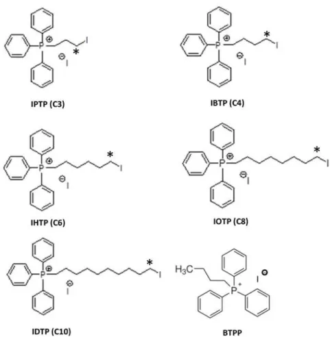
Mitochondria-targeted electrophiles were synthesized containing an iodo-leaving group with alkyl linkers of varying carbon lengths, including 3-, 4-, 6-, 8- and 10-carbons [iodopropyl tri-phenylphosphonium (IPTP), iodobutyl tritri-phenylphosphonium (IBTP), iodohexyl triphenylpho-sphonium (IHTP), iodooctyl triphenylphotriphenylpho-sphonium (IOTP), iododecyl triphenylphotriphenylpho-sphonium (IDTP), respectively]. These analogs were synthesized as previously described [31]. Briefly, tri-phenylphosphine was heated at 100°C for 45 minutes with 1,3-diiodopropane, 1,4-diiodobutane, 1,6-diiodohexane, 1,8-diiodooctane or 1,10-diiododecane, respectively. After the reaction was complete, diethyl ether was added and the precipitated solid was filtered and dried. All com-pounds were characterized by1H-NMR,13C-NMR and MS in order to ensure purity of>99.9%
before their use in biological evaluations.
Cell Culture and Treatments
Breast cancer cell lines. MDA-MB-231 (MB231) and MDA-MB-468 (MB468) human breast adenocarcinoma cells were cultured in DMEM (Mediatech, Manassas, VA) supple-mented with 10% fetal bovine serum (FBS; Atlanta Biologicals, Atlanta, GA). Cultures were maintained in 5% CO2and humidified in a 37°C incubator. MCF10A, an immortalized human
Cell Treatments. Cells were plated at a cell density of 2×10⁵cells/well in 6-well plates and allowed to acclimate for 16h. For breast cancer cells, medium was changed to low FBS-containing medium (0.5%) for 16h prior to treatment. For MCF10A cells, supplemented MEGM medium was diluted 1:20 with non-supplemented MEGM for 16h prior to treatment. Cells were then treated with MTSE in 1mL of fresh medium for times indicated in the figure legend. At the end of treatment, cell lysates were prepared and protein adducts were visualized by Western blot analysis as described below under“Immunoblot Analysis.”
Cell Fractionation
MB231 were plated in 6-well plates and allowed to grow until 90% confluent. Cells were divid-ed into crude mitochondrial and cytoplasmic fractions by differential centrifugation. Briefly, cells were washed 2 times with phosphate-buffered saline (PBS), and each well from a 6-well plate was scraped into 167μL of MIB-1 (mitochondrial isolation buffer-1: HEPES, 50mM;
su-crose 100mM; KCl 100mM; EGTA, 1mM; pH 7.2 with KOH). All 6 wells were combined to make one sample. Digitonin (5mg/mL in MIB-1) was added to a final concentration of 50μg/
106cells and samples mixed by inversion for 1 min. Samples were centrifuged at 1000 x g for 10min at 4°C, and half of supernatant was transferred to a new tube. The pellet was homoge-nized with a micropestle with the remaining supernatant and centrifuged again at 1000 x g for 10min at 4°C. Supernatant was collected, pooled with the initial supernatant, and centrifuged at 14,000 x g for 10min at 4°C in order to pellet the mitochondrial fraction. Supernatant (con-taining cytoplasm components) was collected and used for analysis. Pellet con(con-taining mitoplast enriched fraction was washed once in MIB-2 (mitochondrial isolation buffer-2: prepared as above for MIB-1 with omission of EGTA) by resuspending pellet and repeating previous cen-trifugation step. Supernatant was discarded and mitoplast fraction used for analysis. Mitochon-drial enrichment procedure was confirmed by Western blot analysis of the mitochonMitochon-drial protein, the voltage-dependent anion channel. Protein adducts were visualized by Western blot analysis as described below.
Immunoblot Analysis
Cells were washed with PBS and lysed in a buffer containing 10mM Tris-HCl, pH 7.4, and 1% Triton X-100 and protease inhibitor cocktail (Roche). Soluble proteins were resolved using SDS-PAGE and transferred to nitrocellulose membranes (Bio-Rad). Protein levels were quanti-fied using the method of Bradford (Bio-Rad), and equivalent amounts of protein were loaded. Uniform protein loading was confirmed using Ponceau S staining of membranes and showed no significant differences in protein levels on blots among samples. Membranes were blocked in 5% milk (w/v) in Tris Buffered Saline (pH 7.4) containing 0.5% Tween 20 (TBS-T), and then incubated with anti-TPP (1:10,000) primary antiserum for 3h at room temperature. After washing with TBS-T, membranes were incubated with HRP-conjugated anti-rabbit IgG sec-ondary antibody. Membranes were developed using SuperSignal West Dura chemilumines-cence substrate (Pierce) and imaged using a CCD camera imaging system.
Metabolic Assessment
described previously [33–35]. The concentrations of these compounds were optimized prior to the assay. In our calculations, the oligomycin-insensitive OCR was attributed to proton leak.
To determine the effects of MTSEs, cells were plated on XF24 plates and treated with the in-dicated concentrations of IBTP or BTPP for 4h in 0.5% FBS-containing medium. After treat-ment, the medium was removed and replaced with XF assay medium (DMEM, containing 5mM glucose, 0.5% FBS, 5mM HEPES without bicarbonate) and equilibrated 1h before OCR measurement. For 24h post-treatment measurements, cells were plated on 6-well plates and treated with the indicated concentrations of IBTP or BTPP for 4h. After the incubation, the cells were harvested immediately by trypsinization and replated into XF24 plates for an addi-tional 20h in complete medium containing 10% FBS (total 24h). The medium was removed and replaced with assay medium and equilibrated 1h before OCR measurement. For 72h post-treatment measurements, cells were plated on 6-well plates and treated with the indicated concentrations of IBTP or BTPP for 4h. The medium was replaced with complete medium con-taining 10% FBS, and incubated for 48h. The cells were harvested after 48h, replated in XF24 plates and allowed adhere for an additional 20h in complete medium (total duration 72h). The medium was replaced with assay media and incubated 1h before measurement of OCR.
Glycolytic function was measured using a glycolytic“stress test.”For these experiments, cells were cultured and treated similarly to the mitochondrial stress test, except that prior to measure-ment medium was changed to DMEM without glucose, bicarbonate or HEPES, and containing 0.5% FBS. ECAR was monitored using the XF analyzer, and sequential injections of glucose (10mM), oligomycin (1μg/mL), and 2-deoxyglucose (2DG; 100mM) were used to determine
gly-colytic function parameters. Initially, ECAR was measured in the assay medium without glucose. The ECAR measured in the absence of glucose represents non-glycolytic acidification. Glucose was then added, which stimulates glycolysis and represents glycolytic rate or glycolytic flux in the cells. Oligomycin was injected to inhibit mitochondrial ATP production, and increase glyco-lytic rate in order to meet the energy demands of the cells. This increase represents the maxi-mum glycolytic capacity of the cells. The difference in ECAR between maximaxi-mum glycolytic capacity and glycolytic rate represents glycolytic reserve, which defines the maximum ability of the cells to induce glycolysis to meet the energy demands in the absence of mitochondrial ATP production. Both OCR and ECAR measurements were normalized to total protein per well.
Cell Proliferation
Proliferation of MB231 cells was measured by using CyQuant cell proliferation assay kit from Molecular Probes (Eugene, OR). The basis for the CyQuant assay is the use of a proprietary green fluorescent dye (CyQuant GR dye) that exhibits strong fluorescence when bound to cel-lular nucleic acids. Cells (200,000/well) in 6-well plates were treated with different concentra-tions of IBTP for 24h in medium containing 0.5% FBS containing medium at 37°C in a CO2
incubator. Treatment was stopped by removing the media completely and replenishing with fresh media with 0.5% FBS without IBTP and incubated for another 48h. After incubation, media was completely removed and cells were frozen at−80°C then thawed and lysed accord-ing to the manufacturer’s protocol. The lysates (100μl) were diluted with an equal volume of
CyQuant GR dye, and fluorescence was measured after 5 min using Victor X3 multi-well plate reader with excitation 485 nm and emission 530 nm.
Cell Attachment and Migration
To determine the effect of IBTP on cell migration, the cells were treated as described above. After the treatment, medium was changed to fresh 0.5% FBS-containing medium to remove compounds and scratches were made on the cell layer. The cells were allowed to migrate for 5h into cell free zone. Images of the scratches were captured at 0h and 5h. The length across the cell free zone before and after 5h of incubation was measured using ImageJ software (NIH, Be-thesda, USA).
To determine the effect of IBTP on cell attachment, the cells were trypsinized after treat-ment, counted and the viability was assessed by trypan blue exclusion assay. The cells from each treatment group were then transferred to a 100-mm tissue culture dish and incubated for 24h. At the end of the incubation, the media was collected and total number of viable cells that failed to attach to the substratum was counted.
Statistical analysis
Results are reported as means ± SEM, and n = 3 or more determinations as indicated in the Figure legends. Data were analyzed by one-way analysis of variance (ANOVA) followed by post-hoc analysis for significant results among the groups by pairwise t-tests using MegaStat Software. The minimum level of significance was set at p<0.05.
Results
Post-translational modification of proteins by MTSEs in breast cancer
cells
We hypothesized that MTSEs may represent a novel class of compounds which act by forming adducts with key metabolic proteins and ultimately inhibiting mitochondrial bioenergetics pathways. Therefore, analogs of IBTP were synthesized with varying chain lengths in order to optimize the hydrophobicity and accessibility of the electrophilic carbon adjacent to the iodo-leaving group (Fig. 1). Compounds include iodopropyl-, iodobutyl-, iodohexyl-, iodooctyl-, and iododecyl-triphenylphosphonium (TPP), and a non-electrophilic analog with a 4-carbon alkyl chain (BTPP) that can be used as a control for effects due to the TPP moiety independent of alkylation activity.
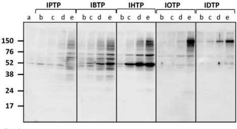
In order to characterize protein adduct formation by MTSEs in breast cancer cells, MB231 cells were treated with MTSE compounds over a dose-range from 0–5μM, and adduct
Accumulation of protein adducts in mitochondria in breast cancer cells
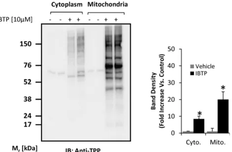
Because triphenylphosphonium compounds accumulate within the mitochondrion, IBTP treat-ment is expected to result in more protein adducts in the mitochondrial fraction than the cyto-plasmic fraction, although this has not been demonstrated in cancer cells. MB231 cells were treated with vehicle, BTPP or IBTP over a dose range up to 15μM, and proteins containingtriphenylphosphonium adducts were detected (S1 Fig.). No adducts were formed in the BTPP-treated cells, as expected. Adduct formation with proteins in cytosolic or mitochondrial compart-ments was determined by treatment of MB231 cells with 10μM IBTP or vehicle for 4h, followed
by isolation of the mitochondrial and cytoplasmic fractions. The quality of mitochondria-enriched fractions was determined by Western blot analysis to confirm the presence of the voltage-dependent anion channel in the mitochondria fractions, and its absence in the cyto-plasmic fraction (S2 Fig.). Differences in protein adduct formation in these fractions were deter-mined by Western blot analysis.Fig. 4shows an enrichment of protein adducts of approximately 2.3 fold in the mitochondrion when compared with the cytosolic fraction, clearly indicating that IBTP concentrates within the mitochondria and forms protein adducts.
Effects of IBTP on cellular metabolism
As IBTP accumulates within the mitochondrion and forms protein adducts, we sought to de-termine the effects of IBTP on the metabolic parameters of MB231 cells over time. MB231 cells
Fig 1. Structures of mitochondria-targeted electrophiles.The compounds were synthesized as described under“Materials and Methods.”
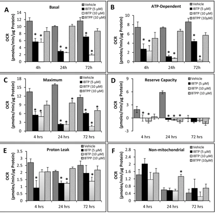
were treated with different doses of IBTP or BTPP for 4h, at which point various bioenergetic parameters were measured using the“mitochondrial stress test.”(Fig. 5A, 4h). In order to avoid differences in proliferation due to treatments for longer time points, medium was changed after treatment (4h), and cells were allowed to recover for 24h or 72h. A defined num-ber of cells were then transferred and analyzed in the mitochondrial stress test (Fig. 5B, 24h; Fig. 5C, 72h). Treatment of MB231 cells with 5 and 10μM IBTP significantly inhibited the
basal OCR at all the time points measured; however, by 72h post-treatment cells treated with 5μM IBTP began to recover. In contrast, the non-electrophilic TPP compound BTPP did not
affect the basal OCR significantly at any time point studied (Fig. 6A). However, maximal OCR (24h and 72h post-treatment) and reserve capacity (24h post-treatment) were significantly de-creased and non-mitochondrial OCR (24h post-treatment) was significantly inde-creased after
Fig 2. Dose-dependent modification of proteins by MTSEs of different chain length in MB231 cells. MB231 cells were treated with the indicated doses for 4h. At the end of treatment, cell lysates were prepared and protein adducts were visualized by Western blot analysis using an antibody directed against TPP. a= Vehicle;b= 0.5μM;c= 1μM;d= 2μM;e= 5μM. IPTP = 3 carbons, IBTP = 4 carbons, IHTP = 6 carbons,
IOTP = 8 carbons, IDTP = 10 carbons. The values are mean±SE of 3–5 samples obtained from two separate experiments;*p<0.05 compared to no treatment.
BTPP exposure. Thus, the triphenylphosphonium moiety has distinct effects alone in the ab-sence of an electrophilic component, and these effects persist 24–72h post-treatment. There were no significant effects on ATP-linked OCR in cells treated with the non-reactive analog, BTPP (Fig. 6, panels A and B). After IBTP treatment, basal OCR, ATP-linked OCR, maximal OCR, and reserve capacity were all significantly decreased at 4h-, 24h-, and 72h post-treatment (Fig. 6, panels A-D). Proton leak was decreased by IBTP, and the effect recovers over time at the 5μM IBTP dose, but not at 10μM (Fig. 6, panel E). There was no significant change in
non-mitochondrial OCR with IBTP (Fig. 6, panel F).
Fig 3. Time-dependent modification of proteins by MTSEs of different chain lengths in MB231 cells. MB231 cells were treated with 5μM of indicated MTSEs for the indicated times. At the end of treatment, cell
lysates were prepared and protein adducts were visualized by Western blot analysis using an antibody directed against TPP (upper panel). Lane densities were quantified and plotted in the lower panel. Values are mean±SE of 3–5 samples obtained from two separate experiments;*p<0.05 compared to no treatment.
Due to the decreased mitochondrial metabolism observed in response to IBTP, we investi-gated the possibility of compensatory metabolic activity in the glycolysis pathway. Glycolytic activity was measured using extracellular flux analysis with a glycolysis stress test, as described previously [36]. The cells were cultured and treated similarly to the previous experiment for OCR, with the exception that the media was unbuffered and did not contain glucose (see Mate-rials and Methods), allowing for the measurement of extracellular acidification rate (ECAR) in response to glucose. There were no significant differences in baseline) non-glycolytic) ECAR across groups at each time point, therefore, the baseline reading of ECAR was taken and de-fined as 100% for each group in subsequent analyses. Glycolysis was stimulated by the addition of glucose (Fig. 7A,“G”arrows), the glycolytic reserve capacity was determined by inhibiting mitochondrial ATP production at Complex V with oligomycin (“O”arrows), and specificity for glycolysis was determined by inhibiting flux through glycolysis with 2-deoxyglucose (“2DG”arrows). ECAR measured was specific for glycolysis because addition of 2DG returned ECAR levels to those observed in the absence of glucose. Glycolysis rate values are shown in Fig. 7B(obtained after addition of glucose), and the results indicate that no significant changes were found in any parameters at 4h, but that at 24h and 72h glycolysis was induced by IBTP (5 or 10μM). Significant differences emerged in the glycolytic reserve capacity (Fig. 7C), which
was decreased by IBTP treatment (24 and 72h post-treatment), but not BTPP. These results suggest that MB231 cells treated with IBTP for 4h, followed by 24h or 72h recovery increases glycolytic flux at the expense of the glycolytic reserve. ATP levels measured at the 24h post-treatment time point were not significantly changed by BTPP or IBTP (data not shown), indi-cating that the maintenance of cellular ATP can be attributed to increased glycolytic flux.
Effect of IBTP on cell proliferation
Next, we sought to determine the effects of IBTP on cell proliferation of MB231 cells compared with non-tumorigenic MCF10A cells. Both cell lines were treated with concentrations ranging
Fig 4. Mitochondrial protein modification by IBTP in breast cancer cells.MB231 cells were treated with 10μM IBTP or EtOH vehicle for 4h, and cell fractions were obtained as described in the Materials and
Methods. At the end of treatment, cell lysates were prepared and protein adducts were visualized by Western blot analysis using an antibody directed against TPP. The values are mean±SE of 3–5 samples obtained from two separate experiments;*p<0.05 compared to vehicle.
Fig 5. Effect of IBTP treatment on mitochondrial respiration of MB231 cells. Panel A:Cells plated on XF24 plates were treated with the indicated concentrations of IBTP or BTPP for 4h in 0.5% FBS-containing medium. After treatment, the medium was removed and replaced with XF assay medium (DMEM, containing 5mM glucose, 0.5% FBS, 5mM HEPES without bicarbonate) and equilibrated 1h before OCR measurement. Panel B:Cells plated on 6-well plates were treated with the indicated concentrations of IBTP or BTPP for 4h. After the incubation, the cells were harvested immediately by trypsinization. The harvested cells were replated in XF24 plates and allowed adhere for an additional 20h in complete medium containing 10% FBS (total 24h). The medium was removed and replaced with assay medium and equilibrated 1h before OCR measurement.Panel C: After 4h of IBTP or BTPP treatment, the medium was replaced with complete medium containing 10% FBS, and incubated for 48h. The cells were harvested after 48h, replated in XF24 plates and allowed adhere for an additional 20h in complete medium. The medium was replaced with assay media and incubated 1h before measurement of OCR (total duration 72h).
from 500nM to 10μM for 4h or 24h, followed by recovery for an additional 48h. The effect of
IBTP on cell proliferation was analyzed by quantifying the number of cells in each well. Expos-ing MB231 or MCF10A cells to different doses of IBTP for short durations (4h), did not signifi-cantly affect proliferation (data not shown). However, prolonged incubation with IBTP for 24h caused significant inhibition of cancer cell proliferation (Fig. 8). IBTP inhibited the prolifera-tion of MB231 cells dose-dependently, as demonstrated by decreased cell number, ranging from 29% (500nM) to 60% (10μM) compared to vehicle-treated cells. Under identical
condi-tions, the nonreactive analog BTPP did not cause significant inhibition of the proliferation in
Fig 6. Bioenergetic parameters in MB231 cells. Panels A-F:Bioenergetic parameters were calculated from the OCR traces inFig. 5, panels A-C. Values are mean±SE obtained from 10–15 wells in two separate experiments;*p<0.05 compared to BTPP; #p<0.05 compared to vehicle.
MB231 cells. Significant inhibition of proliferation was also observed in another triple negative breast cancer cell line MB468 (data not shown). On the other hand, IBTP treatment did not in-hibit cell proliferation significantly in MCF10A cells. Interestingly, we have previously reported that the basal OCR of MB231 cells is approximately two-fold higher than the OCR of MCF10A cells [3], suggesting that differences in mitochondrial function may render MB231 cells more susceptible to the inhibitory effects of IBTP on proliferation.
Fig 7. Effect of IBTP treatment on glycolysis of MB231 cells.Cells were plated, cultured, and treated as described forFig. 5and a glycolysis stress test was performed as described in the Materials and Methods.Panel A:ECAR is represented as a function of time, and the third untreated time point was used to define 100% ECAR for each group. Arrows indicate times of injection for glucose (G), oligomycin (O), and 2-deoxyglucose (2DG).Panel B:Rate of glycolytic flux in MB231 cells with and without BTPP or IBTP treatment. Glycolysis was defined as the %ECAR after addition of glucose (10mM).Panel C: Glycolytic reserve of MB231 cells with and without BTPP or IBTP treatment. Glycolytic reserve represents the difference in ECAR between glycolysis and the maximum glycolytic capacity observed after addition of oligomycin (1μM). Values are mean±SE obtained from 10–15 wells in two separate experiments; *p<0.05 compared to BTPP; #p<0.05 compared to vehicle.
Effect of IBTP on cell adhesion and migration
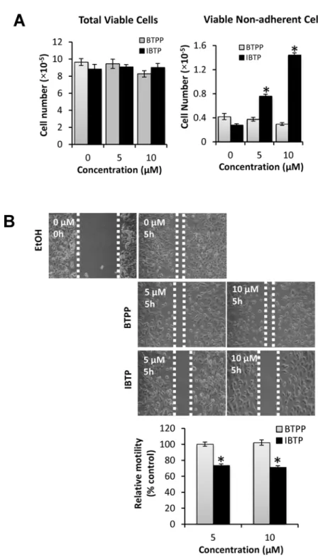
Cancer cells are recognized for their ability to migrate away from the primary tumor and ad-here at distal sites to form new colonies during metastasis. Tad-herefore, we next examined the ef-fect of IBTP on MB231 cell attachment and ability to migratein vitro. To study the effect of IBTP on cell attachment, the cells were treated with different doses of IBTP for 4h. After treat-ment, cells were trypsinized, counted, and the viability was noted. All the cells from each well were transferred to new 100-mm plates and allowed to attach and grow for an additional 24h. At the end of the incubation, the media was collected and the number of viable cells in the media was determined, representing the number of viable cells that failed to attach to the sub-stratum.Fig. 9, panel A represents total viable cells present in each group. IBTP or BTPP treat-ment for 4h had no effect on the viability of tumor cells (Fig. 9, Panel A:“Total Viable Cells”). However, treatment with IBTP dose-dependently inhibited the ability of MB231 cells to attach to the substratum, as indicated by increased number of viable cells floating in the medium (Fig. 9, Panel A:“Viable Non-Adherent Cells”). However, BTPP did not exhibit any significant inhibition and was comparable to vehicle treated groups.
To investigate the effect of IBTP on the migration of cancer cells, we used the scratch assay method (Fig. 9, panel B). After treatment with different doses of IBTP or BTPP, cell-free zones were created in the cell monolayer using a sterile pipet tip. After incubation for 5h, the extent to which the cells migrate into the cell-free zone was measured. Cells treated with vehicle alone migrated into the cell-free zone almost completely (Fig. 9B: EtOH), and similar results were ob-served with BTPP treatment (Fig. 9B: BTPP). However, IBTP treatment (either 5 or 10μM),
clearly inhibited the migration of cells into the cell-free zone as seen from increased cell-free areas compared to either vehicle or BTPP treated group (Fig. 9B: IBTP).
Discussion
Patients with breast tumors that are negative for estrogen, progesterone, and Her2/neu recep-tors (triple-negative) have few treatment options and are particularly susceptible to recurrence following initial therapy, resulting in high mortality rates for patients with this tumor subtype [37,38]. Our group and others have shown that triple negative breast cancer cells exhibit
Fig 8. Effects of IBTP on proliferation of human breast cancer cells or non-tumorigenic breast epithelial cells.MB231 or MCF10A cells were treated with the indicated concentrations of IBTP (black diamonds) or BTPP (gray squares) for 24h. The number of cells in each group at the end of the experiment was measured. The values are mean±SE of duplicates from two independent experiments. (*p<0.05 compared to BTPP).
Fig 9. Effects of IBTP on cell attachment and migration in human breast cancer cells.MB231 cells were treated with the indicated concentrations of IBTP. At the end of the treatment, the cells were either assayed for the cells ability to attach the substratum or migration by scratch assay.Panel A:To determine the effect of IBTP on cell attachment, viable cells were counted (“Total Viable Cells”) and replated onto a 100mm tissue culture dish for 24h. At the end of the incubation, the media was collected and number of viable cells that failed to attach to the substratum was counted (“Viable non-adherent Cells”). The values represent the mean±SE of triplicates from two independent experiments. (*p<0.05 compared to BTPP).Panel B:To determine the effect of IBTP or BTPP on migration, cells were treated with the indicated concentration of compounds for 4h. A scratch assay was performed as described in the Materials and Methods, where cells were allowed to migrate into the cell-free zone for 5h. The values represent the mean±SE of three separate images obtained from triplicate wells. (*p<0.05 compared to BTPP).
higher mitochondrial metabolism than non-tumorigenic cells [3,7], in addition to being highly glycolytic [3,39]. The enhanced mitochondrial metabolism suggests that mitochondria-targeted compounds might provide a novel treatment strategy. In our previous study, MB231, as well as tumorigenic, metastatic MCF10A subclones (MCF10CA a.1 and MCF10A d.1α) ex-hibited higher energetic parameters under ambient O2levels (21%). The mitochondrial
ener-getic parameters were further increased as O2approached physiological levels (4–5% O2), and
this was shown to be hypoxia-inducible factor 1-α(HIF-1α)-dependent [3]. We have also pre-viously shown that chemo-resistant glioma cells are more invasive and have higher mitochon-drial bioenergetics than chemo-sensitive glioma cells [40]. However, it is not the case that all cancer types exhibit higher mitochondrial metabolism. There are a limited number of known tumor types which have been shown to have decreased mitochondrial metabolism due to mu-tations in Krebs cycle enzymes or electron transport complex subunits (e.g., pheochromocyto-ma, paragangliopheochromocyto-ma, familial renal cell carcinopheochromocyto-ma, glioma) [41–43]. Nevertheless,
mitochondria-driven tumors have been shownin vivousing13C-glucose metabolic analysis [44], and evidence of upregulated mitochondrial function has been shownin situin human breast cancer tissue [45]. It is likely that drugs which target highly energized mitochondria may be useful in these tumor types, and will provide a novel strategy for specific cancers having aug-mented mitochondrial bioenergetic profiles.
Oxidation or other post-translational modifications of protein thiols can modulate the activ-ity of enzymes and proteins, and thereby can impact important cellular functions [16]. IBTP has previously been used as a research tool to investigate the status of reduced thiols in cell cul-ture models, isolated mitochondria, and isolated tissues from animal models of oxidative stress [27,30]. We have previously demonstrated biological effects of IBTP in intact cultured endo-thelial cells [46]. IDTP has also previously been synthesized as a thiol probe, but its activity has been less well characterized [27,30].
In the current study, we have developed and characterized a novel series of mitochondria-targeted soft electrophiles (MTSEs) and demonstrate mitochondrial protein thiol modification of these compounds in breast cancer cells (Fig. 4). MTSEs localize to mitochondria due to the presence of a lipophilic, delocalized cationic triphenylphosphonium (TPP) moiety [30]. In the present study, we report that treatment with MTSEs containing a 6-carbon or less alkyl spacer between the iodo-and phosphonium groups results in a concentration- and time-dependent accumulation of TPP-protein adducts in triple negative breast cancer cells (Figs.2and3). As the lipophilicity increases with chain length, TPP compounds more readily accumulate within the mitochondria [47]. However, when the MTSE chain length was increased to 8 or 10 car-bons, fewer protein adducts were observed irrespective of the dose or duration of incubation tested. At high concentrations, this may be due to destabilization of the membrane barrier or disruption of the lipid bilayer that may limit accumulation of MTSEs within the mitochondria. At lower concentrations the greater hydrophobicity and consequent greater adsorption to the matrix-facing surface of the TPP compounds, and the penetration of the longer iodoalkyl chain into the lipid bilayer may limit their access to many of the soft nucleophiles in the mitochondri-al matrix. These studies demonstrate that MTSEs containing 6 carbons or less are likely to be more effective for reacting with a wider range of mitochondrial matrix protein targets, and therefore of greater utility in anti-cancer applications.
Isocitrate dehydrogenase andα-ketoglutarate dehydrogenase also produce NADH which can then be oxidized by Complex I of the electron transport chain (ETC). Inhibition of Krebs cycle enzymes by IBTP would be expected to decrease mitochondrial oxygen consumption by limit-ing the production of NADH, thereby inhibitlimit-ing electron flux through the ETC. Another possi-ble explanation for the decreased mitochondrial oxygen consumption in response to IBTP is that inhibition of key metabolic proteins may cause a shift in energy production toward other metabolic pathways. In fact, our data demonstrate that IBTP treatment of cancer cells results in increased reliance on glycolysis at the expense of the glycolytic reserve capacity (Fig. 6). It is likely that dual treatment of cancer cells with IBTP and an inhibitor of glycolysis would cause energetic failure and subsequent cell death, and further studies are warranted to explore this possibility.
IBTP was also shown to have preliminary anti-cancer properties since it inhibited prolifera-tion, attachment and migration of cancer cells. These effects were specific to the MTSE and re-quire protein adduct formation, as BTPP did not have similar effects. Previous studies from our group have shown that the soft electrophile 15d-PGJ2also inhibits cancer cell adhesion
and migration due at least in part to focal adhesion disassembly and extensive F-actin reorgani-zation at very low concentrations [49]. It is likely that the inhibitory effects of IBTP on cancer cell migration are mediated by modification of mitochondrial proteins, but the mechanism is not yet clear. A recent report demonstrates that mitochondrial superoxide production with preserved mitochondrial function increases cancer cell migration [50]. We have previously shown in other cell types that IBTP also increases reactive oxygen species [46], but in this study we show that IBTP compromises mitochondrial function and inhibits cancer cell migration, suggesting involvement of a different mechanism. Zhaoet al. recently reported that mitochon-drial function has an important role in breast cancer cell migration, and that blocking mito-chondrial ATP production with oligomycin A inhibits breast cancer cell migration [51]. Our results that IBTP inhibits mitochondrial ATP production and migration in MB231 cells are consistent with this potential mechanism. Further studies are necessary to determine which protein target(s) are involved in IBTP’s effects on cancer cell migration.
In order to attribute these effects to the electrophilic nature of the compounds, we used BTPP as a mitochondria-targeted non-electrophilic control. BTPP localizes to the mitochon-drion in a similar manner to IBTP [30], but does not form stable protein adducts on cysteine residues like IBTP. BTPP also does not affect the malignant phenotypes of breast cancer cells suggesting that electrophilic modification of specific mitochondrial proteins is involved in the activity of MTSEs. Interestingly, BTPP also caused some changes in mitochondrial function; however, to significantly lesser extents than MTSEs. We have previously reported this type of partial effect with TPP control molecules in studies investigating the ability of IBTP to attenu-ate HO-1 induction by lipid electrophiles [46]. Presumably, the transient inhibition by BTPP alone represents changes in mitochondrial function that are the result of accumulation of the triphenylphosphonium moiety within the mitochondrion, whereas the longer and more pro-nounced inhibition observed with IBTP treatment is the result of thiol modification. This is consistent with reports that ion fluxes alter respiration in multiple cell types. For example, mi-tochondrial Ca2+is a positive effector of ATP synthesis [52]; however, Ca2+overload results in mitochondrial dysfunction and ultimately cell death [52]. In addition, opening of the mito-chondrial ATP-sensitive potassium channels and increased potassium flux has been shown to change matrix volume [53], induce Complex I-dependent ROS generation [54] [55], and inhib-it minhib-itochondrial permeabilinhib-ity transinhib-ition [56].
metabolism. These compounds are not overtly cytotoxic at the time points examined in this study, suggesting that they may prove to be useful for prevention of tumor recurrence and in-duction of cytostasis. This is an important aspect, because the main cause of death, particularly with triple negative breast cancers, is recurrence and subsequent metastasis. Therefore, devel-opment of agents such as MTSEs represents a novel treatment strategy for triple negative breast cancer where few treatment options currently exist.
Supporting Information
S1 Fig. Representative dose-dependent modification of proteins by IBTP or BTPP in MB231 cells.MB231 cells were treated with EtOH vehicle, IBTP (1–15μM), or BTPP (10 or
15μM) for 4h. At the end of treatment, cell lysates were prepared and protein adducts were
vi-sualized by Western blot analysis using an antibody directed against TPP. (TIFF)
S2 Fig. Representative crude separation of mitochondrial and cytosolic fractions.MB231 cells were treated with EtOH vehicle (C), 10μM IBTP (IB), or 10μM BTPP (BT). Mitochondrial
and cytosolic fractions were obtained as described in the Materials and Methods. Relative puri-ty was confirmed by the absence of the voltage-dependent anion channel (VDAC) in the cyto-solic fractions, and its presence in the mitochondrial fractions.
(TIFF)
Author Contributions
Conceived and designed the experiments: PKV J-YO ARD PGO RAJS MPM SEV AL. Per-formed the experiments: PKV J-YO FZ MRS HG PGO. Analyzed the data: PKV J-YO ARD PGO SEV AL. Contributed reagents/materials/analysis tools: PGO RAJS MPM SEV AL. Wrote the paper: PKV J-YO MRS SEV AL.
References
1. Warburg O. On respiratory impairment in cancer cells. Science. 1956; 124:269–70. PMID:13351639
2. Cheng G, Zielonka J, McAllister DM, Mackinnon AC Jr., Joseph J, Dwinell MB, et al. Mitochondria-targeted vitamin E analogs inhibit breast cancer cell energy metabolism and promote cell death. BMC Cancer. 2013; 13:285. doi:10.1186/1471-2407-13-285PMID:23764021
3. Diers AR, Vayalil PK, Oliva CR, Griguer CE, Darley-Usmar V, Hurst DR, et al. Mitochondrial bioenerget-ics of metastatic breast cancer cells in response to dynamic changes in oxygen tension: effects of HIF-1alpha. PLoS One. 2013; 8:e68348. doi:10.1371/journal.pone.0068348PMID:23840849
4. Li Z, Lopez M, Hardy M, McAllister DM, Kalyanaraman B, Zhao M. A (99m)Tc-labeled triphenylpho-sphonium derivative for the early detection of breast tumors. Cancer Biother Radiopharm. 2009; 24:579–87. doi:10.1089/cbr.2008.0606PMID:19877888
5. Griguer CE, Oliva CR. Bioenergetics pathways and therapeutic resistance in gliomas: emerging role of mitochondria. Curr Pharm Des. 2011; 17:2421–7. PMID:21827418
6. Griguer CE, Oliva CR, Gillespie GY. Glucose metabolism heterogeneity in human and mouse malig-nant glioma cell lines. J Neurooncol. 2005; 74:123–33. PMID:16193382
7. Cheng G, Zielonka J, Dranka BP, McAllister D, Mackinnon AC Jr., Joseph J, et al. Mitochondria-targeted drugs synergize with 2-deoxyglucose to trigger breast cancer cell death. Cancer Res. 2012; 72:2634–44. doi:10.1158/0008-5472.CAN-11-3928PMID:22431711
8. Millard M, Gallagher JD, Olenyuk BZ, Neamati N. A selective mitochondrial-targeted chlorambucil with remarkable cytotoxicity in breast and pancreatic cancers. J Med Chem. 2013; 56:9170–9. doi:10.1021/ jm4012438PMID:24147900
9. Reily C, Mitchell T, Chacko BK, Benavides G, Murphy MP, Darley-Usmar V. Mitochondrially targeted compounds and their impact on cellular bioenergetics. Redox Biol. 2013; 1:86–93. PMID:23667828
redox cell signalling mechanisms. Biochem J. 2010; 426:31–41. doi:10.1042/BJ20091293PMID: 19916962
11. Lopachin RM, Gavin T, Decaprio A, Barber DS. Application of the Hard and Soft, Acids and Bases (HSAB) theory to toxicant—target interactions. Chem Res Toxicol. 2012; 25:239–51. doi:10.1021/ tx2003257PMID:22053936
12. Schopfer FJ, Cipollina C, Freeman BA. Formation and signaling actions of electrophilic lipids. Chem Rev. 2011; 111:5997–6021. doi:10.1021/cr200131ePMID:21928855
13. Baker LM, Baker PR, Golin-Bisello F, Schopfer FJ, Fink M, Woodcock SR, et al. Nitro-fatty acid reaction with glutathione and cysteine. Kinetic analysis of thiol alkylation by a Michael addition reaction. J Biol Chem. 2007; 282:31085–93. PMID:17720974
14. Ahn YH, Hwang Y, Liu H, Wang XJ, Zhang Y, Stephenson KK, et al. Electrophilic tuning of the chemo-protective natural product sulforaphane. Proc Natl Acad Sci U S A. 2010; 107:9590–5. doi:10.1073/ pnas.1004104107PMID:20439747
15. Roberts DW, Aptula AO, Patlewicz GY. Chemistry-based risk assessment for skin sensitization: quanti-tative mechanistic modeling for the S(N)Ar domain. Chem Res Toxicol. 2011; 24:1003–11. doi:10. 1021/tx100420wPMID:21671633
16. Wall SB, Oh JY, Diers AR, Landar A. Oxidative modification of proteins: an emerging mechanism of cell signaling. Front Physiol. 2012; 3:369. doi:10.3389/fphys.2012.00369PMID:23049513
17. Forman HJ, Davies KJ, Ursini F. How do nutritional antioxidants really work: nucleophilic tone and para-hormesis versus free radical scavenging in vivo. Free Radic Biol Med. 2014; 66:24–35. doi:10. 1016/j.freeradbiomed.2013.05.045PMID:23747930
18. Oh JY, Giles N, Landar A, Darley-Usmar V. Accumulation of 15-deoxy-delta(12,14)-prostaglandin J2 adduct formation with Keap1 over time: effects on potency for intracellular antioxidant defence induc-tion. Biochem J. 2008; 411:297–306. doi:10.1042/bj20071189PMID:18237271
19. Sanchez-Gomez FJ, Cernuda-Morollon E, Stamatakis K, Perez-Sala D. Protein thiol modification by 15-deoxy-Delta12,14-prostaglandin J2 addition in mesangial cells: role in the inhibition of pro-inflammatory genes. Mol Pharmacol. 2004; 66:1349–58. PMID:15317873
20. Lu J, Holmgren A. Selenoproteins. J Biol Chem. 2009; 284:723–7. doi:10.1074/jbc.R800045200 PMID:18757362
21. LoPachin RM, Gavin T, Geohagen BC, Das S. Neurotoxic mechanisms of electrophilic type-2 alkenes: soft soft interactions described by quantum mechanical parameters. Toxicol Sci. 2007; 98:561–70. PMID:17519395
22. Landar A, Oh JY, Giles NM, Isom A, Kirk M, Barnes S, et al. A sensitive method for the quantitative measurement of protein thiol modification in response to oxidative stress. Free Radic Biol Med. 2006; 40:459–68. PMID:16443161
23. Cooper CE, Patel RP, Brookes PS, Darley-Usmar VM. Nanotransducers in cellular redox signaling: modification of thiols by reactive oxygen and nitrogen species. Trends Biochem Sci. 2002; 27:489–92. PMID:12368076
24. Hansen JM, Go YM, Jones DP. Nuclear and mitochondrial compartmentation of oxidative stress and redox signaling. Annu Rev Pharmacol Toxicol. 2006; 46:215–34. PMID:16402904
25. Nulton-Persson AC, Starke DW, Mieyal JJ, Szweda LI. Reversible inactivation of alpha-ketoglutarate dehydrogenase in response to alterations in the mitochondrial glutathione status. Biochemistry. 2003; 42:4235–42. PMID:12680778
26. Kil IS, Park JW. Regulation of mitochondrial NADP+-dependent isocitrate dehydrogenase activity by glutathionylation. J Biol Chem. 2005; 280:10846–54. PMID:15653693
27. Venkatraman A, Landar A, Davis AJ, Ulasova E, Page G, Murphy MP, et al. Oxidative modification of hepatic mitochondria protein thiols: effect of chronic alcohol consumption. Am J Physiol Gastrointest Liver Physiol. 2004; 286:G521–7. PMID:14670822
28. Taylor ER, Hurrell F, Shannon RJ, Lin TK, Hirst J, Murphy MP. Reversible glutathionylation of complex I increases mitochondrial superoxide formation. J Biol Chem. 2003; 278:19603–10. PMID:12649289
29. Chen YR, Chen CL, Pfeiffer DR, Zweier JL. Mitochondrial complex II in the post-ischemic heart: oxida-tive injury and the role of protein S-glutathionylation. J Biol Chem. 2007; 282:32640–54. PMID: 17848555
30. Lin TK, Hughes G, Muratovska A, Blaikie FH, Brookes PS, Darley-Usmar V, et al. Specific modification of mitochondrial protein thiols in response to oxidative stress: a proteomics approach. J Biol Chem. 2002; 277:17048–56. PMID:11861642
32. Ferrick DA, Neilson A, Beeson C. Advances in measuring cellular bioenergetics using extracellular flux. Drug Discov Today. 2008; 13:268–74. doi:10.1016/j.drudis.2007.12.008PMID:18342804
33. Wu M, Neilson A, Swift AL, Moran R, Tamagnine J, Parslow D, et al. Multiparameter metabolic analysis reveals a close link between attenuated mitochondrial bioenergetic function and enhanced glycolysis dependency in human tumor cells. Am J Physiol Cell Physiol. 2007; 292:C125–36. PMID:16971499
34. Dranka BP, Benavides GA, Diers AR, Giordano S, Zelickson BR, Reily C, et al. Assessing bioenergetic function in response to oxidative stress by metabolic profiling. Free Radic Biol Med. 2011; 51:1621–35. doi:10.1016/j.freeradbiomed.2011.08.005PMID:21872656
35. Hill BG, Dranka BP, Zou L, Chatham JC, Darley-Usmar VM. Importance of the bioenergetic reserve capacity in response to cardiomyocyte stress induced by 4-hydroxynonenal. Biochem J. 2009; 424:99–107. doi:10.1042/BJ20090934PMID:19740075
36. Wu M, Neilson A, Swift AL, Moran R, Tamagnine J, Parslow D, et al. Multiparameter metabolic analysis reveals a close link between attenuated mitochondrial bioenergetic function and enhanced glycolysis dependency in human tumor cells. American journal of physiology Cell physiology. 2007; 292: C125–36. PMID:16971499
37. Dent R, Trudeau M, Pritchard KI, Hanna WM, Kahn HK, Sawka CA, et al. Triple-negative breast cancer: clinical features and patterns of recurrence. Clin Cancer Res. 2007; 13:4429–34. PMID:17671126
38. Lin NU, Claus E, Sohl J, Razzak AR, Arnaout A, Winer EP. Sites of distant recurrence and clinical out-comes in patients with metastatic triple-negative breast cancer: high incidence of central nervous sys-tem metastases. Cancer. 2008; 113:2638–45. doi:10.1002/cncr.23930PMID:18833576
39. McCleland ML, Adler AS, Shang Y, Hunsaker T, Truong T, Peterson D, et al. An integrated genomic screen identifies LDHB as an essential gene for triple-negative breast cancer. Cancer Res. 2012; 72:5812–23. doi:10.1158/0008-5472.CAN-12-1098PMID:23139210
40. Oliva CR, Nozell SE, Diers A, McClugage SG 3rd, Sarkaria JN, Markert JM, et al. Acquisition of temozo-lomide chemoresistance in gliomas leads to remodeling of mitochondrial electron transport chain. J Biol Chem. 2010; 285:39759–67. doi:10.1074/jbc.M110.147504PMID:20870728
41. Astuti D, Latif F, Dallol A, Dahia PL, Douglas F, George E, et al. Gene mutations in the succinate dehy-drogenase subunit SDHB cause susceptibility to familial pheochromocytoma and to familial paragan-glioma. Am J Hum Genet. 2001; 69:49–54. PMID:11404820
42. Tomlinson IP, Alam NA, Rowan AJ, Barclay E, Jaeger EE, Kelsell D, et al. Germline mutations in FH predispose to dominantly inherited uterine fibroids, skin leiomyomata and papillary renal cell cancer. Nat Genet. 2002; 30:406–10. PMID:11865300
43. Yan H, Parsons DW, Jin G, McLendon R, Rasheed BA, Yuan W, et al. IDH1 and IDH2 mutations in glio-mas. N Engl J Med. 2009; 360:765–73. doi:10.1056/NEJMoa0808710PMID:19228619
44. Marin-Valencia I, Yang C, Mashimo T, Cho S, Baek H, Yang XL, et al. Analysis of tumor metabolism re-veals mitochondrial glucose oxidation in genetically diverse human glioblastomas in the mouse brain in vivo. Cell Metab. 2012; 15:827–37. doi:10.1016/j.cmet.2012.05.001PMID:22682223
45. Whitaker-Menezes D, Martinez-Outschoorn UE, Flomenberg N, Birbe RC, Witkiewicz AK, Howell A, et al. Hyperactivation of oxidative mitochondrial metabolism in epithelial cancer cells in situ: visualizing the therapeutic effects of metformin in tumor tissue. Cell Cycle. 2011; 10:4047–64. doi:10.4161/cc.10. 23.18151PMID:22134189
46. Ricart KC, Bolisetty S, Johnson MS, Perez J, Agarwal A, Murphy MP, et al. The permissive role of mito-chondria in the induction of haem oxygenase-1 in endothelial cells. Biochem J. 2009; 419:427–36. doi: 10.1042/BJ20081350PMID:19161347
47. Ross MF, Da Ros T, Blaikie FH, Prime TA, Porteous CM, Severina II, et al. Accumulation of lipophilic dications by mitochondria and cells. Biochem J. 2006; 400:199–208. PMID:16948637
48. Marley K, Mooney DT, Clark-Scannell G, Tong TT, Watson J, Hagen TM, et al. Mass tagging approach for mitochondrial thiol proteins. J Proteome Res. 2005; 4:1403–12. PMID:16083293
49. Diers AR, Dranka BP, Ricart KC, Oh JY, Johnson MS, Zhou F, et al. Modulation of mammary cancer cell migration by 15-deoxy-delta(12,14)-prostaglandin J(2): implications for anti-metastatic therapy. Bio-chem J. 2010; 430:69–78. doi:10.1042/BJ20091193PMID:20536428
50. Porporato PE, Payen VL, Perez-Escuredo J, De Saedeleer CJ, Danhier P, Copetti T, et al. A mitochon-drial switch promotes tumor metastasis. Cell Rep. 2014; 8:754–66. doi:10.1016/j.celrep.2014.06.043 PMID:25066121
51. Zhao J, Zhang J, Yu M, Xie Y, Huang Y, Wolff DW, et al. Mitochondrial dynamics regulates migration and invasion of breast cancer cells. Oncogene. 2013; 32:4814–24. doi:10.1038/onc.2012.494PMID: 23128392
53. Kowaltowski AJ, Seetharaman S, Paucek P, Garlid KD. Bioenergetic consequences of opening the ATP-sensitive K(+) channel of heart mitochondria. Am J Physiol Heart Circ Physiol. 2001; 280: H649–57. PMID:11158963
54. Costa AD, Jakob R, Costa CL, Andrukhiv K, West IC, Garlid KD. The mechanism by which the mito-chondrial ATP-sensitive K+ channel opening and H2O2 inhibit the mitomito-chondrial permeability transition. J Biol Chem. 2006; 281:20801–8. PMID:16720572
55. Wall SB, Smith MR, Ricart K, Zhou F, Vayalil PK, Oh JY, et al. Detection of electrophile-sensitive pro-teins. Biochim Biophys Acta. 2014; 1840:913–22. doi:10.1016/j.bbagen.2013.09.003PMID:24021887