The Brazilian Journal of
INFECTIOUS DISEASES
w w w . e l s e v i e r . c o m / l o c a t e / b j i d
Original article
Brazilian guidelines for the management of candidiasis – a
joint meeting report of three medical societies: Sociedade
Brasileira de Infectologia, Sociedade Paulista de Infectologia
and Sociedade Brasileira de Medicina Tropical
夽
Arnaldo Lopes Colombo
a,∗,1, Thaís Guimarães
b,1, Luis Fernando Aranha Camargo
a,1,
Rosana Richtmann
c,1, Flavio de Queiroz-Telles
d,1, Mauro José Costa Salles
e,1,
Clóvis Arns da Cunha
f,1, Maria Aparecida Shikanai Yasuda
g,1, Maria Luiza Moretti
h,1,
Marcio Nucci
i,1aUniversidade Federal de São Paulo (UNIFESP), São Paulo, SP, Brazil
bHospital do Servidor Público Estadual de São Paulo (HC-FMUSP), São Paulo, SP, Brazil cInstituto de Infectologia Emilio Ribas/Pro-Matre-Santa Joana, São Paulo, SP, Brazil dHospital de Clínicas, Universidade Federal do Paraná (UFPR), Curitiba, PR, Brazil eSanta Casa de Misericórdia de São Paulo, São Paulo, SP, Brazil
fUFPR, Curitiba, PR, Brazil
gUniversidade de São Paulo (USP), São Paulo, SP, Brazil
hUniversidade Estadual de Campinas (UNICAMP), São Paulo, SP, Brazil iUniversidade Federal do Rio de Janeiro (UFRJ), Rio de Janeiro, RJ, Brazil
a r t i c l e
i n f o
Article history:
Received 14 February 2013 Accepted 16 February 2013 Available online 18 May 2013
Keywords: Candidiasis Candidemia Treatment Antifungals
a b s t r a c t
Candidainfections account for 80% of all fungal infections in the hospital environment, including bloodstream, urinary tract and surgical site infections. Bloodstream infections are now a major challenge for tertiary hospitals worldwide due to their high prevalence and mortality rates. The incidence of candidemia in tertiary public hospitals in Brazil is approx-imately 2.5 cases per 1000 hospital admissions. Due to the importance of this infection, the authors provide a review of the diversity of the genusCandidaand its clinical relevance, the therapeutic options and discuss the treatment of major infections caused byCandida. Each topography is discussed with regard to epidemiological, clinical and laboratory diagnostic and therapeutic recommendations based on levels of evidence.
© 2013 Elsevier Editora Ltda. All rights reserved.
夽
This article was originally published in Braz J Infect Dis. 2012;16(Suppl. 1):S1–34.
∗ Corresponding author at: Division of Infectious Diseases, Universidade Federal de São Paulo, Rua Botucatu, 740, São Paulo, SP, 04023-062,
Brazil.
E-mail address:colomboal@terra.com.br(A.L. Colombo). 1 On behalf of the Consenso Brasileiro de Infecc¸ões por Candida.
1413-8670/$ – see front matter © 2013 Elsevier Editora Ltda. All rights reserved.
Introduction
Importance of genus Candida in contemporary medicine
Among the fungi of medical interest, yeasts of the genus Can-didaare of great importance because of the high frequency that they colonize and infect human hosts.Candidaspecies are found in the gastrointestinal tract in 20–80% of healthy adults. Approximately 20–30% of women have vaginalCandida colonization.1 These commensal micro-organisms become pathogenic when there are changes in the mechanisms of host defense or when anatomical barriers secondary to burns are compromised or invasive medical procedures occur. Changes in host defense mechanisms may be due to physiological changes in childhood (prematurity) and aging but are more often associated with degenerative diseases, malignancies, congenital or acquired immunodeficiencies and immunosup-pression induced by drugs and medical procedures.2
In the medical community, oral candidiasis and vaginitis caused byCandidaaccount for a significant number of clini-cal complaints brought to colleagues of different specialties. Candidais the predominant genus among the yeasts of the autochthonous microbiota of the oral cavity and other seg-ments of the gastrointestinal tract. The prevalence of oral cavity colonization by yeasts in normal individuals varies, but most authors report rates of approximately 20–40% in the gen-eral population.3Among the 20 species ofCandidaof medical importance,Candida albicansis the most prevalent yeast in the oral cavity (accounting for more than 90% of isolates), along with other sites of colonization by this fungus. If there is a dis-ruption of local defense mechanisms, metabolic dysfunction or the presence of diseases associated with immunosuppres-sion, the colonized subject can develop infection and disease.1 Currently, oral candidiasis is the most prevalent opportunis-tic infection among patients living with AIDS; it is considered a marker of the progression of the immunological deterio-ration that affects this population. Among treatment-naïve patients infected with human immunodeficiency virus (HIV) or those with no response to highly active anti-retroviral ther-apy, episodes of oral candidiasis usually become recurrent and may progress to esophagitis.4
Vulvovaginal candidiasis is the second leading cause of infectious leucorrhea. It is responsible for approximately 13 million cases of vaginitis documented annually in North American patients. Surveys reveal that 75% of women expe-rience an episode of vaginal candidiasis during childbearing years, with the estimation that 5% of these women have recurrent episodes.5 Candida vulvovaginitis can be sporadic or recurrent, and infections are termed primary or sec-ondary according to the presence or absence of comorbidities associated with this condition. Primary vulvovaginitis is idiopathic and accounts for the vast majority of cases. Secondary vulvovaginitis can have different causes, includ-ing hormonal imbalances, metabolic disorders, medications (i.e., antibiotics, contraceptives) and diseases associated with immunosuppression.6
In the hospital environment,Candidainfections account for 80% of all fungal infections, including bloodstream, uri-nary tract and surgical site infections. Pulmouri-nary infections
caused byCandidaare poorly documented in clinical practice.7 Bloodstream infections are now a major challenge for tertiary hospitals worldwide due to their high prevalence and mor-tality rates.8 The incidence of candidemia in tertiary public hospitals in Brazil is approximately 2.5 cases per 1000 hos-pital admissions, a rate considered two to ten times higher than those registered in European and American hospitals and similar to the rates in neighboring countries.9–11In addi-tion to infecaddi-tion in the bloodstream, urinary candidiasis is common in hospitalized patients. This laboratory finding is controversial, as it may reflect different clinical possibilities that range from a simple contamination of biological material at the time of collection to a colonization of the urinary tract, sepsis or localized invasive disease caused byCandida spp.In most cases, candiduria involves colonization but not urinary infection.12
Diversity of the genus Candida and its clinical relevance
The genusCandidahas become recognized as thenomen con-servandum, first at the International Botanical Congress held in Montreal in 1959. This genus consists of approximately 200 species, of which about 20 have been linked to cases of human mycosis.2Most of the yeasts have no known sexual form, and identification at the species level is obtained by analyzing their micromorphological characteristics and biochemical profiles. Morphological characterization of the majority of isolates of this genus consists of the observation of its capacity to pro-duce blastoconidia, pseudo-hyphae (sometimes true hyphae) and eventually chlamydospores (C. albicansandCandida dublin-iensis). In fact, Candida spp.have great genetic diversity and distinct morphological and biochemical characteristics but traditionally have been classified in the same genus.13
Despite the large number of Candida species already described, the main species of clinical interest areC. albicans, Candida parapsilosis,Candida tropicalis,Candida glabrata,Candida krusei,Candida guilliermondiiandCandida lusitaniae. However, several cases of superficial and invasive diseases and emerg-ing species ofCandidahave been described, involving isolates ofC. dubliniensis,Candida kefyr,Candida rugosa,Candida famata, Candida utilis, Candida lipolytica, Candida norvegensis,Candida inconspicua, among others.14 Recently, molecular tools have been used in the revision of the taxonomy. These tools are essential for the characterization of some species as agents of emerging infections in the human host, includingC. dublin-iensis,Candida pseudorugosa,Candida metapsilosisandCandida orthopsilosis; these last two were associated with the complex “psilosis”, formerly characterized asC. parapsilosisgenotypes I, II and III.15,16
resistance to azoles have been reported in patients who have prolonged exposure to these drugs; additionally, few isolates resistant to echnocandins have been also reported.18 Resis-tance to amphotericin B is considered anecdotal.19
C. dubliniensishas been recognized as a new species whose morphological and biochemical characteristics are very sim-ilar to those of C. albicans. Molecular tests are needed to differentiate the two species. This new species was first described in Ireland, where 17–35% of patients with HIV infec-tion have oral colonizainfec-tion or infecinfec-tion withC. dubliniensis.20 In a Brazilian study that evaluated 548 yeast samples stored in a mycology yeast collection, it was determined that 2% of samples originally identified as C. albicans were actu-allyC. dubliniensis.21This emerging species seems to be less pathogenic thanC. albicans, but it has a high probability of developing resistance to azoles.22
C. parapsilosis is an important agent of candidemia and is responsible for 15–30% of candidemias in most series published in Brazil.9,23In the Northern Hemisphere, the occur-rence is higher among children and premature newborns, but C. parapsilosisin Brazil can be found in all age groups.24The frequency ofC. parapsilosisvaries between public and private hospitals in Brazil but is prevalent in the public setting.25,26 Characteristically,C. parapsilosisgrows in glucose solution, has great capacity to produce “biofilm” and often colonizes the skin of health professionals. Several studies have reported out-breaks of candidemia due toC. parapsilosis associated with the presence of a central venous catheter (CVC) and the use of parenteral nutrition.27Clinical isolates of this species are usually sensitive to amphotericin B and triazoles.22However, data generated by the SENTRY – a global candidemia surveil-lance network – identified some samples of C. parapsilosis resistant to fluconazole.28High minimum inhibitory concen-tration (MIC) values for echinocandins have been described against clinical isolates ofC. parapsilosis. However, in most cases, these values are still within the range of susceptibility to this class of drugs.29In comparative clinical trials performed with caspofungin, micafungin and anidulafungin, the three echinocandins available for clinical use, their therapeutic results for infections caused byC. parapsilosiswere similar to those obtained with infections caused byC. albicans.30–32Aside from a clinical study conducted by Moura-Duarte et al. that observed a higher number of cases of persistent candidemia due toC. parapsilosisin patients treated with caspofungin than those treated with amphotericin B, the rate of therapeutic success obtained for infections caused byC. parapsilosiswas similar to the rate for C. albicans infections.30 Thus far, in this context, although some authors suggest that there is a possibility of rebound infections caused byC. parapsilosisin patients exposed to echinocandins, data from clinical trials indicate that echinocandins have good efficacy inC. parap-silosisinfections.33–35 An important aspect to be considered regardingC. parapsilosis is the recent change in the taxon-omy: due to the sequencing of different essential genes of clinical isolates ofC. parapsilosis, Tavanti et al. characterized the genetic heterogeneity of this taxon. As a result, “com-plex psilosis” was reclassified to include three species: C. parapsilosis,C. orthopsilosisandC. metapsilosis.15The biological differences that may be presented by species within the “com-plex psilosis” are still not completely understood. However,
the isolates from the three species may exhibit differences in patterns of susceptibility to antifungal agents and biofilm production.16,36
C. tropicalisis a potential opportunistic agent when the host is neutropenic and when there is suppression of bacterial flora due to antibiotic use and damage to the gastrointesti-nal mucosa.C. tropicalisis the second or third most common etiologic agent of candidemia in patients with cancer, par-ticularly leukemia, and less frequently in patients with solid tumors.37 In Brazil, unlike countries in Europe and in the United States, C. tropicalisaccounts for a substantial num-ber of documented cases of candidemia in non-neutropenic patients or patients with cancer.9,23,25,26,38,39Clinical isolates of this species are susceptible to amphotericin B and most of the azoles. However, some authors have documented the occurrence (usually <5%) of isolates resistant to fluconazole. Considering that this species has a strong phenomenon of partial inhibition of growth inin vitrotests (trailing), there is some doubt as to whether the rates ofin vitroresistance to fluconazole are overestimated.40
C. glabratahas emerged as an important hospital pathogen, representing the second or third most common species among the agents of candidemia reported in medical centers in Europe and the United States.41In Latin America, data gen-erated from case series documented until 2005 show that the isolation of C. glabrata candidemia accounted for no more than 5–8% of all episodes of fungemia in public hospitals.9,42 Recently, data from cohorts of private hospitals and medi-cal centers that perform large numbers of organ transplants, where the practice of prophylaxis with fluconazole in high risk patients seems to be more common, indicate that the preva-lence ofC. glabrataamong the causative agents of fungemia reaches more than 10% of the cases.43 Clinical isolates of C. glabrata are less susceptible to fluconazole. Most series documented that 50% of C. glabrata strains have reduced susceptibility to fluconazole and that 10–20% of strains are resistant to this drug.44Consequently, increases in the rates of colonization/infection by C. glabratahave been observed in different groups of patients exposed to fluconazole.45 In addition to therapeutic issues with azoles in infections associated with C. glabrata, Pfaller et al. observed that iso-lates ofC. glabrata may have lowerin vitro susceptibility to amphotericin B and suggested the need for higher doses of polienic for the treatment of invasive infections caused by this agent.46 Another epidemiologic aspect of this pathogen is its high prevalence in elderly patients. In a multicenter study, which evaluated samples of candidemia in 17 med-ical centers in the state of Iowa, it was observed that C. glabratais more prevalent in elderly patients and accounted for 25% of all fungemias documented in patients over 65 years.47
Invasive infections caused byC. guilliermondiiare still infre-quent, although there are several case reports, especially in patients with cancer.50Despite the lack of information avail-able in the literature, there are reports ofin vitroresistance of clinical samples ofC. guilliermondiito amphotericin B, triazoles and echinocandins. The clinical relevance of thesein vitrodata is still debated; thus, clinical and laboratory monitoring of patients treated with these drugs is recommended to identify treatment failure.51
C. lusitaniaeis infrequently a causative agent of invasive disease but has been reported as a candidemia agent in immunocompromised patients. From a total of 86 reported cases of invasive disease by this species, 70 were identified in patients with cancer. Often, clinical isolates ofC. lusitaniae have primary or secondary resistance to amphotericin B, but they are very sensitive to all triazoles.52
The epidemiological and therapeutic peculiarities pre-sented by different species ofCandida spp.justify the need to identify yeast at the species level when these micro-organisms are associated with systemic diseases. This procedure is fun-damental for choosing the best therapeutic approach to be administered to patients. In summary, it is important to note thatC. kruseiisolates are completely resistant to fluconazole and that, more often than other species (exceptC. krusei),C. glabratasamples can be resistant to or can require higher doses of azoles for successful treatment. Likewise, higher doses of amphotericin B should be used in the treatment of invasive infections caused byC. kruseiandC. glabrata. Finally, clinical isolates ofC. lusitaniaemay be resistant to amphotericin B.28,46 In this context, it is important to recognize that, for the clin-ician, the support of mycological diagnostics is essential for the prevention, control and treatment ofCandidainfections. Full identification of yeast species is necessary; this infor-mation is essential not only for the definition of therapeutic choice but also for the control of hospital infection rates at dif-ferent sites and during the investigation of outbreaks.1In this sense, it is important to know the wide range of manual and automated commercial systems available that allow rapid and accurate identification of yeasts of clinical interest.53These guidelines suggest that all medical centers that treat patients at risk for developing invasive fungal infections must have a microbiology laboratory able to identify the main fungal species of medical interest. There is no technical, medical or administrative element that supports the clinical staff of tertiary hospitals for working in medical centers without the basic support of mycological diagnosis.
With regard to susceptibility testing, in view of discussions concerning the existing clinical validation of cutoff points for different therapeutic classes and the difficulty of access to this test for most medical centers in Brazil, it is not possible to recommend its universal use. Therefore, the best scientific evi-dence available on clinical-laboratory susceptibility tests was generated byin vitroassays performed withCandidaspecies and fluconazole.44,54
Thus, the indication for antifungal susceptibility testing has been evaluated in two different scenarios: during epidemi-ological investigation and while assisting the clinician at the bedside. In the first scenario, susceptibility tests are needed for surveillance studies of species distribution and for moni-toring MICs for different antifungal drugs in several hospital
facilities. This allows us to identify and characterize temporal trends and the geographic emergence of pathogens resistant to different drugs, thus supporting a safe indication of empir-ical therapy.55
While at the bedside, there are four indications for per-forming susceptibility testing with azole: (a) to evaluate the susceptibility to antifungal agents in patients with hematoge-nous candidiasis with poor response to the drug in use, information that, along with species identification, is impor-tant for guiding a possible change in regimen; (b) to evaluate the susceptibility to fluconazole in a sample ofCandida spp. isolated from invasive infections in the event that this triazole was started empirically; (c) to shorten the time therapy started with echinocandin or a lipid formulation of amphotericin B, introducing sequential therapy with oral fluconazole (de-escalation); and (d) for superficial infections withC. glabrata or otherCandidastrains that may be resistant to fluconazole and to assess the possiblein vitroactivity of a new oral triazole, such as voriconazole.56
If the medical center decided to make the clinical results of in vitroantifungal susceptibility tests available, testing should be performed by reference laboratories using standardized methodology from regulatory authorities such as the CLSI and EUCAST, or using methods known to be equivalent to these tests, such as E-TEST and Vitek-2.57–60
Therapeutic options for infections caused by Candida spp.
During the last decade, the traditional therapeutic com-pounds, consisting mainly of polienic, imidazole and first-generation triazoles, have been expanded with the devel-opment and validation of new systemic antifungal agents. Among the new antifungal agents active against Candida spp. developed in the last decade, we highlight the second-generation triazoles and a novel class of antifungal agents, the echinocandins.
Polienic
infusion) and particularly with the liposomal formulation (one-hour infusion). The most serious adverse effects are related to the nephrotoxicity of conventional amphotericin B, including the deterioration of renal, cardiac and hematopoi-etic functions. Of these, renal failure is the most common, occurring in 12–80%, depending on the criteria adopted for renal failure and the population evaluated.62Among the var-ious alternatives to reduce nephrotoxicity, hydration with 500 mL of isotonic saline solution produces better results without compromising effectiveness, but it can be limited in critically ill patients.63 Among the lipid formulations of amphotericin B, the liposomal formulation causes a lower incidence of nephrotoxicity.64,65
Amphotericin B is fungicidal and is active against vari-ousCandidaspecies. Secondary resistance is rare. There are data suggesting that amphotericin B MICs forC. glabrataand C. krusei are higher, requiring the use of higher doses of polienic. There is evidence that primary and/or secondary resistance to amphotericin B can occur with clinical isolates ofC. lusitaniae.66,67
Azoles
The azoles are a therapeutic class of great clinical util-ity because of their broad spectrums of action (especially voriconazole and posaconazole), their safety and the avail-ability of oral and intravenous formulations (fluconazole and voriconazole). This therapeutic class can be divided into two groups: the imidazoles and triazoles. The first imidazole with topical action, clotrimazole, was launched in 1960, and it is still being used for superficial candidiasis. In turn, the triazole compounds are subdivided into first-generation (itracona-zole and flucona(itracona-zole) and second-generation (voricona(itracona-zole and posaconazole) compounds. Isavuconazole, a new second-generation triazole, is still under clinical investigation.68
The azole derivatives are characterized by their selective inhibition of the production of ergosterol, a steroid found in the fungal cell membrane. Their mode of action is the inhibition of fungal 14-␣-demethylase, a cytochrome p450-dependent enzyme. Its catalyzing process is essential for the conversion of lanosterol into ergosterol, other actions that can contribute to the antifungal activity have been described, such as inhibition of the yeast transformation into mycelium, the decrease in fungal cell adhesion and the accumulation of steroids that are potentially toxic to fungal cells once the conversion of lanosterol into ergosterol is blocked.69,70 Mech-anisms of resistance related to drug efflux, as described with C. glabrata, invariably lead to cross-resistance. Mutations in the gene ERG-11 and changes in the target enzyme 14-␣ -demethylase, as described with C. krusei and fluconazole, may not cause cross-resistance, as the second-generation tri-azoles (voriconazole and posaconazole) have higher avidity for the target enzyme.71Recently, there has been discussion regarding harmonization of the breakpoints of susceptibility to fluconazole, and the MIC value limit for susceptible strains was decreased to 2g/mL forC. albicans,C. parapsilosisandC. tropicalis.72Based on this change, higher rates of resistance to fluconazole are expected.73
Because the triazoles are cleared via the hepatic metabolism, many drug interactions are possible.
Ketoconazole
Ketoconazole was the first imidazole developed for oral ther-apy of fungal infections. It has a wide spectrum of action against agents of dermatomycoses, endemic mycoses (includ-ing paracoccidioidomycosis and histoplasmosis) and isolates ofCandida spp. Given its limited efficacy in systemic fungal infections in immunocompromised hosts and its toxicity (hep-atotoxicity and depression of steroidogenesis), this drug was replaced by fluconazole and itraconazole in most indications (first-generation triazole).69
Itraconazole
Itraconazole is a soluble triazole that is available in capsule form. Its intravenous formulation and oral solution, both in cyclodextrin, are not currently available in Brazil. Although it can be used for infections caused byCandida, the primary indication is for mild to moderate endemic mycoses, such as paracoccidioidomycosis, histoplasmosis, coccidioidomyco-sis, blastomycococcidioidomyco-sis, chromoblastomycococcidioidomyco-sis, phaeohyphomyco-sis and sporotrichophaeohyphomyco-sis, in addition to dermatomycophaeohyphomyco-sis.74,75 Because it is well tolerated in long-term use, and consider-ing its excellent availability in keratinized and subcutaneous tissues, itraconazole can be used in chronic mucocutaneous candidiasis and onychomycosis. It is considered as an alterna-tive drug in cases of oral and vaginal candidiasis. Considering that only the capsule formulation is available in Brazil, itra-conazole is not indicated for treatment of hematogenous candidiasis and other invasive forms of mycosis.76
Fluconazole
Fluconazole is a water-soluble triazole for parenteral (200 mg) and oral use (100 mg and 150 mg) that has antifungal activ-ity against dermatophytes,Cryptococcus neoformansand most Candida spp., except forC. krusei, which has primary resistance, andC. glabrata, which has a lower susceptibility to fluconazole, particularly when isolated from patients with prior exposure to this antifungal. Fluconazole has an excellent safety profile, good absorption in the gastrointestinal tract and distribution in different compartments of the body, including the cen-tral nervous system and the eyes. Fluconazole is effective in the treatment of superficial and deep infections byCandida spp., including cases of oroesophageal candidiasis, hematoge-nous candidiasis and candiduria and its complications.77Most cases of toxicity to fluconazole are related to drug-induced hepatitis and are often asymptomatic. GI intolerance is not fre-quent, and leukopenia and thrombocytopenia are rare. Unlike ketoconazole, there is no blockage in hormonal synthesis with fluconazole. The dose should be reduced patients with creati-nine clearance <50 mL/min.78
Voriconazole
oral formulation has good bioavailability and allows for safe sequential therapy and therapeutic levels in different tissues, including the central nervous system. Dose adjust-ments are needed in cases of moderate hepatic impairment, and the risks-benefits should be measured in severe forms of liver failure. Renal elimination of the active form is minimal, with no need for dose adjustment when using the oral formulation. However, the use of the intravenous form must be evaluated on a case-by-case basis in patients with creatinine clearance under 50 mL/min, as the excipi-ent (cyclodextrin) can be accumulated in patiexcipi-ents with renal failure. Regarding safety, the main adverse effects are tran-sient visual disturbances (up to 30% of patients) reversible with discontinuation of the drug, elevations of transaminases and bilirubin, skin reactions and photosensitivity (up to 25%); with use, it is recommended to avoid sun exposure and/or to use sunscreen.79
In the treatment of esophageal candidiasis, voriconazole has clinical efficacy similar to fluconazole. Although its use is most important in invasive aspergillosis, in a study with non-neutropenic patients with candidemia or invasive can-didiasis, voriconazole exhibited similar efficacy and less renal toxicity compared to conventional amphotericin B followed by fluconazole.80,81
Posaconazole
Posaconazole is a triazole whose chemical structure has been modified from the itraconazole molecule. This azole has a broad antifungal spectrum that actsin vitroandin vivoagainst isolates ofCandida spp., includingC. kruseiand some isolates ofC. glabrataresistant to fluconazole,Aspergillus spp.,Fusarium spp., dematiaceous fungi and some agents of mucormycosis. To date, posaconazole is only available in an oral solution that is administered three to four times per day. The absorp-tion can decrease in certain condiabsorp-tions, such as when the patient is receiving a proton pump inhibitor. An oral for-mulation in tablet form with a single daily administration and improved absorption and an intravenous formulation are under development. While the main indication is prophy-laxis of fungal infections in patients with acute myelogenous leukemia and myelodysplastic syndrome receiving remission-inducing therapy as well as transplant recipients of allogeneic hematopoietic stem cells with chronic graft-versus-host dis-ease, the triazole treatment is also indicated as a rescue treatment in several fungal infections, including oropharyn-geal candidiasis. However, its unique availability in an oral suspension formulation may be a limitation for patients who are clinically unstable and/or with problems swallowing and absorbing drugs that require oral treatment.82This drug is not yet available for clinical use in Brazil.
Echinocandins
Echinocandins are a new class of antifungal exclusively for parenteral use that are classified as inhibitors of the enzyme complex 1,3--d-glucan synthase, which synthesizes 1,3--d -glucan, an essential polysaccharide component of the fungal cell wall. The echinocandins are rapidly fungicidal for Can-didaspecies and fungistatic forAspergillusspecies.83Currently, three drugs represent this therapeutic class: caspofungin, micafungin and anidulafungin.
By acting on an exclusive structure of fungal cells (the cell wall), the echinocandins are currently among the most safe and well-tolerated drugs. When present, the adverse effects are mild, such as fever, phlebitis at the infusion site and transient elevation of liver enzymes. In addition to fever, other symptoms mediated by histamine release may rarely occur, including rash, facial swelling, pruritus, sensa-tion of warmth and bronchospasm. Given the small hepatic metabolism of these drugs, few (caspofungin and micafungin) or no drug interactions (anidulafungin) occur with the use of these drugs.83
Caspofungin
Caspofungin has been available for clinical use in Brazil for almost a decade. Its formulation is available in vials of 50 mg and 70 mg. The dose needed for invasive candidiasis is 70 mg, followed by 50 mg daily. The elimination of the drug occurs by spontaneous hydrolysis and acetylation in the liver; it does not undergo oxidative metabolism by the cytochrome complex P450-dependent enzyme, which explains its low interference with other drugs metabolized in the liver. This antifungal has no renal elimination; therefore, dose adjustment in patients with renal failure is not indicated. In cases of moderate hepatic failure, it is recommended to use a low dosage (35 mg/day in adults). There are no clinical data regarding its use in patients with severe hepatic impairment. It has good distribution in dif-ferent body fluids and tissues, and its concentration is limited in the cerebrospinal fluid, urine and eyes.84Caspofungin has a large plasma protein binding capacity. This drug should not be used in pregnant women, and there is little clinical infor-mation regarding pediatric indications; however, case series suggest that it is an effective and safe choice even in this group.85 Caspofungin has been evaluated in patients with candidemia and/or invasive candidiasis in a randomized trial comparing conventional amphotericin B, which had the same success rate and lower toxicity.30
Anidulafungin
This echinocandin is available in vials of 100 mg. Among the few randomized clinical trials available for this drug, two studies have validated its clinical use in esophageal candidia-sis and invasive candidiacandidia-sis/candidemia, both in comparison to fluconazole. In the candidemia/invasive candidiasis study, anidulafungin was one of the few antifungal drugs that yielded the best therapeutic result versus the comparator (flucona-zole) in a clinical study involving patients with candidemia.32 Experiences with anidulafungin in the pediatric popula-tion, in which the safety and efficacy of caspofungin and micafungin have been demonstrated, are very limited.86,87 This echinocandin has less hepatic metabolism and may be indicated for patients with moderate or severe hepatic impair-ment without any need for dose adjustimpair-ment.88
Micafungin
Table 1 – Pharmacological aspects of systemic antifungals.
Name Tissue distribution Drug interactions Adverse events
Amphotericin B and lipidic formulations
Broad
High concentrations in lungs, liver, spleen
Low concentration in CNS
Cyclosporine, aminoglycosides, foscarnet, pentamidine, antineoplastic (renal toxicity)
Infusion reactions (fever, chills, hypotension, thrombophlebitis) Renal toxicity (< lipidic formulation)
Hypokalemia Anemia
Itraconazole Broad
Low concentrations in saliva, urine and CSF
Hepatic metabolism
Inhibitors of gastric acidity (↓absorption of itraconazole) Rifampicin, carbamazepine, phenytoin, phenobarbital (↓
serum)
Cyclosporine, terfenadine, astemizole, cisapride, warfarin, digoxin, lovastatin, simvastatin (↑
serum)
Nausea, vomiting Increase in transaminases
Fluconazole Broad
High concentrations in CNS, aqueous humor and prostate Urinary clearance (active metabolites)
Rifampicin, phenytoin, carbamazepine (↓level of fluconazole)
Nausea, vomiting Transient Increase in transaminases
Voriconazole Broad
High concentrations in CNS, liver and adrenal cortex Liver metabolism
Terfenadine, astemizole, cisapride, ergot alkaloids, quinidine, tacrolimus, cyclosporine, omeprazole (↑serum) Sirolimus (↑concentration of voriconazole)
Rifampicin, carbamazepine and phenobarbital (↓concentrations of voriconazole)
Transient visual disturbances Transient Increase in transaminases Photosensitivity
Caspofungin Broad
Low concentrations in CNS and urine
Cyclosporine (↑caspofungin concentration)
Rifampin, efavirenz, nevirapine, phenytoin, dexamethasone, carbamazepine (↓caspofungin concentration)
Reactions related to infusion (fever, chills, rash, thrombophlebitis) Transient increase in transaminases
Anidulafungin Broad
Low concentrations in CNS and urine
Not described
Micafungin Broad
Low concentrations in CNS and urine
Itraconazole, sirolimus and nifedipine
(↑serum)
and caspofungina.31,89Unlike other echinocandins, micafun-gin does not require a loading dose for treatment initiation.90
Dosage and drug interactions of antifungals
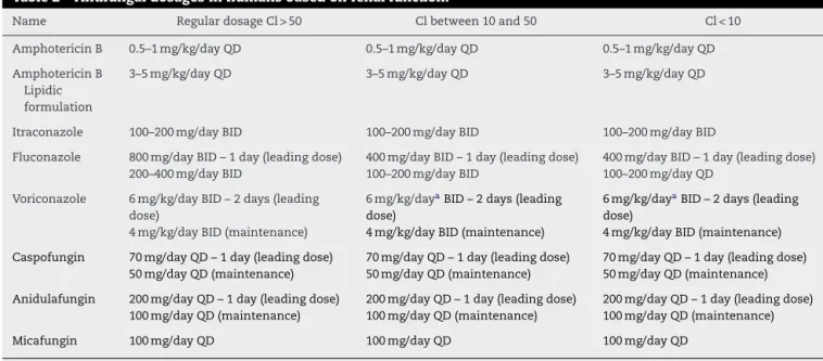
Tables 1 and 2show the pharmacological aspects and antifun-gal dosages for systemic use.
Below, we discuss the treatment of major infections caused byCandida. The recommendations for therapy are indicated for adult patients and were based on levels of evidence accord-ing to the strength of the recommendation and the quality of evidence from the American Society of Infectious Diseases, adapted from the Canadian Ministry of Health,91as shown in Table 3.
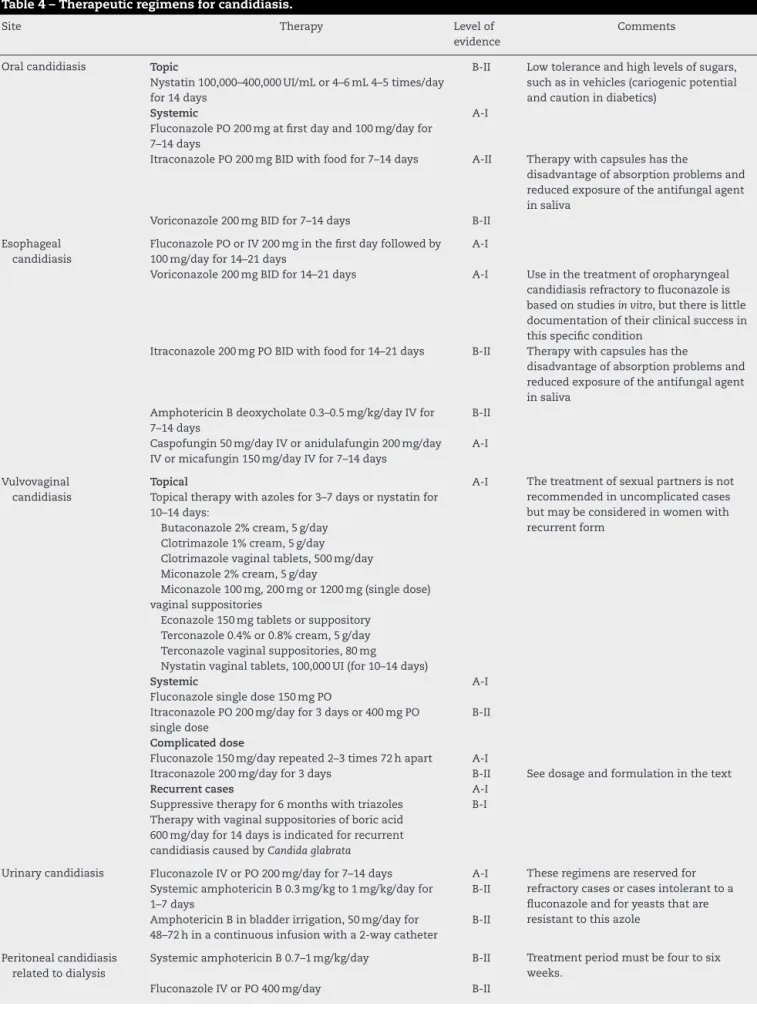
Each topography was discussed with regard to epidemi-ological, clinical and laboratory diagnostic and therapeutic recommendations. The therapeutic options for treating can-didiasis are summarized inTable 4.
Treatment
Oral candidiasis
Epidemiological aspects
Table 2 – Antifungal dosages in humans based on renal function.
Name Regular dosage Cl > 50 Cl between 10 and 50 Cl < 10
Amphotericin B 0.5–1 mg/kg/day QD 0.5–1 mg/kg/day QD 0.5–1 mg/kg/day QD
Amphotericin B Lipidic formulation
3–5 mg/kg/day QD 3–5 mg/kg/day QD 3–5 mg/kg/day QD
Itraconazole 100–200 mg/day BID 100–200 mg/day BID 100–200 mg/day BID
Fluconazole 800 mg/day BID – 1 day (leading dose) 200–400 mg/day BID
400 mg/day BID – 1 day (leading dose) 100–200 mg/day BID
400 mg/day BID – 1 day (leading dose) 100–200 mg/day QD
Voriconazole 6 mg/kg/day BID – 2 days (leading dose)
4 mg/kg/day BID (maintenance)
6 mg/kg/dayaBID – 2 days (leading dose)
4 mg/kg/day BID (maintenance)
6 mg/kg/dayaBID – 2 days (leading dose)
4 mg/kg/day BID (maintenance)
Caspofungin 70 mg/day QD – 1 day (leading dose) 50 mg/day QD (maintenance)
70 mg/day QD – 1 day (leading dose) 50 mg/day QD (maintenance)
70 mg/day QD – 1 day (leading dose) 50 mg/day QD (maintenance)
Anidulafungin 200 mg/day QD – 1 day (leading dose) 100 mg/day QD (maintenance)
200 mg/day QD – 1 day (leading dose) 100 mg/day QD (maintenance)
200 mg/day QD – 1 day (leading dose) 100 mg/day QD (maintenance)
Micafungin 100 mg/day QD 100 mg/day QD 100 mg/day QD
Cl, creatinine clearance (mL/min).
a Avoid the use of IV voriconazole in patients with creatinine clearance <50 mL/min (toxicity risk). There are no restrictions for use of the oral
formulation in cases of renal failure.
Table 3 – Strength of recommendation and quality of evidence.
Category Definition
Strength of recommendation
A Strong evidence to support recommendation B Moderate evidence to support recommendation C Poor evidence to support recommendation
Quality of evidence
I Evidence of≥1 randomized controlled clinical trial II Evidence of≥1 well-designed clinical trial, not
randomized, cohort or case–control studies (preferably more than one center), or multiple sets of results of uncontrolled studies
III Evidence based on expert opinion or clinical experience, descriptive studies or committee reports
associated with oral candidiasis in adult patients are AIDS, diabetes and exposure to antibiotics and/or corticosteroids for different conditions. Therefore, all adult patients presenting with oral candidiasis without obvious cause should be inves-tigated for HIV infection.94
C. albicansaccounts for approximately 90% of the isolates causing oroesophageal candidiasis, but C. tropicalis, C. kru-sei, C. glabrata,C. parapsilosisand C. dubliniensiscan also be detected.95 In AIDS patients unresponsive to antiretroviral therapy, episodes of oropharyngeal candidiasis become recur-rent, requiring prolonged use or repeated cycles of therapy with triazoles. In this scenario, there is an increase in episodes of candidiasis byCandida non-albicansisolates resistant to flu-conazole or even in the risk of selecting resistant strains ofC. albicansto this drug.96
Clinical and laboratory diagnosis
Clinical manifestations are varied and depend on the host’s immune status and the extent of oral candidiasis. The largest
clinical experience of infectious disease is in the form of pseudomembranous candidiasis. The most common symp-toms are oral discomfort, burning pain and the presence of removable white plaque under erythematous mucosa. These conditions make feeding difficult, and they can com-promise the regularity of oral drug treatments.97 However, other clinical presentations are known. Erythematous can-didiasis presents itself as erythematous infiltrate with reduced papillae when present on the tongue. Patients using dental prostheses with oral candidiasis have chronic erythema and discomfort in the region of the prosthesis. Angular cheilitis caused byCandida spp.manifests as discomfort, erythema, and fissures in the angular region of the lips.98
The clinical presentation is usually very characteristic of this condition, particularly when it is pseudomembranous. However, clinical diagnosis should be confirmed by laboratory investigation as follows: (a) by direct mycological examination, with scrapes of lesions in KOH preparations or by Gram stain-ing, where the specimen is analyzed by the presence of fungal elements consistent withCandida spp. and/or (b) by culturing in selective fungal medium (preferably chromogenic medium to identify different species), where the yeast is isolated and the agent is forwarded to complete identification.99
Culture is particularly important in cases of recurrent can-didiasis in patients with AIDS, in cases of poor response to conventional therapy or when an injury that is suggestive of candidiasis arises in patients receiving any antifungal drug. In these situations, the identification of the agent species and testing for susceptibility to antifungal agents are necessary recommendations for optimizing a new therapeutic indica-tion in view of the possibility of infecindica-tion by strains ofCandida spp. resistant to one or all triazoles.100,101
Therapeutic recommendations
Table 4 – Therapeutic regimens for candidiasis.
Site Therapy Level of
evidence
Comments
Oral candidiasis Topic
Nystatin 100,000–400,000 UI/mL or 4–6 mL 4–5 times/day for 14 days
B-II Low tolerance and high levels of sugars, such as in vehicles (cariogenic potential and caution in diabetics)
Systemic
Fluconazole PO 200 mg at first day and 100 mg/day for 7–14 days
A-I
Itraconazole PO 200 mg BID with food for 7–14 days A-II Therapy with capsules has the
disadvantage of absorption problems and reduced exposure of the antifungal agent in saliva
Voriconazole 200 mg BID for 7–14 days B-II
Esophageal candidiasis
Fluconazole PO or IV 200 mg in the first day followed by 100 mg/day for 14–21 days
A-I
Voriconazole 200 mg BID for 14–21 days A-I Use in the treatment of oropharyngeal candidiasis refractory to fluconazole is based on studiesin vitro, but there is little documentation of their clinical success in this specific condition
Itraconazole 200 mg PO BID with food for 14–21 days B-II Therapy with capsules has the
disadvantage of absorption problems and reduced exposure of the antifungal agent in saliva
Amphotericin B deoxycholate 0.3–0.5 mg/kg/day IV for 7–14 days
B-II
Caspofungin 50 mg/day IV or anidulafungin 200 mg/day IV or micafungin 150 mg/day IV for 7–14 days
A-I
Vulvovaginal candidiasis
Topical
Topical therapy with azoles for 3–7 days or nystatin for 10–14 days:
A-I The treatment of sexual partners is not recommended in uncomplicated cases but may be considered in women with recurrent form
Butaconazole 2% cream, 5 g/day Clotrimazole 1% cream, 5 g/day Clotrimazole vaginal tablets, 500 mg/day Miconazole 2% cream, 5 g/day
Miconazole 100 mg, 200 mg or 1200 mg (single dose) vaginal suppositories
Econazole 150 mg tablets or suppository Terconazole 0.4% or 0.8% cream, 5 g/day Terconazole vaginal suppositories, 80 mg
Nystatin vaginal tablets, 100,000 UI (for 10–14 days) Systemic
Fluconazole single dose 150 mg PO
A-I
Itraconazole PO 200 mg/day for 3 days or 400 mg PO single dose
B-II
Complicated dose
Fluconazole 150 mg/day repeated 2–3 times 72 h apart A-I
Itraconazole 200 mg/day for 3 days B-II See dosage and formulation in the text
Recurrent cases A-I
Suppressive therapy for 6 months with triazoles B-I Therapy with vaginal suppositories of boric acid
600 mg/day for 14 days is indicated for recurrent candidiasis caused byCandida glabrata
Urinary candidiasis Fluconazole IV or PO 200 mg/day for 7–14 days A-I These regimens are reserved for refractory cases or cases intolerant to a fluconazole and for yeasts that are resistant to this azole
Systemic amphotericin B 0.3 mg/kg to 1 mg/kg/day for 1–7 days
B-II
Amphotericin B in bladder irrigation, 50 mg/day for 48–72 h in a continuous infusion with a 2-way catheter
B-II
Peritoneal candidiasis related to dialysis
Systemic amphotericin B 0.7–1 mg/kg/day B-II Treatment period must be four to six weeks.
Table 4 – (Continued)
Site Therapy Level of
evidence
Comments
Postoperative peritoneal candidiasis
Systemic amphotericin B 0.7–1 mg/kg/day B-II
Fluconazole IV or PO 400 mg/day B-II
Echinocandins B-I
Respiratory tract candidiasis
Upon confirmation of a diagnosis of pneumonia, the choice of antifungal should be made as discussed in the section on acute disseminated candidiasis; there may be choice between echinocandins, fluconazole or amphotericin B formulations
B-II The finding of a positive culture for Candida spp.in respiratory tract samples should be taken as evidence of
colonization of this site, where the risk of pneumonia is generally low
Hematogenous candidiasis
Non-neutropenic patients
Anidulafungin IV 200 mg at first day followed by 100 mg/day IV
A-I
Caspofungin 70 mg IV at first day followed by 50 mg/day IV
A-I
Micafungin EV 100 mg/day A-I Considered for sequential therapy to
complete the minimum period of 14 days of treatment after the definition of the agent and upon favorable documentation of clinical response to treatment with echinocandins. Medical centers with rates of incidence exceeding 10% of fluconazole-resistant strains should not use fluconazole in any patient before the identification of the agent
Fluconazole IV 800 mg/day at first day followed by 400 mg/day
B-I
Amphotericin B liposomal formulation 3 mg/kg/day B-I A liposomal formulation and amphotericin B are alternatives for patients who are not responsive to echinocandins, who are intolerant to the therapeutic class or who develop endocarditis or meningitis Amphotericin B in lipidic complex from 3 mg/kg/day to
5 mg/kg/day
B-II The duration of antifungal therapy should be at least 14 days after negative cultures and the disappearance of signs and symptoms related to hematogenous candidiasis
Hematogenous candidiasis
Neutropenic patients
Echinocandins A-I The doses and treatment time should
meet the same criteria established for non-neutropenic patients
Amphotericin B liposomal formulation B-I
Amphotericin B in lipidic complex B-II
Evidence of endophthalmitis
Fluconazole B-III
Voriconazole B-III
Antifungal therapy is recommended for a period of four to six weeks, with monitoring by an ophthalmologist for further characterization of the treatment time and treatment response
Evidence of endocarditis
Amphotericin B in lipidic complex (1st choice) B-II
Echinocandins (alternative) B-II
Fluconazole (sequential use) B-II
Chronic disseminated candidiasis B-II
Amphotericin B deoxycholate 0.6–0.7 mg/kg/day B-II Fluconazole should be used whenCandida species are susceptible and the patient is clinically stable, always after a long period of treatment with formulations of amphotericin or echinocandin
Table 4 – (Continued)
Site Therapy Level of
evidence
Comments
Fluconazole 6 mg/kg/day in stable and non-neutropenic patients, with no previous use of fluconazole
B-II
Echinocandins in regular dosage The antifungal should be used until
complete resolution of the abscesses identified in imaging
recurrence.92 Topical therapy is recommended for patients without HIV/AIDS (B-I) and for the initial episodes of crypto-coccosis in patients with HIV/AIDS (A-I).
Topical therapy (uncomplicated infection)
Nystatin 100,000–400,000 IU/mL and 4–6 mL four to five times a day for 14 days (B-II) should be administered. Successful treat-ment depends on the time of contact with the oral mucosa for at least two minutes. It is worth mentioning that this drug has a low tolerance and high sugar content as a vehicle. It also has cariogenic potential and should be used with caution in diabetic patients.98
In the U.S. and Europe, an oral clotrimazole solution is avail-able for use three to five times a day for 14 days (B-II). In these countries, topical therapy is the rule in mild and/or early candidiasis, even in patients with AIDS.102Unfortunately, in Brazil, clotrimazole is not available in formulations suitable for oral use. In this context, in view of the difficulties in handling nystatin, topical therapy is restricted to only a few patients.
Systemic therapy
The best therapeutic option for systemic candidiasis is oral fluconazole; the other options are considered only in patients unresponsive or intolerant to this drug (A-I).100Fluconazole 200 mg PO in the first day and 100 mg/day for 7–14 days (A-I).
In patients with oropharyngeal candidiasis refractory to fluconazole, the options are as follows:
• Itraconazole 200 mg orally BID with food for 7–14 days (A-II).103,104 Considering that in Brazil we do not have an oral solution, capsules have the disadvantage of impaired absorption and less exposure of the antifungal agent in saliva (B-III).
• Voriconazole 200 mg BID for 7–14 days. This drug has been
validated in comparative clinical trials with fluconazole in patients with esophageal candidiasis (A-I).105Its use in oral therapy for oropharyngeal candidiasis refractory to fluconazole is based on in vitro studies, but with limited documentation of their clinical success for this specific con-dition (B-II).
• Posaconazole 200 mg PO on the first day followed by 100 mg orally QD for 13 days for primary therapy (A-I) or 400 mg TID for 3 days, followed by 400 mg QD for 25 days for refractory cases (B-II). This drug has been validated for this indica-tion in two clinical trials: a randomized comparison with fluconazole and an open study for refractory cases.106,107Its
indication should be reserved for cases of poor response to fluconazole (B-I). This drug is not available in Brazil.
• Amphotericin B deoxycholate 0.3–0.5 mg/kg/day IV for 7–14 days (B-II).108This drug should be reserved for cases refrac-tory to fluconazole (B-II).
• Caspofungin 50 mg/day IV or anidulafungin 200 mg/day IV
or micafungin 150 mg/day IV for 7–14 days. These drugs have been validated in clinical trials comparing flucona-zole in patients with esophageal candidiasis (A-I).109–111The use of these drugs should be reserved for treatment of esophageal candidiasis refractory to fluconazole (B-I).
Given that oral candidiasis is related to the imbalance between the colonizing agent and the local or systemic defense mechanisms, we should try to act toward control of the underlying disease and/or removal of the predisposing conditions. Otherwise, the trend favors chronicity of the pro-cess, as it occurs in patients with prostheses and AIDS that is unresponsive to antiretroviral therapy.
Esophageal candidiasis
Epidemiological aspects
Esophageal candidiasis is considered a form of semi-invasive candidiasis that primarily affects patients with AIDS, can-cer, diabetes, previous esophageal diseases, malnutrition and alcoholism, along with those in therapies using corticoste-roids, antibiotics, H2 receptor antagonists and proton-pump inhibitors.92 In clinical practice, most cases of esophageal candidiasis occur in AIDS patients, followed by lower frequen-cies of diabetics and critically ill patients exposed to multiple antibiotic cycles.99
Clinical and laboratory diagnosis
Candida esophagitis can be oligosymptomatic, but its main clinical manifestations include dysphagia, odynophagia and retroesternal burning. In children, nausea, vomiting and dehydration are the main signs. Although the presence of concomitant oral and esophageal candidiasis is common, par-ticularly in AIDS patients, the absence of oral candidiasis does not exclude esophagitis diagnosis. Complications include bleeding, perforation and stenosis.101
lesions. Apart from the morphological findings, it is rec-ommended to perform a scrap (brush) to obtain a sample for microscopic examination and culturing, in addition to a mucosal biopsy.99
The microscopic examination of fungal elements is per-formed with a sample obtained by scraping on a slide with KOH or by Gram stain. The culture is performed with a sample obtained by scraping or biopsy. A biopsy should be processed with hematoxylin–eosin staining and silver methenamine (Grocott).99
The definitive diagnosis of esophageal candidiasis is made when, in addition to the clinical and morphological endo-scopic findings, we identify fungal elements on microendo-scopic examination and/or observe the presence of fungal elements in tissue, confirming invasion by the pathogen. From an aca-demic point of view, the isolated identification ofCandidain culture but no fungal elements by microscopic examination and biopsy may represent colonization of the gastrointestinal tract and not infection.101
Therapeutic recommendations
Systemic therapy is recommended for cases of esophageal candidiasis (B-II). This starts with empirical systemic therapy (A-I) with fluconazole 200 mg PO or IV in the first day, followed by 100 mg QD for 14–21 days (A-I). When endoscopy is not per-formed at the time of diagnosis, it should be perper-formed if no improvement occurs within 3–5 days.95
In patients with esophageal candidiasis refractory to flu-conazole, the options are as follows:
• Voriconazole 200 mg BID for 14–21 days. This drug was validated in a comparative clinical trial with fluconazole in patients with esophageal candidiasis (A-I).105Its use in the treatment of esophageal candidiasis refractory to flu-conazole may have a compromised result due to eventual cross-resistance; however, it is a good indication for suscep-tibility tests, if available (B-II).
• Itraconazole 200 mg PO BID with food for 14–21 days (A-II).103,104Given that there is no oral formulation in Brazil and cross-resistance is commonly observed across triazoles, treatment with capsules presents problems with absorption and lesser exposure of the drug to the saliva. These factors can compromise treatment success.
• Posaconazole 200 mg PO on the first day followed by 100 mg PO QD for 13 days for primary therapy (A-I), or 400 mg BID for 3 days followed by 400 mg QD for 25 days for refractory cases (B-II). This drug was validated for this indication in two clin-ical trials: one controlled and randomized with fluconazole and another open-label for refractory cases.106,107 Its use for esophageal candidiasis refractory to fluconazole may be compromised by an eventual cross-resistance; however, it is a good indication for susceptibility tests, if available (B-II). This drug is not available in Brazil.
• Amphotericin B deoxycholate 0.3–0.5 mg/kg/day IV for 7–14 days (B-II).108
• Caspofungin 50 mg/day IV or anidulafungin 200 mg/day IV or micafungin 150 mg/day IV for 7–14 days. These drugs were validated in comparative clinical trials with fluconazole in patients with esophageal candidiasis (A-I).109–111
Vulvovaginal candidiasis
Epidemiological aspects
Vaginal candidiasis is highly prevalent in women during their childbearing life; approximately 75% have at least one episode lifelong, and 5–10% can develop a recurrence (defined as at least four episodes of vaginitis by Candida spp. within one year).112
The most frequent predisposing factors for vaginal candidi-asis include exposure to high levels of estrogens (birth control, pregnancy and hormone replacement), uncontrolleddiabetes mellitus, use of topical and systemic antibiotics and inadequate hygiene habits. Most women with recurrent vaginal candidia-sis do not have underlying diseases associated with systemic immunosuppression, and recurrence may be secondary to a deficiency in the local immune response to the agent.113
Vulvovaginal candidiasis is usually classified as compli-cated or uncomplicompli-cated, pending on the severity of the clinical presentation and basic conditions of the host. Uncomplicated forms of vaginitis account for more than 90% of cases and have an excellent response to short oral or topical therapy. Patients with more complicated vaginitis require a prolonged antimycotic therapy.114
C. albicansis the most frequent cause of vaginitis, account-ing for approximately 74–95% of cases, followed byC. glabrata in approximately 14.5% of cases. Thenon-albicansspecies are more common in recurrent forms and may be found in 10–20% of these patients.C. glabratais the species most frequently identified in these cases.115,116
Clinical and laboratory diagnosis
Considering that 30% of women may haveCandida coloniza-tion and there is a wide differential diagnosis for infectious leukorrhea, the diagnosis ofC. vulvovaginitisshould be based on clinical and laboratory findings.117
Candidiasis involves the vulva and the vaginal lumen, causing intense itching, burning, local discomfort, dysuria, vaginal discharge and dyspareunia. Clinical examination revealed swelling and redness of the vulva and/or vagina, vaginal discharge that looks like milk and, eventually, vulvar carved cracks.118
Clinical diagnosis must be performed by the following tests:117
• Direct microscopic examination with the addition of KOH (10%) or Gram stain to search for fungal elements, comple-mented by evaluation of the vaginal pH (infection usually occurs with a pH between 4 and 4.5);
• Culture in specific material. To decrease costs, some authors recommend prompt culture only for complicated or recur-rent vulvovaginal candidiasis.
Therapeutic recommendations
in 80–90% of patients who completed therapy (A-I). Generally, higher concentrations and doses of topical medications are effective over a period of 3 days. Lower doses of the same for-mulations require more prolonged therapy.102The options for topical therapy are numerous and include the following:
• Butaconazole 2% cream, 5 g/day;
• Clotrimazole cream 1%, 5 g/day;
• Clotrimazole vaginal tablets, 500 mg/day;
• Miconazole 2% cream, 5 g/day;
• Miconazole, 100 mg, 200 mg or 1200 mg (single dose), vaginal suppositories;
• Econazole, 150 mg, tablet or suppository;
• Terconazole 0.4% or 0.8% cream, 5 g/day;
• Terconazole, 80 mg, vaginal suppositories;
• Nystatin, 100,000 IU vaginal tablets (10–14 days).
There are formulations containing combination therapy with other agents that will not be commented upon in the text:
• Systemic therapy: the use of oral triazoles is a safe and effi-cient alternative to topical therapy. There is a large amount of clinical experience in treating vulvovaginal candidiasis with fluconazole 150 mg QD, single dose (A-I).102 Another option to this drug is itraconazole 200 mg QD for 3 days or 400 mg single dose (B-II).120Systemic therapy with triazoles is not indicated in pregnant women. The treatment of sex-ual partners is not recommended in uncomplicated cases but may be considered in recurrent cases.121
Complicated vulvovaginal candidiasis.
• Moderate and severe cases and/or immunocompromised patients: prolonged topical and systemic therapy should be administered to these patients. Topical therapy is recom-mended for at least 7–14 days using any of the formulations listed above (A-I).102In case of systemic therapy, the follow-ing drugs can be considered:
◦ Fluconazole 150 mg/day, repeated two or three times 72 h apart (A-I);
◦ Itraconazole 200 mg/day for 3 days (B-II).
Recurrent vulvovaginal candidiasis.
• If the diagnosis of recurrent vulvovaginal candidiasis is made and if there is no identification of or possibility to con-trol or remove the triggering factors, suppressive therapy with triazoles for six months is an effective control measure for recurrent episodes (A-I).122
• In such patients, attack therapy can be administered with any of the topical formulations listed above for 7–14 days (A-I) or fluconazole 150 mg/day each 72 h (days 1, 4 and 7) or until complete symptoms remission; this is the preferred regimen in clinical practice. Once the initial episode is con-trolled, maintenance therapy with fluconazole 150 mg/day once a week for six months is indicated (A-II).122
• Although the largest clinical experience of suppressive ther-apy for recurrent candidiasis was with fluconazole (A-I), there are published trials that suggest maintenance therapy
with clotrimazole 500 mg suppositories twice a week or itra-conazole (200 mg PO twice a week or 200 mg PO BID monthly) (B-I).123,124
• Cases of vulvovaginal candidiasis caused byC. glabratamay not respond to fluconazole. In these cases, vaginal suppos-itories of boric acid 600 mg/day for 14 days are indicated (B-I).125
Urinary candidiasis
Epidemiological aspects
The term candiduria refers to the growth of Candida spp. in urine cultures collected by appropriate techniques; this finding is not necessarily accompanied by signs and/or symptoms of urinary tract infection (UTI). Candiduria is very frequent among patients exposed to risk factors; up to 20% of hospitalized patients may have candiduria dur-ing their hospitalization, particularly intensive care unit (ICU) patients.126,127This laboratory finding fosters dilemmas regarding its interpretation, as it can represent a simple con-tamination of the urine collection, candiduria asymptomatic cystitis or pyelonephritis, primary renal candidiasis, uretero-pelvic fungus ball or disseminated candidiasis with renal manifestations.
Among hospitalized patients, the factors most often related to the development of candiduria are advanced age, female gender, broad-spectrum antibiotics, the use of corticosteroids and immunosuppressive drugs, the pres-ence of urinary tract abnormalities, diabetes, delayed vesical catheterization, postoperative of major surgery and malignancies.127,128
Series of cases from Brazil confirm that the three most prevalent species isolated from urine in hospitalized patients areC. albicans,C. tropicalisandC. glabrata. These studies mea-sure prevalences ranging from 35.5 to 70% for C. albicans, 4.6–52.5% forC. tropicalisand 7–8.8% forC. glabrata.129–132
Clinical and laboratory diagnosis
In outpatients not exposed to the risk factors mentioned, in most cases, the identification ofCandidain urine reflects inadequate collection or processing of the sample and con-sequent contamination of the culture. In patients exposed to risk factors for UTI byCandida, the finding of candiduria may signify colonization or infection. In these patients, the count-ing of colonies is highly variable and directly dependent on the methodology used to collect material. Thus, the isolation ofCandidain the urine may occur even in the absence of dis-ease, and there is considerable controversy regarding the value of colony counts obtained in culture, a procedure with low specificity and sensitivity in differentiating between patients colonized and infected by this agent.12
value for the interpretation of quantitative urine cultures for the recognition of patients with infection of the lower UTI or pyelonephritis.135
Therapeutic recommendations
• The best therapeutic approach for patients with candiduria should be defined on individual basis, considering clinical and epidemiological data to classify each patient into one of the following conditions: (1) no prior risk factors for can-diduria, (2) exposure to risk factors but unlikely to be a case of disseminated candidiasis, or (3) exposure to risk factors for candiduria with septicemia without defining etiology and possible/probable systemic dissemination.102,12
• The therapeutic approach suggested for these three differ-ent scenarios are the following. (1) No prior risk factors for candiduria: in this category, we have patients without underlying diseases who did not undergo catheterization and who have no history of previous use of corticoste-roids and antibiotics. They should not receive systemic antifungal agents. It is recommended to request a new collection of material and, if yeasts are found, to inves-tigate the possibility of fungal genital mucositis in the vagina or the glans (C-III).136(2) Predisposed to candiduria, but unlikely to be disseminated candidiasis: this cate-gory includes asymptomatic outpatients or inpatients who underwent catheterization and/or other predisposing fac-tors for candiduria. In these patients, the initial approach is the removal of the predisposing factors with subsequent clinical and laboratory follow-up (C-III). In the vast major-ity of patients, candiduria resolves after the introduction of these measures. Patients with symptoms of cystitis and with positive urine for yeasts should be treated with anti-fungal agents (B-III).102,136 (3) Predisposed to candiduria with probable systemic dissemination: critically ill patients with risk factors for systemic fungal infection and who evolve with candiduria and signs of sepsis should be inves-tigated for invasive candidiasis (blood) and should begin the use of systemic antifungal drugs. This means that the patient is not merely colonized (C-III).102
• If there are indications for treatment, treatment regimens include the following:
– Fluconazole, oral or intravenous dose of 200 mg/day for 7–14 days (A-I).137
– Amphotericin B, systemic dose of 0.3 mg/kg to 1 mg/kg/day for 1–7 days (B-II) or amphotericin B, bladder irrigation, 50 mg/day for 48–72 h with continuous infusion in a two-way tube (B-II). These schemes are reserved for cases refractory infections or those intol-erant to fluconazole, along with yeasts resistant to this azole.102,138
– In case of suspicion of systemic candidiasis, the patient should be treated according to the recommendations for hematogenous candidiasis.102
– Clinical experience with candiduria and echinocandins or voriconazole is restricted; pharmacological data suggest that the urinary concentrations of both antifungals are reduced.139
– In the clinical management of patients with can-diduria, it is important to consider the removal of
the catheterization system, taking into account that this measure may resolves approximately 40% of cases, besides reducing the recurrence of infection (B-I).139If it is not possible to remove the system, it is at least recom-mended to change it.140
Peritoneal candidiasis related to dialysis
Epidemiological aspects
Peritoneal dialysis is a modality of renal replacement therapy that currently accounts for only 10–20% of dialysis modalities. It can be performed continuously with an oriented procedure performed at home or intermittently, which has been com-pletely abandoned. Among the complications of peritoneal dialysis, infection ranks second place after cardiovascular events, and fungal infections account for 2–14% of peritoni-tis cases.141The overall mortality in most series ranges from 10 to 25% of cases, and there are a few reports of up to 50% deaths.142Among the fungal peritonitis diseases, 80–90% are caused byCandida, particularly isolates ofC. albicans,C. para-psilosisandC. tropicalis.143The risk factors for the occurrence of fungal peritonitis in patients on peritoneal dialysis are not completely known.144 The basic conditions most commonly reported in patients with fungal peritonitis include diabetes, the prior occurrence of peritonitis by other agents and the previous use of antibiotics.145
Clinical and laboratory diagnosis
Diagnosis is made through clinical signs and symptoms of peritonitis, which are represented by abdominal pain, dis-tention, and fever associated with clouding of the dialysis fluid, whose cell count increases due to the neutrophil count (>100 leukocytes/mm3). Etiologic evidence is obtained by
iden-tification of yeasts in bacterioscopic examination of the peritoneal fluid, with growth ofCandida spp.in culture.141,145
Therapeutic recommendations
The guidelines for the treatment of fungal peritonitis are based on case reports and open-label studies of limited groups of patients. Among the key recommendations for the treat-ment of this complication, the authors suggest that the early removal of the dialysis catheter is essential to the success of the therapy (B-II).146
The largest experience in the treatment of fungal peritoni-tis is with fluconazole or amphotericin B (B-II). Many authors recommend starting with amphotericin and completing treat-ment with fluconazole after clinical improvetreat-ment (B-II).146
Some authors suggest the use of intraperitoneal flucona-zole concomitantly with the systemic use of amphotericin B (C-III).147The treatment period is usually four to six weeks. It is essential to monitor the patient by abdominal ultrasound to rule out collections and to guide the treatment time (B-III).146 There is little reliable information regarding doses of antifungal agents, but the authors suggest the use of 0.7 mg/kg to 1 mg/kg/day of amphotericin B and 400 mg/day of fluconazole.148