Angiotensin II, progesterone, and prostaglandins are sequential
steps in the pathway to bovine oocyte nuclear maturation
Lucas Carvalho Siqueira
a, Marcos Henrique Barreta
a, Bernardo Gasperin
a,
Rodrigo Bohrer
a, Joabel Tonellotto Santos
a, José Buratini Junior
b,
João Francisco Oliveira
a, Paulo Bayard Gonçalves
a,*
aLaboratory of Biotechnology and Animal Reproduction - BioRep, Federal University of Santa Maria, Santa Maria, RS, Brazil bDepartament of Physiology, State University of São Paulo (UNESP), Botucatu, SP, Brazil
Received 28 April 2011; received in revised form 12 December 2011; accepted 15 December 2011
Abstract
Oocyte meiotic resumption is triggered by the ovulatory gonadotropin surge; in cattle, angiotensin II (AngII) and prostaglan-dins (PG) are key mediators of this gonadotropin-induced event. Here, we tested the hypothesis that progesterone (P4) is also
involved in oocyte meiotic resumption induced by the gonadotropin surge. In Experiment I, P4induced nuclear maturation in a
dose-dependent manner using a coculture of follicular hemisections and cumulus-oocyte complexes. In the second experiment, using an in vivomodel, an injection of mifepristone (MIFE; P4 receptor antagonist) at the antrum of preovulatory follicles
prevented GnRH-induced oocyte meiotic resumptionin vivo. In Experiment III (coculture system similar to that of Experiment I), MIFE prevented stimulatory effects of AngII on resumption of meiosis, but saralasin (AngII receptor antagonist) did not inhibit P4actions. In Experiments IV and V, fibroblast growth Factor 10 (FGF10; known to suppress steroidogenesis in granulosa cells),
blocked AngII-but not P4-induced oocyte meiotic resumption. Therefore, we inferred that AngII is upstream to P4in a cascade
to induce meiotic resumption. Previously, we had reported that AngII acted throughout the PGs pathway to modulate nuclear progression. In Experiment V, indomethacin inhibited resumption of meiosis induced by P4, providing further support to the
AngII-P4sequential effect on meiotic resumption. In conclusion, we inferred that AngII, P4and PGs are sequential steps in the
same pathway that culminates with bovine oocyte maturation. © 2012 Elsevier Inc. All rights reserved.
Keywords:Ovulation; Meiotic resumption; Angiotensin; Steroid; Eicosanoid
1. Introduction
The preovulatory gonadotropin surge triggers a cas-cade of events that culminates with ovulation and nuclear oocyte maturation. Recently, angiotensin II (AngII) has been recognized as one of the earliest mediators of gonadotropin-induced ovulation and oocyte maturation
[1–3]. The positive effect of AngII in these processes is mediated through a Type 2 receptor[1]. Furthermore, the concentration of AngII and expression of its receptors (AT2) within the follicle increased during the interval between the gonadotropin surge and ovulation (Siqueira, et al, unpublished data). Other studies provided additional evidence that AngII regulated secretion of progesterone (P4) and prostaglandins (Pg), hormones involved in ovu-lation[4,5]. In granulosa cell culture, AngII upregulated expression of cyclooxygenase 2 (COX-2), the rate-limit-ing enzyme for PG production[3].
* Corresponding author. Tel:⫹55 55 3220 8752; fax:⫹55 55 3220 8484.
E-mail address:[email protected](P.B. Gonçalves).
Theriogenology 77 (2012) 1779–1787
www.theriojournal.com
During follicle development, bovine oocytes remain arrested at prophase of the first meiotic division, and resume meiosis after the preovulatory LH surge[6], or after removal from the follicular environment[7]. The presence of follicular wall fragments in a coculture system with cumulus-oocyte complexes (COCs) pre-vents meiotic resumption[8]. This coculture system is a good model to study the role of factors that act through follicular cells on oocyte nuclear maturation
[9,10]. Using this coculture system, we reported that AngII acted through a PG pathway to mediate gonad-otropin-induced oocyte meiotic resumption[2].
The cyclooxygenase pathway is a classical mediator of LH-induced ovulation and nuclear oocyte maturation in cattle[11–15]. Progesterone is another key element in the ovulatory cascade and oocyte maturation
[13,14,16]. Indeed, there are indications that PGs are downstream factors to this steroid; in that regard, a gonadotropin surge stimulates an increase in intrafol-licular P4,which acts by binding to its nuclear receptor and increasing abundance of mRNA for COX2 [14]. The role of P4on oocyte nuclear maturation in cattle remains controversial. Nuclear and membrane proges-terone receptors are present in bovine COCs, and reg-ulated duringin vitromaturation in the presence of FSH and LH[16]. Although Sirotkin[17]reported a stimu-latory effect on oocyte meiotic resumption, more recent studies concluded that P4was not necessary to promote nuclear maturation, cumulus expansion, and early em-bryo development[18,19].
Follicular cells secrete factors that prevent oocyte meiotic resumption before the LH surge. The family of fibroblast growth factors (FGFs) is composed of more than 20 factors, largely studied for their roles in em-bryogenesis and oogenesis. Buratini, et al[20]reported that the bovine theca cells and oocytes expressed FGF10. Expression of FGF10 receptor (FGFR2IIIb) was identified in theca[21], granulosa[20], and cumu-lus cells[22]. Furthermore, FGF10 in the granulosa cell culture inhibited steroidogenesis[20]and AT2 expres-sion [23]. Activation of FGF receptors (FGFRs) ap-peared to be involved in inhibition of germinal vesicle breakdown (GVBD) in mice[24]. Conversely, Zhang, et al[25]reported that FGF10 improved bovine oocyte maturation, cumulus expansion and subsequently em-bryo development in medium containing estradiol and in the absence of follicular cells.
The information summarized above provided an im-petus to investigate interactions between FGF10 and factors involved in triggering bovine oocyte meiotic resumption. In the present study, a combination ofin
vivoandin vitroexperiments were conducted to test the hypothesis that P4plays a role in regulation of oocyte meiotic progression induced by gonadotropin surge in concert with AngII and PGs. In anin vitroexperiment, interactions of P4 and AngII with FGF10 (an antis-teroidogenic factor recently described as an important regulator of follicular development) were studied, with regards to their roles in resumption of meiosis.
2. Materials and methods
All experimental procedures were reviewed and ap-proved by the Federal University of Santa Maria Ani-mal Care and Use Committee (23081.004717/2010 –53 CCR/UFSM). All chemicals used were purchased from Sigma Chemical Company (St. Louis, MO, USA), un-less otherwise indicated in the text.
2.1. Preparation of follicular hemisections, oocyte recovery and nuclear maturation
Bovine ovaries at various stages of the estrous cycle were obtained from an abattoir and transported to the laboratory in saline solution (0.9% NaCl) at 30 °C containing 100 IU ml⫺1 penicillin and 50
g ml⫺1 streptomycin sulfate. Procedures for follicle dissection and culture procedures were previously validated in our laboratory[2,9,10]. Briefly, transparent follicles, 2 to 5 mm in diameter, were selected and dissected from ovar-ian stromal tissue, and sectioned into halves. Follicular hemi-sections were washed in TCM 199 containing 0.4% bovine serum albumin (BSA) and randomly dis-tributed into four-well culture dishes (Nunc, Roskilde, Denmark) containing culture medium with the desired treatment. There were eight follicular halves per 200l of medium. Dishes were incubated for 2 h before add-ing COCs.
The COCs were aspirated from follicles 3 to 8 mm in diameter, recovered under a stereomicroscope, and selected according to Leibfred and First[26]. Grades 1 and 2 COCs (n⫽10 to 30) were randomly distributed into treatments and cultured in an incubator at 39 °C in a saturated humidity atmosphere containing 5% CO2in air and 95% air, for either 7, 15, or 24 h, depending on the experiment. The culture medium used was TCM 199 containing Earle’s salts and L-glutamine (Gibco BRL, Grand Island, NY, USA) supplemented with 25 mMHEPES, 0.2 mMpyruvic acid, 2.2 mg ml⫺1
culture period, cumulus cells were removed by vortex-ing for 5 min and oocytes were fixed with 4% para-formldehyde for 15 min, followed by permeabilization of the nuclear membranes with 0.5% Triton X-100. After 2 h, oocytes were fixed, stained with Hoechst (33,342) and mounted under a coverslip with Vectashield (Vector Laboratories, Burlington, Ontario, L7N 3J5, Canada) for nuclear evaluation. Oocytes were classified according to their nuclear chromatin config-uration using a fluorescent microscope as germinal ves-icle (GV), GV breakdown (GVBD), metaphase I (MI), anaphase I (AI), telophase I (TI), and metaphase II (MII). In all experiments, all treatments were repeated three times.
2.2. Cattle, superovulation protocol, and ultrasound-guided intrafollicular injection
The superovulation protocol and intrafollicular in-jection procedures were previously described[2]. Five cycling cows (Bos taurus taurus), multiparous, with body condition scores of 3 and 4 (1 ⫽ thin to 5 ⫽ obese) were submitted to the 9-d “progesterone/FSH-based” superovulation protocol. On day 9 of the pro-gesterone treatment, the number of follicles in each ovary was evaluated by transrectal ultrasonography, and all follicles 5 to 11 mm in diameter were aspirated using a vacuum pump, leaving no more than the three largest follicles in each ovary. On the afternoon of Day 10, after the intravaginal device had been removed, each ovary was examined with transrectal ultrasonog-raphy, a map of the follicles was prepared, and all follicles⬎12 mm in diameter were subjected to intra-follicular injections.
Intrafollicular injections were done with a 7.5 MHz transducer attached to a biopsy guide and a scanner (AquilaVet Scanner; Pie Medical Equipment BV, Maastricht, the Netherlands). A system with two sterile needles was used, as previously described[1]. Briefly, the ovary was manipulated to introduce the needle into the follicle via the ovarian stroma at the base of the follicle. When the ovary and follicle were in position, the outer needle was advanced until the image of its tip became visible on the screen, 3 to 5 mm from the follicle. At this moment, a second operator pushed the inner needle forward until the image of the needle tip was visible within the follicle. Treatments were then injected into the follicle. To obtain the desired final concentration inside the follicle, the dose of each treat-ment was calculated based on the volume of follicular fluid, estimated by the linear regression equation V⫽
⫺685.1⫹120.7 D, where V corresponded to the
esti-mated follicular volume and D to the diameter of the follicle to be injected [1]. The injection volume per follicle ranged from 80 to 110l, approximately 10% of follicular fluid volume. Cows were excluded from the experiment if the injected follicle had a reduction in diameter⬎1 mm within 2 h after injection (evidence of leakage).
2.2.1. Experiment I: progesterone induced oocyte nuclear maturation in vitro
The first experiment was designed to assess the P4 effect on nuclear maturation. Oocytes (n ⫽ 565) cul-tured with follicular hemisections treated were with 0, 10, 100, 1,000 or 10,000 ng/ml of P4. After 22 h of culture, oocytes were considered mature when classi-fied as AI, TI, or MII.
2.2.2. Experiment II: effect of progesterone
antagonist on lh-induced meiotic resumption in vivo
Five cows were primed for superovulation and ma-nipulated to have no more than three follicles⬎12 mm in each ovary at the time of injection. For each cow, follicles in the right ovary were treated to obtain a final concentration in follicular fluid of 1Mof mifepristone (MIFE group; n ⫽ 10), whereas those from the left ovary were treated with 0.9% saline (control group; n⫽10). Immediately after the intrafollicular injections, the cows were given 100g of gonadorelin acetate im (Profertil, Tortuga, Brazil), a GnRH agonist. Fifteen h after GnRH treatment, cows were ovariectomized by colpotomy. The COCs were recovered and processed as described above. Oocytes at GVBD or MI stages were considered as having resumed meiosis.
2.2.3. Experiment III: progesterone mediates AngII-induced meiotic resumption in vitro
The COCs (n⫽ 540) were selected and distributed among the following seven groups for 15 h of culture: positive and negative controls; AngII (10⫺11
M); AngII plus MIFE (1M; P4antagonist); P4(100 ng/ml), P4plus saralasin (10⫺5M; AngII antagonist); and AngII plus sara-lasin. In all groups, except the positive control, follic-ular hemisections and COCs were cocultured. Oocytes in MI or latter stages were considered to have normal resumption of meiosis.
2.2.4. Experiment IV: effect of FGF10 on AngII-induced meiotic resumption in vitro
Control COCs were cultured in medium in the ab-sence (positive control; n⫽84) or presence (negative control; n⫽88) of follicular hemisections for 7 h. Four treatment groups were established; the COCs were cul-tured in the presence of: a) AngII (10⫺11
M; n ⫽ 83) with follicular hemisections; b) AngII and FGF10 (100 ng/ml) with follicular hemisections (AngII⫹FGF10 group; n⫽82); c) FGF10 with follicular hemisections (FGF10⫹cells group; n⫽ 80); and d) FGF10 without follicular hemisections (FGF10 group; n⫽88). Oocyte nuclear chromatin configuration was classified as GV or germinal vesicle breakdown (GVBD).
2.2.5. Experiment V: effect of FGF10 or indomethacin on progesterone-induced meiotic resumption in vitro
Control COCs were cultured for 7 h in the absence (positive control; n⫽85) or presence (negative control; n ⫽ 82) of follicular hemisections. Three treatment groups were established. The COCs were cocultured with follicular cells in the presence of: a) progesterone (100 ng/ml; P4group; n⫽84); b) P4plus FGF10 (100 ng/ml; P4 ⫹ FGF10 group; n ⫽ 80) and c) P4 plus indomethacin (a COX nonselective inhibitor; 10 M, P4⫹indo group; n ⫽ 85). Oocyte nuclear chromatin configuration was classified as GV or germinal vesicle breakdown (GVBD).
2.3. Statistical analysis
Data from Experiments I, III, IV, and V were ana-lyzed using the ANOVA test in a statistical model for categorical data, using the PROC CATMOD (Categor-ical Data Analysis Procedures). All in vitro experi-ments were performed in triplicate. When there were significant differences, independent variables were compared using the contrast test. All data were ana-lyzed using statistical analysis software (SAS; SAS Institute, Inc., Cary, NC, USA). In Experiment II, mei-otic resumption was compared using the generalized linear models from JMP software (SAS Institute, Inc.).
3. Results
3.1. Experiment I: progesterone induced oocyte nuclear maturation in vitro
The hypothesis tested in this experiment was that P4 induces nuclear maturation in bovine oocytes. Bovine COCs, recovered from abattoir-derived ovaries, were cocultured with follicular hemisections for 22 h with
various concentrations of P4. Progesterone induced nu-clear maturation in bovine oocytes cultured with follic-ular cells in a dose-dependent manner (Fig. 1). The MII rate was greatest for oocytes cultured with follicular cells treated with 100 ng/ml of P4(P⬍ 0.01).
3.2. Experiment II: effect of progesterone antagonist on LH-induced meiotic resumption in vivo
Once P4 stimulated nuclear maturation in vitro, whether the LH-induced resumption of meiosis was mediated by progesterone was tested using an in vivo
model. The mean initial diameter of follicles treated with progesterone antagonist (MIFE; 12.8⫾ 0.4 mm) did not differ from those injected with saline (13.1 ⫾ 0.5 mm; P ⬎ 0.05). From the injected follicles, 20 oocytes were recovered and evaluated (10 per group). The ability of the LH surge (induced by the GnRH agonist) to induce resumption of meiosis was impaired when follicles were treated with the progesterone re-ceptor antagonist (MIFE; 70, 10 and 20% were GV, GVBD, and MI, respectively; P⬍ 0.01; Fig. 2B). As expected, the GnRH agonist induced 90% of meiotic resumption in oocytes from saline-treated follicles (10, 10, and 80% were GV, GVBD, and MI).
3.3. Experiment III: progesterone mediated AngII-induced meiotic resumption in vitro
Since the role of AngII in resumption of meiosis and ovulation is well established, we tested the hy-pothesis that AngII is an upstream factor to P4in the cascade of meiotic resumption. Meiotic resumption
Progesterone (ng/mL)
0 10 100 1000 10000
Me
ta
phase I
I
(%)
0 20 40 60
80 4 2 8 3
10 1 . 1 10 2 . 1 11 . 0 3 .
7 x x x
y − −
× + × − + =
01 . 0 <
P
was inhibited when the COCs were cocultured with follicular hemisections (Fig. 2A; positive vs. nega-tive controls). With this model, AngII or P4induced meiotic resumption (61 and 66%, respectively, com-pared with 32% of the negative control; P ⬍ 0.01). However, AngII did not induce resumption of mei-osis when saralasin or MIFE was present in the maturation medium. Independent of the presence of saralasin, most oocytes reached MI in the presence of P4. A further experiment was done, culturing COCs without follicular hemisections for 22 h, with or without MIFE, to exclude a detrimental effect on oocyte maturation. Oocytes treated with MIFE reached a similar rate of nu-clear maturation (88%) to that of oocytes cultured in the control medium (85%).
3.4. Experiment IV: effect of FGF10 on AngII-induced meiotic resumption in vitro
Our hypothesis was that FGF10 has a negative role in the resumption of meiosis induced by AngII. In the absence of follicular cells, the rate of meiotic resump-tion rate was not different between the positive control and FGF10-treated COCs after 7 h of culture (Fig. 3A). Also, FGF10 did not affect the ability of follicular cells to prevent oocytes from resuming meiosis. However, FGF10 inhibited the AngII effect in follicular cells. When oocytes were cultured simultaneously with AngII and FGF10, 32% achieved GVBD, whereas 62% of those cultured only with AngII achieved GVBD (P ⬍
0.01;Fig. 3A).
3.5. Experiment V: effect of FGF10 or indomethacin on P4-induced meiotic resumption in vitro
The role of FGF10 in the oocyte meiotic resumption induced by P4was examined. Using a coculture system of oocytes and follicular hemisections, the P4 effect on the meiosis resumption was not affected by FGF10 (P⬎0.05). However, when indomethacin (a nonselec-tive PG antagonist) was included in the coculture sys-tem of oocytes and follicular hemisections, the P4effect was inhibited, implicating prostaglandins in P4-induced meiosis resumption (Fig. 3B).
4. Discussion
In the present study, we tested the hypothesis that P4 is an intermediate factor between AngII and PGs in the meiotic resumption stimulatory cascade. The main findings were 1) progesterone induced meiosis resump-tion, in a concentration-dependent manner, of bovine cumulus-enclosed oocytes cultured with follicle walls; 2) MIFE inhibited GnRH-induced oocyte meiotic re-sumption in vivo; 3) MIFE inhibited oocyte meiotic resumption induced by AngIIin vitro, whereas an An-gII receptor antagonist did not interfere with the P4 stimulatory effect; 4) P4-induced oocyte meiotic re-sumption was blocked by indomethacin (cox non-se-lective inhibitor)in vitro; and 5) FGF10 inhibited An-gII-but not P4-induced oocyte meiotic resumption in
vitro. Previously, using the same in vitro coculture system of bovine cumulus-enclosed oocytes and follic-ular hemisections, we reported that AngII acted through
GVBD (%
)
0 20 40 60 80 100
Positive control
Negative control
AngII AngII + MIF
P4 + saralasin
AngII + saralasin P4
Follicular cells
Saline MIF a
a
c
c
c
b b
b
b
A B
Fig. 2. Effect of angiotensin II (AngII), progesterone (P4) or progesterone antagonist on the cascade of oocyte meiotic resumptionin vitro(Panel A) orin vivo(Panel B).in vitro, bovine cumulus– oocyte complexes (n⫽540) were cocultured for 15 h with follicular cells and AngII, AngII plus mifepristone (MIFE), P4, P4plus saralasin, and AngII plus saralasin (Experiment III).in vivo, follicles (ⱖ12 mm) were challenged with GnRH and intrafollicular injected with saline (n⫽10) or MIFE (n⫽10). After 15 h, oocytes were obtained by follicular aspiration to access the nuclear maturation stage (Experiment II).a-cWithin a panel, columns without a common superscript differed (P
PGs to mediate LH-induced oocyte meiotic resumption
[2] and that AngII, in synergism with LH, induced P4 and PG synthesis in the bovine dominant follicle (Siqueira, et al unpublished). Based on all of these data, we inferred that AngII is upstream to P4in a pathway to induce oocyte nuclear maturation that is initiated by a gonadotropin surge and stimulates production of PGs. In this study, we used two experimental models already established. In the first approach, spontaneous meiotic progression was inhibited in a coculture system with oocyte and follicular hemisections[8,9]. With this model, P4 stimulated oocyte nuclear maturation in a dose-dependent manner. In the second model, cows were primed for superovulation and, after a GnRH challenge, intrafollicular injections guided by ultra-songraphy were performed in the right (treatment) and
left (control) ovaries [1,2]. In this experiment, MIFE inhibited oocyte meiotic resumption. Progesterone also participates in the oocyte nuclear maturation in pri-mates and swine [27,28]. In monkeys, inhibition of follicular progesterone production by trilostane (steroid synthesis inhibitor) did not reduce gonadotropin-in-duced oocyte maturation, but increased the percentage of degenerated oocytes[27]. In pigs, treatment of COCs with MIFE modified the pattern of expression of P4 receptors in cumulus and reduced progesterone synthe-sis[28].
The reason of the lower positive effect at higher doses of progesterone was unclear. A similar proges-terone dose–response effect was observed in oxytocin secretion in bovine granulosa cells cultured in vitro [29]. Nevertheless, 1,000 and 10,000 ng/ml are supra-physiologic doses; therefore, reduced support for oocyte maturation with these doses may not be physi-ologically relevant. Previous studies demonstrated that progesterone concentrations in follicular fluid in vivo
increased between Time 0 and 3.5 h after GnRH, de-creased between 6 and 18 h, with a second increase in progesterone evident at 24 h[14]. These increases were concomitant to the upregulation of progesterone recep-tor mRNA expression in follicular wall[4]. Neverthe-less, the maximum concentration of progesterone in follicular fluid between the LH surge and ovulation in cattle is 250 ng/ml[14].
Previously, others and we reported that P4(mediated by AngII)[2] and PGs [30,31]are in the pathway of oocyte meiotic resumption. Herein, we confirmed the hypothesis that AngII is upstream to P4in the cascade of resumption of meiosis. In Experiment III, a P4 re-ceptor antagonist prevented AngII stimulatory effects on resumption of meiosis, but saralasin did not inhibit P4 actions. There are indications that the stimulatory effects of AngII on oocyte nuclear maturation are me-diated by PGs [2]. In Experiment V, indomethacin inhibited resumption of meiosis induced by P4, suggest-ing that PGs also mediate this steroid actions. Proges-terone is essential to induce PG secretion during the ovulatory process [5] and we recently demonstrated that AngII has a synergistic action with LH to induce production of P4and PGs by granulosa cells from large dominant follicles (Siqueira, et al, unpublished). Based on these data, we inferred that AngII, P4and PGs are sequential steps from the same pathway.
We previously demonstrated that Ang II has no effect on meiotic resumptionin vitroin the absence of follicular cells[9]. Nevertheless, we also demonstrated that Ang II is indispensable for bovine oocyte meiotic
Follicular cells
GVBD (%)
0 20 40 60 80 100
Positive control
Negative control
P4 P4
FGF10 P4 + INDO a
b b
c c
A
B
Follicular cells
GVBD (%)
0 20 40 60 80 100
Positive control
Negative control
AngII AngII + FGF10
FGF10 FGF10 a
c b
c c
ab A
resumptionin vivo[2]. Cumulus-oocyte complexes ma-tured in vitro in media supplemented with BSA and gonadotrophins, (in similar concentrations to those used in the present experiments) can synthesize proges-terone, reaching concentrations of 40 ng/ml after 16 h of culture [32]. Perhaps these concentrations are not enough to overcome the negative effect of follicular cells. Unfortunately, progesterone secretion by COCs cocultured with follicular hemisections was not mea-sured.
Oocytes remain arrested in germinal vesicle during follicle development and are able to reinitiate meiosis after the LH surge. The coculture of oocytes and fol-licular hemisections efficiently inhibits oocyte meiotic resumption, probably because during the culture pe-riod, cells from 3 to 8 mm follicles produce inhibitory factors. Using the coculture system, we can reproduce the inhibitory effect of the follicle environment and test if LH-induced factors, e.g. Ang II, progesterone and prostaglandins, are able to overcome the negative effect of follicular cells-secreted factor on meiotic resump-tion.
There were no indications that toxicity was respon-sible for the inhibitory effects of the antagonists used. Saralasin, MIFE, and indomethacin are safe for cell viability and function [2,5]. Indeed, in the present study, saralasin did not affected P4-induced meiotic resumption. Also, MIFE (1M) in the absence of fol-licular hemisections did not impair bovine oocyte nu-clear maturation (Experminent III), nor did it affect subsequent embryo development[16]. Recently, it was demonstrated that progesterone signaling is not essen-tial for bovine oocyte meiotic resumptionin vitrousing trilostane[16]. Based on these data, we inferred that the progesterone positive effect on oocyte meiotic resump-tion is mediated through follicular cellsin vivo.
In the present study, FGF10 inhibited the positive effect of AngII, but not of P4 on oocyte meiotic re-sumption. Although cumulus cells also express FGF10 receptors [22], FGF10 did not affect meiotic resump-tion rate in the absence of follicular cells. Therefore, we inferred that FGF10 inhibited meiotic progression by acting on the follicular wall. Indeed, FGF10 may be acting on AngII-induced meiotic resumption by modu-lating steroid production in follicular cells. Type II receptors for AngII (AT2) are responsible for transduc-ing AngII positive signal for resumption of meiosis in oocytes and ovulation[1]. Furthermore, FGF10 down-regulates the expression of AT2 receptors in follicular cells[33]and inhibits steroidogenesis[20]. Activation of FGFR2IIIb (FGF10 receptor) inhibits
gonadotropin-induced progesterone secretion in granulosa cells[34]. Therefore, FGF10 could be exerting its negative effect through downregulation of AT2 expression, and con-sequently, decreasing AngII-stimulated progesterone synthesis or directly inhibiting follicular cell steroido-genesis. The discrepancy between our results and those recently reported by Zhang, et al[25]could be due to differences in culture conditions, such as the presence of estradiol and the absence of follicular cells in the system. Nevertheless, further studies are necessary to elucidate the role of FGF10 on bovine oocyte nuclear maturation.
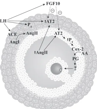
Taken together with other studies from our group, it is possible to propose a model (Fig. 4) in which the gonadotropin surge stimulates a single cascade of events to induce ovulation and nuclear oocyte matura-tion. In this model, gonadotropin surge stimulates An-gII secretion and upregulates AT2 expression in follic-ular cells, whereas AngII increases follicfollic-ular cells secretion of, P4 that stimulates PGs. Ultimately, this sequence of events culminates with ovulation of a fer-tile oocyte.
AngII LH
ACE
AngI
AngII
P4 AT2
Cox-2
?
FGF10
AT2 P4
AA PG
In summary, based on the present work, we con-cluded that P4 in cattle, similar to AngII and PGs, mediated the resumption of meiotic progression in-duced by gonadotropin surge. Indeed, based on our study, we speculated that AngII, P4 and PGs are se-quential steps in the same pathway that culminates with oocyte maturation.
Acknowledgments
The authors thank Adalberto Siqueira for providing animals and Silva Abattoir that kindly provided bovine ovaries. This study was supported by the Brazilian Council of Scientific and Technological Development (CNPq) and CAPES.
References
[1] Ferreira R, Oliveira JF, Fernandes R, Moraes JF, Gonçalves PB. The role of angiotensin II in the early stages of bovine ovula-tion. Reproduction 2007;134:713–19.
[2] Barreta MH, Oliveira JF, Ferreira R, Antoniazzi AQ, Gasperin BG, Sandri LR, et al. Evidence that the effect of angiotensin II on bovine oocyte nuclear maturation is mediated by prostaglan-dins E2 and F2alpha. Reproduction 2008;136:733– 40. [3] Portela VM, Zamberlam G, Gonçalves PB, de Oliveira JF, Price
CA. Role of angiotensin II in the periovulatory epidermal growth factor-like cascade in bovine granulosa cells in vitro. Biol Reprod 2011;85:1167–74.
[4] Jo M, Komar CM, Fortune JE. Gonadotropin surge induces two separate increases in messenger RNA for progesterone receptor in bovine preovulatory follicles. Biol Reprod 2002;67:1981– 88. [5] Bridges PJ, Komar CM, Fortune JE. Gonadotropin-induced expression of messenger ribonucleic acid for cyclooxygenase-2 and production of prostaglandins E and F2alpha in bovine preovulatory follicles are regulated by the progesterone recep-tor. Endocrinology 2006;147:4713–22.
[6] Ayalon D, Tsafriri A, Lindner HR, Cordova T, Harell A. Serum gonadotrophin levels in pro-oestrous rats in relation to the resumption of meiosis by the oocytes. J Reprod Fertil 1972;31: 51– 8.
[7] Pincus G, Enzmann EV. The comparative behavior of mamma-lian eggs in vivo and in vitro: I. The activation of ovarian eggs. J Exp Med 1935;62:665–75.
[8] Richard FJ, Sirard MA. Effects of follicular cells on oocyte maturation. II: Theca cell inhibition of bovine oocyte matura-tion in vitro. Biol Reprod 1996;54:22– 8.
[9] Giometti IC, Bertagnolli AC, Ornes RC, da Costa LF, Caram-bula SF, Reis AM, et al. Angiotensin II reverses the inhibitory action produced by theca cells on bovine oocyte nuclear matu-ration. Theriogenology 2005;63:1014 –25.
[10] Stefanello JR, Barreta MH, Porciuncula PM, Arruda JN, Ol-iveira JF, OlOl-iveira MA, et al. Effect of angiotensin II with follicle cells and insulin-like growth factor-I or insulin on bo-vine oocyte maturation and embryo development. Theriogenol-ogy 2006;66:2068 –76.
[11] Algire JE, Srikandakumar A, Guilbault LA, Downey BR. Pre-ovulatory changes in follicular prostaglandins and their role in ovulation in cattle. Can J Vet Res 1992;56:67–9.
[12] Bridges PJ, Fortune JE. Regulation, action and transport of prostaglandins during the periovulatory period in cattle. Mol Cell Endocrinol 2007;263:1–9.
[13] Nuttinck F, Marquant-Le Guienne B, Clément L, Reinaud P, Charpigny G, Grimard B. Expression of genes involved in prostaglandin E2 and progesterone production in bovine cumu-lus-oocyte complexes during in vitro maturation and fertiliza-tion. Reproduction 2008;135:593– 603.
[14] Fortune JE, Willis EL, Bridges PJ, Yang CS. The periovulatory period in cattle: Progesterone, prostaglandins, oxytocin and ADAMTS proteases. Anim Reprod 2009;6:60 –71.
[15] Nuttinck F, Gall L, Ruffini S, Laffont L, Clement L, Reinaud P, et al. PTGS2-related PGE2 affects oocyte MAPK phosphoryla-tion and meiosis progression in cattle: Late effects on early embryonic development. Biol Reprod 2011;84:1248 –57. [16] Aparicio IM, Garcia-Herreros M, O’Shea LC, Hensey C,
Lon-ergan P, Fair T. Expression, regulation, and function of proges-terone receptors in bovine cumulus oocyte complexes during in vitro maturation. Biol Reprod 2011;84:910 –21.
[17] Sirotkin AV. Involvement of steroid hormones in bovine oocytes maturation in vitro. J Steroid Biochem Mol Biol 1992;41:855–58. [18] Silva CC, Knight PG. Effects of androgens, progesterone and their antagonists on the developmental competence of in vitro matured bovine oocytes. J Reprod Fertil 2000;119:261– 69. [19] Wang HF, Isobe N, Kumamoto K, Yamashiro H, Yamashita Y,
Terada T. Studies of the role of steroid hormone in the regulation of oocyte maturation in cattle. Reprod Biol Endocrinol 2006;4:4. [20] Buratini J Jr., Pinto MG, Castilho AC, Amorim RL, Giometti
IC, Portela VM, et al. Expression and function of fibroblast growth factor 10 and its receptor, fibroblast growth factor re-ceptor 2b, in bovine follicles. Biol Reprod 2007;77:743–50. [21] Berisha B, Sinowatz F, Schams D. Expression and localization
of fibroblast growth factor (FGF) family members during the final growth of bovine ovarian follicles. Mol Reprod Dev 2004; 67:162–71.
[22] Cho JH, Itoh T, Sendai Y, Hoshi H. Fibroblast growth factor 7 stimulates in vitro growth of oocytes originating from bovine early antral follicles. Mol Reprod Dev 2008;75:1736 – 43. [23] Portela VM, Gonçalves PB, Veiga AM, Nicola E, Buratini J Jr.,
Price CA. Regulation of angiotensin type 2 receptor in bovine granulosa cells. Endocrinology 2008;149:5004 –11.
[24] Peluso JJ. N-cadherin mediated cell contact inhibits germinal vesicle breakdown in mouse oocytes maintained in vitro. Re-production 2006;131:429 –37.
[25] Zhang K, Hansen PJ, Ealy AD. Fibroblast growth factor 10 enhances bovine oocyte maturation and developmental compe-tence in vitro. Reproduction 2010;140:815–26.
[26] Leibfried L, First NL. Characterization of bovine follicular oocytes and their ability to mature in vitro. J Anim Sci 1979; 48:76 – 86.
[27] Borman SM, Chaffin CL, Schwinof KM, Stouffer RL, Zelinski-Wooten MB. Progesterone promotes oocyte maturation, but not ovulation, in nonhuman primate follicles without a gonadotro-pin surge. Biol Reprod 2004;71:366 –73.
[29] Voss AK, Fortune JE. Estradiol-17 beta has a biphasic effect on oxytocin secretion by bovine granulosa cells. Biol Reprod 1993; 48:1404 – 09.
[30] Murdoch WJ, Peterson TA, Van Kirk EA, Vincent DL, Inskeep EK. Interactive roles of progesterone, prostaglandins, and col-lagenase in the ovulatory mechanism of the ewe. Biol Reprod 1986;35:1187–94.
[31] Murdoch WJ. Differential effects of indomethacin on the sheep ovary: Prostaglandin biosynthesis, intracellular calcium, apop-tosis, and ovulation. Prostaglandins 1996;52:497–506.
[32] Mingoti GZ, Garcia JM, Rosa-e-Silva AA. Steroidogenesis in cumulus cells of bovine cumulus-oocyte-complexes matured in vitro with BSA and different concentrations of steroids. Anim Reprod Sci 2002;69:175– 86.
[33] Portela VM, Gonçalves PBD, Veiga AM, Nicola E, Buratini J Jr., Price CA. Regulation of angiotensin type 2 receptor in bovine granulosa cells. Endocrinology 2008;149:5004 –11. [34] Parrott JA, Skinner MK. Developmental and hormonal