209
Artigo
ISSN 0102-695X Received 02/20/05. Accepted 08/15/05
* E-mail: salgadoh@fcfar.unesp.br, Tel. + 55-16-33016967
Revista Brasileira de Farmacognosia Brazilian Journal of Pharmacognosy 15(3): 209-211, Jul./Set. 2005
Antimicrobial activity of
Davilla elliptica
St. Hill (Dilleniaceae)
D.C. Michelin
1, S.M. Iha
1, D. Rinaldo
2, M. Sannomiya
2, L.C. Santos
2, W. Vilegas
2,
H.R.N. Salgado
1*1Departamento de Fármacos e Medicamentos, Faculdade de Ciências Farmacêuticas, UNESP, 14801-902,
Araraquara, SP, Brasil,
2Departamento de Química Orgânica, Instituto de Química de Araraquara, UNESP,
14800-900, Araraquara, SP, Brasil
ABSTRACT: Davilla elliptica St. Hill (“lixinha”), family Dilleniaceae, is commonly used in the
Brazilian folk medicine as purgative and stimulant. This work evaluated the antimicrobial activity of the methanol and chloroform extracts of the leaves and barks of D. elliptica using the disc-diffusion method. The results obtained showed that the methanolic extracts of the leaves and barks presented antimicrobial activity against the tested microorganisms.
Keywords: Davilla elliptica, Dilleniaceae, antimicrobial activity.
INTRODUCTION
Davilla elliptica St. Hill (Dilleniaceae) is a native species from Brazil, which is popularly known as ‘lixinha’ (http://www.propp.ufu.br/revistaeletronica/ b/ocorrencia.pdf, 2003). There are properties attributed to D. elliptica with indications such as purgative and stimulant (Rodrigues; Carvalho, 2001). Phytochemical investigations of different parts of Davilla species have revealed the presence of D-tocoferol, and the fl avonoids myricetin, quercetin, myricetin-3-O-D-L-rhamnoside, quercetin-3-O-D-L-rhamnoside, kaempferol (Gurni; Kubitzki, 1981), as well as saponins and mucilage (Matheucci, 1996).
Despite the popular use of D. elliptica as a medicinal plant, there are no data about the antimicrobial effect of leaf extracts. Thus, the interest in this plant is justifi able because of its potential medicinal value. The present study has the aim of evaluating the antimicrobial activity of D. elliptica extracts obtained from the leaves and barks using the disc-diffusion method. It was also made a phytochemical screening of the chloroform and methanol extracts of the leaves and barks of D. elliptica by TLC on Si gel.
MATERIAL AND METHODS
Microorganisms
Eight microbial species taken from international collections were analyzed. The bacteria Bacillus subtilis (ATCC 9372), Bacillus cereus (ATCC 14579), Shigella spp (IAL 1578), Sthaphylococcus epidermidis (ATCC 12226), Proteus mirabilis (CDC 305), Salmonella spp (ATCC 19196), Enterococcus faecalis (ATCC 29212), and the yeast Candida albicans (ATCC 10231).
Plant material
Plant samples were collected in Porto Nacional, State of Tocantins, Brazil, in August 2002. The plant was identifi ed and authenticated by Dra. Solange Lolis of the Universidade de Tocantins. A voucher specimen (No. 4583) was deposited at the Herbarium of the Universidade de Tocantins (HTO), campus of Porto Nacional.
Extract preparation
The air-dried and powdered leaves (2.0 kg) and barks (2.0 kg) of D. elliptica were extracted separately and exhaustively with CHCl3 and MeOH successively at room temperature (48 h for each solvent). Solvents were evaporated at 60 qC under reduced pressure affording the extracts coded as ECHCl3(55.8 g of the barks and 103.5 g of the leaves) and EMeOH (289.8 g of the barks and 373.8 g of the leaves).
Phytochemical screening
The chromatographic analyses were made by TLC on Si gel eluted with different solvent systems: hexane/ethyl acetate (85:15, v:v), chloroform/methanol/ n-propanol/water (5:6:1:4, v:v:v:v) and chloroform/ methanol (85:15, v:v).
The fl avonoids were identifi ed by their intense coloration in ultraviolet light (254 nm) when revealed with the NP/PEG (diphenylaminoborate/polyethyleneglycol) reagent (Wagner et al., 1984) eluted with chloroform/ methanol (85:15, v:v). Authentic standards (Sigma) of the existing fl avonoids in our laboratory (quercetin, myricetin and kaempferol) were also used.
210
D.C. Michelin, S.M. Iha, D. Rinaldo, M. Sannomiya, L.C. Santos, W. Vilegas, H.R.N. Salgado
Rev. Bras. Farmacogn. Braz J. Pharmacogn. 15(3):jul/set. 2005
the reaction with iron salts.
Iodine vapor and solution of CeSO4 were also used (saponnins and terpenes) as well as anisaldehyde/ sulfuric acid solution for the detection of fl avonoids, terpenes, saponnins, gallic acid and catechins (Wagner et al., 1984).
The compounds classes found in the ECHCl3 and EMeOH leaves and barks of D. elliptica are indicated in Table 1.
Disc diffusion method
The dried plant extracts of leaves and barks were dissolved in the same solvent (MeOH and CHCl3) to a fi nal concentration of 30 mg/mL. Then they were sterilized by fi ltration through 0.45 Pm Millipore fi lters. Antimicrobial tests were carried out by the disc diffusion method (Bauer et al., 1966).
The microorganism cultures were grown in Brain Heart Infusion liquid medium at 37 ºC. After 6 h of growth, each microorganism culture, at a concentration of 106 cells/mL, was inoculated on the surface of
Mueller-Test Leaves Barks
ECHCl3 EMeOH ECHCl 3 EMeOH
Catechins - + - +
Tannins - + - +
Gallic acid - + - +
Flavonoids - + - -
Saponnins - - - -
Terpenes + + + -
Table 1. Phytochemical screening of D. elliptica
Table 2. Antimicrobial activities of the methanolic extracts of D. elliptica.
ECHCl3 = chloroformic extract; EMeOH = methanolic extract; absent = (-); present = (+)
Extracts (mg/mL) Positive controls (mg/disc)
EMeOH leaves
EMeOH
barks Cipro. Keto.
Microorganisms
50 75 100 50 75 100 5 40
S. epidermidis - - - - - - 25 NT
B. subtilis 12 13 13 9 9 10 20 NT
B. cereus 11 11 12 9 9 9 20 NT
E. faecalis 9 10 12 - - - 22 NT
Shigella spp 11 12 12 8 8 9 22 NT
P. mirabilis - - - 22 NT
Salmonella spp 12 14 14 - - - 22 NT
C. albicans 11 12 13 8 9 9 NT 16
Diameter of zone (mm), (-) negative; NT: not tested ; Cipro.: ciprofl oxacin; Keto.: ketoconazole
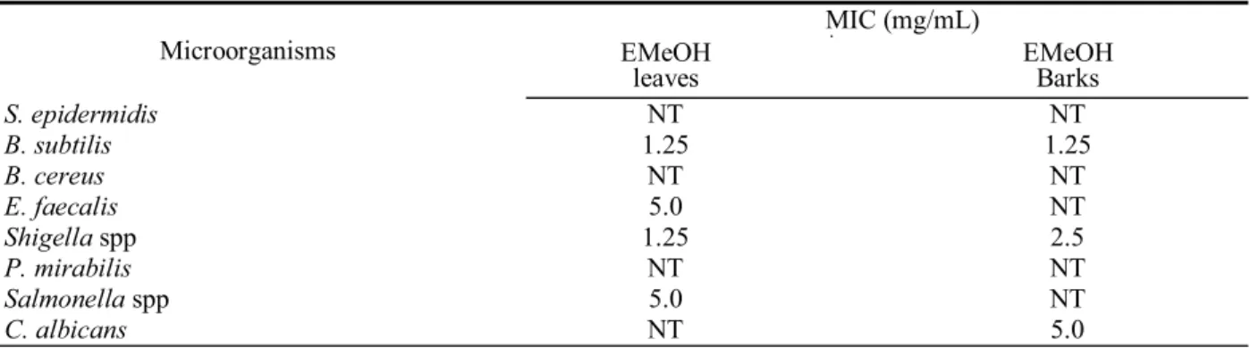
MIC (mg/mL)
Microorganisms EMeOH
leaves EMeOH Barks
S. epidermidis NT NT
B. subtilis 1.25 1.25
B. cereus NT NT
E. faecalis 5.0 NT
Shigella spp 1.25 2.5
P. mirabilis NT NT
Salmonella spp 5.0 NT
C. albicans NT 5.0
Table 3. Minimum inhibitory concentration (MIC) exhibited by the D. elliptica MeOH extracts
211 Antimicrobial activity of Davilla elliptica St. Hill (Dilleniaceae)
Rev. Bras. Farmacogn. Braz J. Pharmacogn. 15(3):jul/set. 2005 Hinton agar plates (100 PL). Subsequently, fi lter papers
discs (6 mm in diameter) saturated with extracts (20 PL) were placed on the surface of each inoculated plate, in Brain Heart Infusion solid medium. The plates were incubated at 35 ºC for 24 h for bacteria and for 48 h for C. albicans. After this period, the zones of growth inhibition around the discs were measured. Overall, cultured microorganisms with halos equal to or greater than 7 mm were considered susceptible to the tested extract.
The negative control was the solvent used and the positive control was ciprofl oxacin (5 Pg/disc) for bacteria and ketoconazole (40 Pg/disc) for C. albicans. All determinations were made in duplicate.
Minimum inhibitory concentration
The minimum inhibitory concentration (MIC) was determined by the dilution method according to National Committee for Clinical Laboratory Standards (NCCLS, 2003). The bacteria were grown in nutrient broth (Brain Heart Infusion liquid medium) for 6 h. After that, 20 PL of 106cells/mL were inoculated in tubes
with nutrient broth supplemented with eight different concentrations (25, 50, 100, 200, 400, 500, 600 and 800PL) of the extracts. After 24 h at 37 ºC, the MIC of each sample was measured by the optical density in the spectrophotometer (620 nm), by comparison of the sample readout with the non inoculated nutrient broth (Nascimento et al., 2000). All determinations were made in duplicate.
RESULTS AND DISCUSSION
A total of 8 microorganisms, which consisted of 7 bacteria and 1 yeast, were tested and the results are summarized in Tables 2 and 3. The ECHCl3 extract of D. elliptica leaves and barks did not show any activity and the results are not shown.
As can be observed in Table 2, the EMeOH of D. elliptica leaves and barks possessed the antimicrobial activity against the microorganisms tested. In the assays against the microorganisms by the agar diffusion method (Table 2), the mean zones of inhibition obtained were between 8 to 14 mm.
Both the EMeOH of D. elliptica leaves and barks were active against B. subtilis, B. cereus, Shigella spp and C. albicans. However, in the extracts of the leaves the observed activity was higher. We also observed the antibacterial activity of the EMeOH leaves against E. faecalis and Salmonella spp (Table 2).
The EMeOH of D. elliptica leaves showed activity against six different species of microorganisms, while the EMeOH of D. elliptica barks showed activity against four different species of microorganisms (Table 2).
The MIC values obtained were ranged between 1.25 to 5.0 mg/mL (Table 3). The best results were
observed for the EMeOH leaves and barks against B. subtilis and EMeOH leaves against Shigella spp with all of them showing MIC at 1.25 mg/mL (Table 3).
Tannins, gallic acid, some catechins and fl avonoids can show antimicrobial activity (Scalbert, 1991; Veluri et al., 2004; Bylka et al., 2004; Harborne et al., 2000). Therefore, the presence of such compounds classes in the EMeOH leaves and barks of the D. elliptica might be responsible for the antimicrobial activity.
ACKNOWLEDGEMENTS
PADC-FCF, FAPESP and CNPq - Brazil
REFERENCES
Bauer AW, Kirby MDK, Sherries JC, Truck M 1966. Antibiotic susceptibility testing by a standardized single disk method. Am J Clin Pathol 45: 493-496.
Bylka W, Matlawska I, Pilewski NA 2004. Natural fl avonoids as antimicrobial agents. J Am Nutraceutical Assoc 7: 24-31.
Gurni AA, Kubitzki K 1981. Flavonoid chemistry and systematics of the Dilleniaceae. Biochem Syst Ecol 9: 109-114.
Harborne JB, Williams CA 2000. Advances in fl avonoids research since 1992. Phytochemistry 55: 481-504. http://www.propp.ufu.br/revistaeletronica/b/ocorrencia.pdf,
acessado em outubro de 2003.
Matheucci LG 1996. Estudo farmacognóstico e farmacológico
de Davilla rugosa Poiret. São Paulo, 75p. Dissertação
de Mestrado - Faculdade de Ciências Farmacêuticas, Universidade de São Paulo.
Nascimento GGF, Locatelli J, Freitas PC, Silva GL 2000. Antibacterial activity of plant extracts and phytochemicals on antibiotic-resistant bacteria. Braz
J Microbiol 31: 247-256.
NCCLS (National Committee for Clinical Laboratory Standards) Performance Standards for Antimicrobial
Disc Susceptibility Tests 2003. Approved Standard
M2-A7, Wayne, Pennsylvania.
Rodrigues VEG, Carvalho DA 2001. Levantamento etnobotânico de plantas medicinais no domínio do cerrado na região do Alto Rio Grande - Minas Gerais. Ciênc Agrotec
25: 102-123.
Scalbert A 1991. Antimicrobial properties of tannins.
Phytochemistry 30: 3875-3883.
Schneider G 1990. Arzneidrogen. Ein kompendium für
pharmazeuten, biologen und chemiker. Mannheim:
Wissenschaftsverlag.
Simões CMO, Schenkel EP, Gosmann G, Mello JCP, Mentz LA, Petrovick PR 2001. Farmacognosia, da planta
ao medicamento. 3. ed, Porto Alegre / Florianópolis:
Ed. Universidade/ UFRGS, Ed. da UFSC.
Veluri R, Weir TL, Bais HP, Stermitz FR, Vivanco JM 2004. Phytotoxic and antimicrobial activities of catechin derivatives. J Agric Food Chem 52: 1077-1082. Wagner HM, Bladt S, Zgainki EM 1984. Plant drug analysis.