Printed version ISSN 0001-3765 / Online version ISSN 1678-2690 http://dx.doi.org/10.1590/0001-3765201720170068
www.scielo.br/aabc | www.fb.com/aabcjournal
Antibiotic activity of
Plectranthus ornatus
Codd., a Traditional Medicinal Plant
FERNANDA R. NASCIMENTO1
, KAMYLLA R.S. ALBUQUERQUE1
,MARCOS R. OLIVEIRA1
, VIRGINIA R. PIZZIOLO1,BEATRIZ G. BRASILEIRO2, GASPAR DIAZ3 and MARISA A.N. DIAZ1
1
Departamento de Bioquímica e Biologia Molecular, Universidade Federal de Viçosa, Avenida Peter Henry Rolfs, s/n, Campus Universitário, 36570-900 Viçosa, MG, Brazil 2
Instituto Federal do Sudeste de Minas Gerais, Avenida Coronel Monteiro de Castro, 550, 36880-000 Muriaé, MG, Brazil 3
Instituto de Ciências Exatas, Universidade Federal de Minas Gerais, Avenida Presidente Antônio Carlos, 6627, Pampulha, 31270-901 Belo Horizonte, MG, Brazil
Manuscript received on February 7, 2017; accepted for publication on July 24, 2017
ABSTRACT
The dichloromethane extract of Plectranthus ornatus Codd., a tradicional medicinal plant, showed
antibiotic activity with minimum inhibitory concentration (MIC) values of 0.4 mg.mL-1 and 100 percent
of biofilm inhibition against Staphylococcus aureus strains isolated from animals with mastitis infections. Based on these antibacterial activities, in addition to ethnopharmacological reports from healing men and
farmers in Brazil, an herbal soap was produced from this active extract and was tested both in vitro and in
vivo. In vivo assays conducted on these herbal soaps led to results similar to those previously conducted with the active extract. These results indicated the great potential of this plant for use as an excipient by preparing herbal antibacterial soaps as an alternative veterinary medicine aimed at controlling bovine
mastitis infections on small Brazilian farms.
Key words: bovine mastitis,herbal soaps, Plectranthus ornatus, Staphylococcus aureus.
Correspondence to: Marisa A.N. Diaz E-mail: [email protected]
INTRODUCTION
Plectranthus ornatus Codd. (family Lamiaceae), is an ornamental and traditional medicinal plant, popularly known in Brazil as “Boldinho” (Lukhoba et al. 2006). It is African native plant which was brought to the Americas by the Portuguese. Ethnopharmacological studies have recommended the use of this plant to treat digestive problems. However, P. ornatus leaves have been used by healing men and farmers, in some regions of Brazil, as an antibiotic for the treatment of skin infections
Staphylococcus aureus. This bacterium produces biofilms, a group of cells that adhere to a surface, and is frequently embedded within a self-produced matrix of an extracellular polymeric substance that causes a dramatic decrease in its susceptibility to the antimicrobial agents. This formation is considered an important virulence factor that is frequently associated with clinical infections (Otto 2008).
The genus Plectranthus is rich in diterpenes and triterpenes. This class of compounds had shown antimicrobial activity (Wellsow et al. 2006, Stavri et al. 2009). The ability of this genus to produce antimicrobial metabolites has led to several phytochemical studies around the word through the isolation of several antimicrobial diterpenes, such as plectrornatin A and two labdane derivatives, as well as plectrornatins B and C (Rijo et al. 2014). The biological activities of the compounds from the genus Plectranthus, associated with their Ethnopharmacological use reported by healing men and farmers, aroused our interest in developing an herbal soap from this plant that can specifically be used by milkers on small farms to wash their hands and milking utensils.
MATERIALS AND METHODS
PLANT MATERIAL AND EXTRACTION
Samples of P. ornathus Codd. were grown in a greenhouse at the Horticultural Department of the Federal University of Viçosa (UFV), Minas Gerais, Brazil. The plants were propagated by cuttings rooted in pre-commercial substrate (Plantmax ®). The plants were harvested 150 days after transplantation and growth. The plants were cut close to the ground, and the aerial parts (leaves) were dried at 40°C for 20 days in an air circulation oven, after which they were grinded into a fine powder. A voucher (39644) was deposited in the herbarium of the Department of Botany of UFV. Forty grams of powder were extracted using petrol, dichloromethane, and ethanol (400 mL) for 1 h at
room temperature, applying the ultrasound method for 10 days for each solvent. The solvents were removed under vacuum at 40°C and stored at 4°C.
MICROORGANISMS TESTING
The bacterial resistant strains (3828, 4075, 4125, and 4158) were isolated from animals with mastitis infections, which were kindly provided by Embrapa Dairy Cattle - Laboratory of Milk Microbiology (Juiz de Fora, Minas Gerais, Brazil), and purchased from the American Type Culture Collection (ATCC, 29213). Bacteria were routinely cultured in brain heart infusion (BHI) at 37°C for 18 h prior to experiments, and cell concentration was adjusted to 106 CFU.mL-1 by optical density at 600 nm. Stock cultures were maintained in BHI containing glycerol at −80°C.
ANTIBACTERIAL SUSCEPTIBILITY TESTING
MINIMAL INHIBITORY CONCENTRATION (MIC) ASSAY
Extract activity on bacterial growth was determined by applying the microdilution method (CLSI 2009). The microorganisms were initially cultured in Petri dishes containing BHI agar (Himedia ®), which were incubated for 24 hours at 37°C. Subsequently, the isolated colonies were subcultured in Mueller-Hinton broth (Himedia®), which was incubated at 37°C for 180 rpm until the culture reached the exponential phase. These colonies were then diluted to an optical density corresponding to 0.5 in the McFarland standard scale (OD620 = 0.10). Microplate holes were filled with 100 µL of
Mueller-Hinton broth (Himedia ®) extracts containing concentrations ranging from 0.1 mg.mL-1 to 10 mg.mL-1 and 106 CFU.mL-1 bacterial suspensions. Whereas the DMSO could be bactericidal a control of microbial growth in this solvent was done with 100 µL of bacterial suspension and 100 µL of Müeller-Hinton broth with DMSO at the highest concentration used in the preparation of the extract. After 24 h at 37°C, 4µL of p-iodonitrotetrazolium (INT, I8377, Sigma®) was added to each well, and the plate was incubated for an additional 2 h at 37°C. A change in the color of the medium from yellow to pink-violet was used as an indication of bacterial growth. The minimal inhibitory concentration of the antibiotics was determined by the same procedure, with concentrations ranging from 0.1 µg.mL-1 a 500 µg.mL-1.
BIOFILM INHIBITORY CONCENTRATION (BIC) ASSAY
Bacterial suspensions were inoculated on microplates containing 180 μL of BHI with different concentrations of the active extracts (MIC, 1/2 MIC, 1/4 MIC, 1/8 MIC, and 1/16 MIC) until reaching the final concentration of 106
CFU.mL-1. After, these concentrations wereincubated at 37°C for 24 h. The supernatant was withdrawn, and the
wells were washed three times with 0.85% saline solution. The remaining bacterial mass was dried at 37°C for 15 min and stained with 200 μL of crystal violet 0.1% for 30 min. Wells were rewashed and dried as previously described, followed by the addition of 300 μL of ethanol and the measurement of absorbance at 630 nm. This test was performed twice in triplicate.
CHEMICAL PROFILING
GC-MS analysis
GC-MS analysis was carried out on a QP2010 Ultra Shimadzu system, employing the following conditions: column fused silica capillary column Rtx-5MS (30 m; 0.25 mm ID; 0.25 film μm). Helium (99.999%) was used as a carrier gas at a constant flow of 1 mL/min, and an injection volume of 1.0 µL was employed (split ratio of 10:1) at an injector temperature of 290°C and an ion source temperature of 200°C. The oven temperature was programmed at 80°C for 5 minutes, then increased to 285°C at a 4°C rate/min and kept at this temperature for 40 minutes. Mass spectra were taken at 70 eV, a scan interval of 0.5 s, and fragments from 35 to 700 Da. The MS transfer line temperature was 290°C.
Identification of phytocomponents
The compounds were identified using the Wiley 7 library database, together with the National Institute of Standards and Technology (NIST) library. The name, molecular weight, molecular formula, and area under peak of the test materials’ components were ascertained.
Production of the herbal soap
without extract was also produced to be used as a reference product.
ANTIBACTERIAL ASSAY OF THE HERBAL SOAP
In vitro
The agar-dilution method was employed in an
in vitro evaluation. The herbal soap (1.0 g) was dissolved in distilled water (50 mL) to obtain a 2% suspension. The suspension was vigorously shaken to dissolve the soap, to disperse the foam, and to homogenize the suspension. Next, 1.0 mL of the soap solution was added to 20 mL of sterile molten culture media in Petri-dishes and allowed to set. One-hundred μL of suspension containing 106 CFU.mL−1 of a resistant 4075 S. aureus strain was then streaked on the plates. After incubation at 37°C for 24 h, inhibition zones were compared to the control to observe the presence or absence of microbial growth.
In vivo
Gloves contaminated with S. aureus from animals with mastitis infection were used to perform the in vivo evaluation (topical test performed according to our institutional ethics, protocol number 773.182). The herbal soap (1.0 g) was dissolved in distilled water (100 mL) to obtain a 1% suspension. This suspension was then vigorously shaken to dissolve the soap, to disperse the foam, and to homogenize it. After, the gloves (12 pairs, 6 for each control soap and herbal soap treatment) were immersed in these solutions for 30 minutes. Before being immersed in the soap, the gloves that the milkers had used to milk the cow’s udder, which had been contaminated with S. aureus, were swabbed, and the sample was placed in bottles with sterile normal saline solution. After being immersed in both the herbal and control soaps, the gloves were again swabbed, and the samples were placed in separate bottles with normal sterile saline solution. Aliquots
from the respective treatments were cultured on an agar plate at 37°C for 24 h to observe the presence or absence of microbial growth.
RESULTS AND DISCUSSION
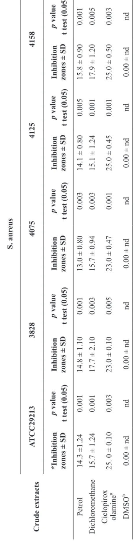
According the results observed in the assays, no differences were observed between the two crude extracts of the plant in relation to the antimicrobial activity of the solvent used in this study. The petrol and dichloromethane crude extracts were active, but the dichloromethane was slightly more active than the petrol extract (Table I).
By analyzing the MIC values obtained for the petrol and dichloromethane crude extracts (Table II), it could be concluded that, these values are lower than some values previously found in extract with antimicrobial activity (Duarte et al. 2004, Virtuoso et al. 2005). Based on the MIC values, extracts can have strong (0.05 to 0.5 mg.mL˗1), moderate (0.6 to 1.5 mg.mL˗1), or weak activity (> 1.5 mg.mL˗1) (Aligiannis et al. 2001). Using these criteria, the extracts of P. ornatus can be considered to be between the strong and moderate inhibitors for the strains used in this study. However, when compared to the positive control, the MIC values were still low.
BIC values were between 2 x MIC (Supra MIC), MIC, and ½ MIC (Sub MIC) (Table III).
The results showed that active extracts are able to inhibit the formation of biofilms. Concentrations corresponding to the Supra MIC and MIC of dichloromethane extracts were enough to inhibit approximately 100% of biofilms formed by S. aureus ATCC 29313, 3828, 4075, and 4158. According to table III, the best BIC value (100% inhibition), obtained for the dichloromethane extract against S. aureus, can indicate that this extract inhibit bacterial film formation in the initial phase of adhesion and formation of biofilms. This value was similar to those found for antibiotic substances reported in prior literature (Nostro et al. 2007). The same was observed for Supra MIC and MIC of the crude petrol extract on S. aureus 3828 and ATCC 29313. Concentrations corresponding to Sub MIC of both extracts were enough to inhibit approximately 30-80% of the biofilms formed by S. aureus ATCC 29313, 3828, 4075, 4128, and 4158.
MIC and BIC results justify the choice of the
dichloromethane extract as an antiseptic agent
in the form of an herbal soap.
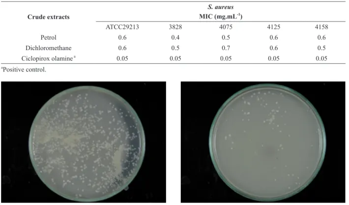
The in vitro results demonstrated that the herbal soap obtained from a dichloromethane crude extract reduces the bacterial load to 89 ± 3.0 CFU (Figure 1 a, b). These results are in agreement with those observed by small farmers.
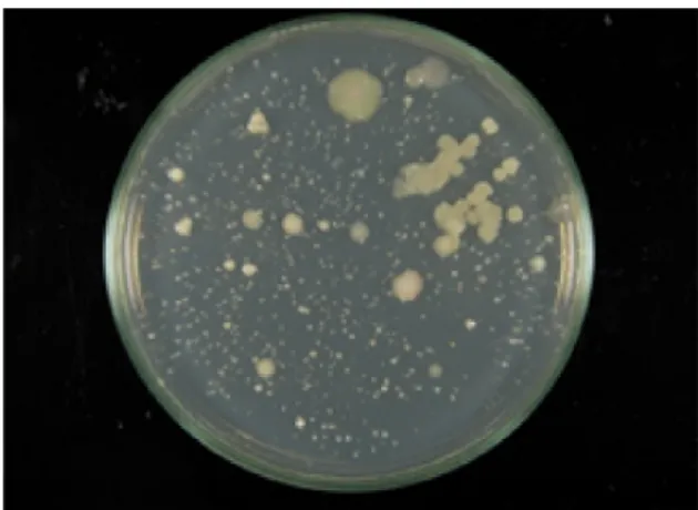
According to the results observed in the in vivo evaluation (Figure 2 a), microbial growth was observed in the Petri dishes after immersing the milker’s gloves in the control soap (Figure 2 b). By contrast, in figure 2 (c) microbial growth was not observed in the Petri dishes after immersing the milker’s gloves in the 1 % suspension of herbal soaps with an active extract of P. ornathus for 30 min.
The chemical constituents in the petrol and dichloromethane extracts of the aerial parts of
P. ornatus belong to different chemical groups, according to the GC/MS analysis of the plant extracts.
T
ABLE I
Staphylococcus aureus
inhibition zones of extracts fr
om Plectrantus orn a tus leaves (mm). Crude extracts S. aur eus A TCC29213 3828 4075 4125 4158
*Inhibition zones ± SD
p
value
t test (0.05)
Inhibition zones ± SD
p
value
t test (0.05)
Inhibition zones ± SD
p
value
t test (0.05)
Inhibition zones ± SD
p
value
t test (0.05)
Inhibition zones ± SD
p
value
t test (0.05)
Petrol
14.3 ±1.24
0.001
14.8 ± 1.10
0.001
13.0 ± 0.80
0.003
14.1 ± 0.80
0.005
15.8 ± 0.90
0.001
Dichloromethane
15.7 ± 1.24
0.001
17.7 ± 2.10
0.003
15.7 ± 0.94
0.003
15.1 ± 1.24
0.001
17.9 ± 1.20
0.005
Ciclopirox olamine
a
25. 0 ± 0.10
0.003
23.0 ± 0.10
0.005
23.0 ± 0.47
0.001
25.0 ± 0.45
0.001
25.0 ± 0.50
0.003
DMSO
b
0.00 ± nd
nd
0.00 ± nd
nd
0.00 ± nd
nd
0.00 ± nd
nd
0.00 ± nd
nd
a Positive control; b Negative control; * Inhibition zones are the mean inc
luding border (7
TABLE II MIC values (mg.mL-1
) of extracts from Plectrantus ornatus leaves against Staphylococcus aureus strains.
Crude extracts
S. aureus MIC (mg.mL-1
)
ATCC29213 3828 4075 4125 4158
Petrol 0.6 0.4 0.5 0.6 0.6
Dichloromethane 0.6 0.5 0.7 0.6 0.5
Ciclopirox olamine a
0.05 0.05 0.05 0.05 0.05
a
Positive control.
a - Control (soap without dichloromethane extract of P. ornathus).
b - Herbal soap with dichloromethane extract of P. ornathus
(89±3.0 CFU).
Figure 1 - In vitro antibacterial activities of herbal soap produced with the active extract of P. ornathus. Tests were performed in triplicate.
TABLE III
BIC values obtained from the actives extracts of Plectrantus ornatus on Staphylococcus aureus strains.
Crude extracts MIC
Concentration
S. aureus % of inhibitionb
ATCC29213 3828 4075 4125 4158
Petrol
Supra MIC* 100 100 70 80 50
MIC** 100 100 50 50 50
Sub MIC*** 50 50 50 30 50
Dichloromethane
Supra MIC* 100 100 100 100 100
MIC** 100 100 100 50 100
Sub MIC*** 50 50 80 50 50
Ciclopirox olaminea 100 100 100 100 100
a
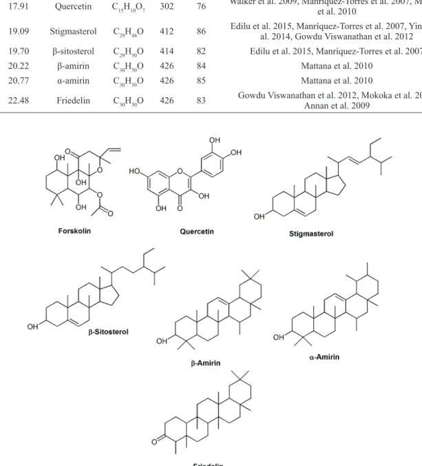
This analysis revealed the presence of compounds like diterpenes, triterpenes, and flavonoids, which are known to exhibit antimicrobial activities that inhibit bacterial growth (Table IV). Therefore, we can assume that the antimicrobial activity observed in crude extracts may well be associated with these types of compounds (Figure 3) (Rijo et al. 2011, Roberto et al. 2007).
CONCLUSIONS
According to the results obtained in this study, the herbal soap from P. ornatus can be used as an antiseptic agent in pre and post-dipping without drawbacks of disinfectants formulated based on iodine or sodium hypochlorite. These can also be used as adjuvant, such as disinfectants for disease
control. These herbal soaps demonstrated a high level of inhibition against S. aureus from cows’ udders and indicates the potential of this plant as an excipient in the production of antiseptic soaps to fight bovine mastitis infections, especially on small farms. Our results validate the use of this plant by small farms to control this disease.
ACKNOWLEDGMENTS
The authors are grateful to Maria Aparecida V.P. Brito (Embrapa/CNPGL, Juiz de Fora, Minas Gerais), who kindly provided the bacterial strains. We thanks the Conselho Nacional de Desenvolvimento Científico e Tecnológico (CNPq) [grant numbers 470153/2011-3].
a - Milker’s gloves immersing in the soap solution.
b - Result immersing the milker’s gloves in the soup control (without active extract).
c - Result after the milker’s gloves had been immersed in the herbal soap (with active extract).
TABLE IV
Main compounds identified by GC-MS in dichloromethane extract of P. ornatus leaves that present previously antibacterial activity.
No. RT (min) Name of the compound
Molecular Formula MW
Peak area (%)
References
1 16.08 Forskolin C22H34O7 410 80 Rijo et al. 2011
2 17.91 Quercetin C15H10O7 302 76 Walker et al. 2009, Manríquez-Torres et al. 2007, Mattana et al. 2010
3 19.09 Stigmasterol C29H48O 412 86 Edilu et al. 2015, Manríquez-Torres et al. 2007, Yinusa et al. 2014, Gowdu Viswanathan et al. 2012 4 19.70 β-sitosterol C29H50O 414 82 Edilu et al. 2015, Manríquez-Torres et al. 2007
5 20.22 β-amirin C30H50O 426 84 Mattana et al. 2010
6 20.77 α-amirin C30H50O 426 85 Mattana et al. 2010
7 22.48 Friedelin C30H50O 426 83 Gowdu Viswanathan et al. 2012, Mokoka et al. 2013, Annan et al. 2009
REFERENCES
ALIGIANNIS N, KALPOUTZAKIS E, MITAKU S AND CHINOU IB. 2001. Composition and antimicrobial activity of the essential oils of two Origanum species. J Agric Food Chem 49: 4168-4170.
ANNAN K, ADU F AND GBEDEMA SY. 2009. Friedelin: A bacterial resistance modulator from Paulinia Pinnata L. J Sci Tech (Ghana) 29: 152-159.
BRASILEIRO BG, PIZZIOLO VR, RASLAN DS, JAMAL CM AND SILVEIRA D. 2006. Antimicrobial and cytotoxic activities screening of some brazilian medicinal plants used in Governador Valadares district. Braz J Pharm Sci 42: 195-202.
CLSI - CLINICAL AND LABORATORY STANDARDS INSTITUTE. 2009. Methods for dilution antimicrobial susceptibility tests for bacteria that grow aerobically; approved standard eighth edition. Document M07-A8. Wayne, PA: CLSI.
DIAZ MAN AND PIZZIOLO VR. 2012. Processo de fabricação e formulação de sabonete para fins cosmecêuticos contendo
óleo de semente de Macaúba (Acronomia aculeata) e o
produto obtido. Brazil Patent BR 1005633-5.
DIAZ MAN, ROSSI CC, MENDONÇA VR, SILVA DM, RIBON ABO, AGUILAR AP AND MUÑOZ GD. 2010. Screening of medicinal plants for antibacterial activities on Staphylococcus aureus strains isolated from bovine mastitis. Braz J Pharmacog 20: 724-728.
DUARTE MCT, FIGUEIRA GM, PEREIRA B, MAGALHÃES PM AND DELARMELINA C. 2004. Atividade
antimicrobiana de extratos hidroalcólicos de espécies da
coleção de plantas medicinais CPQBA/UNICAMP. Braz J Pharmacog 14: 6-8.
EDILU A, ADANE L AND WOYESSA D. 2015. In vitro
antibacterial activities of compounds isolated from roots of Caylusea abyssinica. Ann Clin Microbiol Antimicrob 14: 15. (DOI 10.1186/s12941-015-0072-6).
GOWDU VISWANATHAN MB, ANANTHI JDJ AND KUMAR PS. 2012. Antimicrobial activity of bioactive compounds and leaf extracts in Jatropha tanjorensis. Fitoterapia 83: 1153-1159.
JUE SG, DAWSON GW AND BROGDEN RN. 1985. Ciclopirox olamine 1% cream. Drugs 29: 330-341. LUKHOBA CW, SIMMONDS MS AND PATON AJ.
2006. Plectranthus: a review of ethnobotanical uses. J Ethnopharmacol 103: 1-24.
MANRÍQUEZ-TORRES JJ, ZÚÑIGA-ESTRADA A, GONZÁLEZ-LEDESMA M AND TORRES-VALENCIA JM. 2007. The antibacterial metabolites and proacacipetalin from Acacia cochliacantha. J Mex Chem Soc 51: 228-231. MARINHO ML, ALVES MS, RODRIGUES MLC,
ROTONDANO TEF, VIDAL IF, SILVA WW AND ATHAYDE ACR. 2007. A utilização de plantas medicinais
em medicina veterinária: um resgate do saber popular. Rev Bras Pl Med 9: 64-69.
MATTANA CM, SATORRES SE, SOSA A, FUSCO M AND ALCARÁZ LE. 2010. Antibacterial activity of extracts of
Acacia aroma against resistant and methicillin-sensitive Staphylococcus. Braz J Microbiol 41: 581-587. MOKOKA TA, MCGAW LJ, MDEE LK, BAGLA VP,
IWALEWA EO AND ELOFF JN. 2013. Antimicrobial activity and cytotoxicity of triterpenes isolated from leaves of Maytenus undata (Celastraceae). BMC Complement Altern Med 13: 1.
NASCIMENTO GGF, LOCATELLI J, FREITAS PC AND SILVA GL. 2000 Antibacterial activity of plant extracts and phytochemicals on antibiotic-resistant bacteria. Braz J Microbiol31: 247-256.
NOSTRO A, ROCCARO AS, BISIGNANO G, MARINO A, CANNATELLI MA, PIZZIMENTI FC AND BLANCO AR. 2007. Effects of oregano, carvacrol and thymol on
Staphylococcus aureus and Staphylococcus epidermidis biofilms. J Med Microbiol 56: 519-523.
OTTO M. 2008. Staphylococcal biofilms. In Bacterial biofilms (p. 207-228). Springer Berlin Heidelberg.
RIJO P, DUARTE A, FRANCISCO AP, SEMEDO-LEMSADDEK T AND SIMÕES MF. 2014. In vitro
Antimicrobial Activity of Royleanone Derivatives Against Gram‐Positive Bacterial Pathogens. Phytother Res 28: 76-81. RIJO P, RODRÍGUEZ B, DUARTE A AND FATIMA SIMOES M. 2011. Antimicrobial properties of Plectranthus ornatus extracts, 11-acetoxyhalima-5, 13-dien-15-oic acid metabolite and its derivatives. Nat Prod J 1: 57-64. ROBERTO L, KENTOPFF MR, MACHADO MIL, SILVA
MGV, MORAIS SM AND BRAZ-FILHO R. 2007. Diterpenos tipo abietano isolados de Plectranthus barbatus
Andrews. Quim Nova 30: 1882-1886.
STAVRI M, PATON A, SKELTON BW AND GIBBONS S. 2009. Antibacterial diterpenes from Plectranthus ernstii. J Nat Prod 72: 1191-1194.
VIRTUOSO S, DAVET A, DIAS JFG, CUNICO MM, MIGUEL MD, OLIVEIRA AB AND MIGUEL OG. 2005. Estudo preliminar da atividade antibacteriana das cascas de Erythrina velutina Willd., Fabaceae (Leguminosae) Braz J Pharmacog 15: 137-142.
WALKER CI, ZANOTTO CZ, CERON CS, POZZATTI P, ALVES SH AND MANFRON SH. 2009. Atividade
farmacológica e teor de quercetina de Mirabilis jalapa L.
Lat Am J Pharm 28: 241-246.
WELLSOW J, GRAYER RJ, VEITCH NC, KOKUBUN T, LELLI R, KITE GC AND SIMMONDS MS. 2006. Insect-antifeedant and antibacterial activity of diterpenoids from species of Plectranthus. Phytochemistry 67: 1818-1825. YINUSA I, GEORGE NI, SHUAIBU UO AND AYO RG.