Antimicrobial Photodynamic Therapy: Photodynamic
Antimicrobial Effects of Malachite Green on
Staphylococcus
,
Enterobacteriaceae, and
Candida
J.C. Junqueira, Ph.D.,1 M.A. Ribeiro,1R.D. Rossoni,1J.O. Barbosa,1 S.M.R. Querido, Ph.D.,2and A.O.C. Jorge, Ph.D.1
Abstract
Objective:This study investigatedin vitrothe photodynamic antimicrobial effects of the photosensitizer mala-chite green on clinical strains ofStaphylococcus, Enterobacteriaceae, andCandida.Materials and Methods: Thirty-six microbial strains isolated from the oral cavity of patients undergoing prolonged antibiotic therapy, including 12 Staphylococcus, 12 Enterobacteriaceae, and 12 Candida strains, were studied. The number of cells of each microorganism was standardized to 106cells/mL. Twenty-four assays were carried out for each strain according to the following experimental conditions: gallium-aluminum-arsenide laser and photosensitizer (n¼6, LþPþ), laser and physiologic solution (n¼6, LþP), photosensitizer (n¼6, LPþ), and physiologic solution (n¼6, LP). Next, cultures were prepared on brain–heart infusion agar for the growth of Staphylococcusand En-terobacteriaceae, and on Sabouraud dextrose agar for the growth ofCandida,and incubated for 48 h at 378C. The results are reported as the number of colony-forming units (CFU/mL) and were analyzed with analysis of variance and the Tukey test.Results:TheStaphylococcus, enterobacterial, andCandidastrains were sensitive to photodynamic therapy with malachite green (LþPþ). A reduction of *7 log10 for Staphylococcus, 6 log10 for enterobacteria, and 0.5 log10for the genusCandida. Significant statistical differences were observed between the LþPþgroups and the control groups (LP).Conclusion:TheStaphylococcus, Enterobacteriaceae, andCandida strains studied were sensitive to photodynamic therapy with malachite green.
Introduction
P
hotodynamic antimicrobial therapy consists of the combination of a photosensitizer and visible light, which is able selectively to destroy microbial cells. The antimicrobial effects of this therapy have been known for >100 years.However, only recently has this therapy begun to be studied in detail in the search for alternative treatments for antibiotic-resistant pathogens. Numerous in vitro studies have dem-onstrated that photodynamic therapy is highly effective in the destruction of viruses and protozoans, as well as gram-positive and gram-negative bacteria and fungi.1–4
In photodynamic antimicrobial therapy, the photosensi-tizer is activated by exposure to light of a specific wave-length in the presence of oxygen. The transfer of energy from the activated photosensitizer to available oxygen results in the formation of toxic oxygen species, such as singlet oxygen and free radicals. These reactive chemical species destroy proteins, lipids, nucleic acids, and other cell components.
Photodynamic therapy exerts no genotoxic or mutagenic effects, a fact preventing the development of microbial re-sistance.5
Photosensitizers possess structures similar to those of chlorophyll and hemoglobin (i.e., the molecules contain a heterocyclic ring). In addition, these agents should be bio-logically stable, photochemically active, and minimally toxic to tissues of the organism. The photosensitizers used include hematoporphyrin derivatives, phenothiazines (toluidine blue and methylene blue), cyanines, phytotherapeutic agents, and phthalocyanines.6Phenothiazine dyes have been extensively studied and have been shown to be effective photodynamic agents for the reduction of bacteria7–12 and fungi.13–16 Stu-dies investigating new photosensitizers are currently being conducted. Garcez et al.17 tested azulene to eliminate En-terococcus faecalisfrom root canals. Prates et al.18reported the
use of malachite green as a photosensitizer in photodynamic therapy. This dye is a member of the triarylmethane family, like crystal violet, and shows strong absorption of red light in
1Department of Biosciences and Oral Diagnosis, School of Dentistry of Sa˜o Jose´ dos Campos, Sa˜o Paulo State University/UNESP, Sa˜o Jose´
dos Campos; and2Department of Biological Sciences, Faculty of Pindamonhangaba, Pindamohnangaba, Sa˜o Paulo, Brazil. ªMary Ann Liebert, Inc.
Pp. S67–S72
DOI: 10.1089/pho.2009.2526
the visible spectrum. These authors demonstrated the effi-cacy of this photosensitizer againstAggregatibacter actinomy-cetemcomitans.
Microorganisms such as Staphylococcus,
En-terobacteriaceae, and Candida are found in the oral cavity and are usually present in small numbers. However, pro-longed systemic administration of antimicrobials or immu-nosuppressive therapy may result in an increase of the numbers of these microorganisms.19,20 According to Smith et al.,21
the genusStaphylococcusis commonly isolated from the oral cavity in a specific group of subjects, such as chil-dren, elderly individuals, patients with end-stage systemic diseases, patients with rheumatoid arthritis, and patients with malignant hematologic diseases. Colonization of the oropharynx with methicillin-resistant S. aureus strains is frequently observed in many of these patients.
Isolation of species of the family Enterobacteriaceae from the oral cavity has been demonstrated in various studies. Barbosa et al.22evaluated the subgingival occurrence of
en-teric bacilli and Pseudomonas in patients with periodontal disease, with these microorganisms being detected in 31.2% of the patients studied. The most frequently isolated species wereP. aeruginosaandS. marcescens. Baydas et al.23identified
enterobacteria in the saliva from 19 (76%) of 25 patients with the habit of nail biting and in nine (26.5%) of 34 control patients. The isolates were identified asE. coli,E. aerogenes,
E. cloacae,andE. gergoviae.
Oral infections with yeast of the genusCandidaare highly frequent in patients infected with HIV. Oropharyngeal can-didiasis is the most common manifestation of HIV infection and is observed in 84% of patients.14AlthoughC. albicansis the main etiologic factor of fungal infections, nonalbicans species are also common, with the most important species being, in decreasing order,C. parapsilosis(20–40% of infec-tions), C. tropicalis (10–30%), C. krusei (10–35%), and C. glabrata(5–40%). Currently emerging species includeC. lu-sitaniaeandC. guilliermondii.24
Many studies have demonstrated the efficacy of pheno-thiazine dyes such as methylene blue and toluidine blue in photodynamic therapy for the reduction of Staphylococcus,
Enterobacteriaceae, andCandida.6,8,13,15,26However, the use of malachite green as a photosensitizer has been investigated only withAggregatibacter actinomycetemcomitans.18Therefore, the objective of the present study was to evaluatein vitrothe sensitivity ofStaphylococcus, Enterobacteriaceae, andCandida
strains isolated from the human oral cavity to photodynamic antimicrobial therapy by using malachite green as photo-sensitizer.
Materials and Methods
Microorganisms
Thirty-six microbial strains, including 12 Staphylococcus, 12 Enterobacteriaceae, and 12Candidastrains, were studied. The staphylococcal strains included S. aureus(n¼3), S.
epi-dermidis (n¼3), S. schleiferi (n¼3), S. capitis (n¼1), S.
hae-molyticus (n¼1), and S. lentus (n¼1). The following enterobacteria were selected: Enterobacter cloacae (n¼3),
Klebsiella pneumoniae (n¼3), Klebsiella oxytoca (n¼3), and
Escherichia coli (n¼3). For the genus Candida, C. albicans (n¼3), C. tropicalis (n¼3), C. parapsilosis (n¼2), C. krusei (n¼2), andC. glabrata(n¼2) were used.
All strains were obtained from the Laboratory of Micro-biology and Immunology, Sa˜o Jose´ dos Campos Dental School, UNESP. The strains were isolated from the oral cavity of patients undergoing prolonged antibiotic therapy for a minimum period of 45 days for the treatment of pul-monary tuberculosis.
Twenty-four assays were carried out for each strain, ac-cording to the following experimental conditions: gallium-aluminum-arsenide laser and photosensitizer (n¼6, LþPþ), laser and physiologic solution as control with light (n¼6, LþP), photosensitizer (n¼6, LPþ), and physiologic so-lution as control (n¼6, LP), for a total of 864 assays.
Preparation of the microbial suspension
Microbial suspensions containing 106cells/mL were pre-pared. For this purpose,Staphylococcus strains were seeded onto Mannitol agar (Difco, Detroit, MI), enterobacterial strains were cultured on MacConkey agar (Difco), and Can-dida strains were seeded onto Sabouraud dextrose agar (Difco). All strains were incubated for 24 h at 378C. Next, the
Staphylococcus and enterobacterial strains were cultured in brain–heart infusion broth (Difco) for 18 h at 378C, and the
Candidastrains were cultured in Sabouraud broth (Difco) for 16 h at 378C.
The cultures were centrifuged at 1,300gfor 10 min, and the supernatant was discarded. The sediment was re-suspended in 5 mL sterile physiologic solution (0.85% NaCl). This procedure was repeated, and the number of viable cells in suspension was determined with a spectrophotometer (B582; Micronal, Sa˜o Paulo, Brazil).
Photosensitizer
Malachite green (4-dimethylaminophenol) was used as photosensitizer for the sensitization of the microorganisms studied. The malachite green solution at a concentration of 0.1% was prepared by dissolving the powder (Synth, Sa˜o Paulo, Brazil) in physiologic solution (0.85% NaCl) and was filtered through a sterile membrane (0.22-mm pore diameter; MFS, Dublin, CA).
Low-power laser
The light source used was a gallium-aluminum-arsenide laser (Easy Laser; Clean Line, Taubate´, Brazil) with a wave-length of 660 nm (visible red) and output power of 35 mW. The wavelength of the laser corresponds to the high-absorption spectrum of the photosensitizers used. The laser beam irradiated an area of 0.38 cm2, and the irradiation time
was 4.45 min, resulting in an energy dosage of 26 J/cm2and energy of 10 J. The temperature monitoring at the bottom of the well was made with an Infrared Thermometer (MX4; Raytek, Sorocaba, Brazil), and no increase of temperature was observed after the laser irradiation.
In vitro photosensitization
groups described earlier. Three samples per time were pre-pared, shaken and irradiated, resulting in the time interval of 13.35 min between the first-irradiated and the last-irradiated well. The strains were irradiated under aseptic conditions under a laminar-flow hood.
After irradiation, serial dilutions were prepared for each strain, and 0.1-mL aliquots of the dilutions were seeded in duplicate onto plates containing brain–heart infusion agar (Difco) for the growth ofStaphylococcus and enterobacteria, and Sabouraud dextrose agar (Difco) for the growth of yeast. The plates were incubated for 48 h at 378C. The number of colony-forming units (CFU/mL) was then determined, and the results were submitted to statistical analysis.
The experiment was carried out in the dark, and the plates were covered with a matte black screen with an orifice whose diameter corresponded to the size of the well entrance to prevent the spreading of light. Only 24 wells per microtiter plate were used to avoid light scattering at the well walls, resulting in an overdose at the other already irradiated wells.
Statistical analysis
Statistical analysis was performed with the Minitab pro-gram by using analysis of variance and the Tukey test, with the level of significance set at 5%.
Results
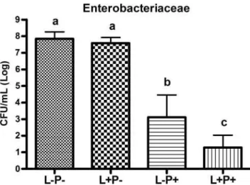
Figures 1–3 show the results of descriptive analysis, analysis of variance, and Tukey test obtained for the 12
Staphylococcus, 12 Enterobacteriaceae, and 12Candidastrains studied, respectively. Similar results were observed for the
Staphylococcus and enterobacterial strains. Figures 1 and 2 show that the strains studied were sensitive to photody-namic therapy with malachite green (LþPþ). Comparison of the photosensitizer alone (LPþ) showed microbial reduc-tion ofStaphylococcusand enterobacteria mediated by mala-chite green. In addition, none of the species was sensitive to the use of laser therapy alone (LþP).
Results different from those obtained for Staphylococcus
and enterobacteria were observed for the Candida strains studied. As shown in Fig. 3, all laser-treatedCandidagroups (LþPþand LþP) had lower mean CFU counts than did the groups not submitted to laser therapy (LPþand LP). However, a significant difference was observed only for the LþPþ group when compared with the LPþand LP groups. Similar results were observed for the LPþgroup when compared with the control group (LP), suggesting that the use of a photosensitizer alone has no toxic effect on clinicalCandidastrains.
Mean CFU counts (log) obtained by analysis of the pho-tosensitivity of eachStaphylococcus, enterobacterial, and Can-didaspecies studied are shown in Tables 1–3, respectively. As
FIG. 1. Mean CFU/mL ofStaphylococcussubmitted to the
following treatments: physiologic solution as control (LP), laser and physiologic solution (LþP), photosensi-tizer (LPþ), and laser and photosensitizer (LþPþ). Differ-ent letters (a, b, c) indicate a significant difference between groups (Tukey test,p<0.05).
FIG. 2. Mean CFU/mL of Enterobacteriaceae submitted to
the following treatments: physiologic solution as control (LP), laser and physiologic solution (LþP), photosensi-tizer (LPþ), and laser and photosensitizer (LþPþ). Differ-ent letters (a, b, c) indicate a significant difference between groups (Tukey test,p<0.05).
FIG. 3. Mean CFU/mL of Candida submitted to the
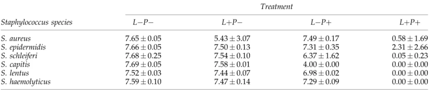
shown in Table 1,S. epidermidiswas the staphylococcal species least sensitive to photodynamic therapy with malachite green. Species of the family Enterobacteriaceae showed similar re-sults under the different experimental conditions (Table 2). Analysis of the genusCandidashowed that all the species had the same sensitivity to photodynamic therapy (Table 3).
Discussion
The present results demonstrate that photodynamic ther-apy with malachite green was effective in microbial reduc-tion of allStaphylococcus and enterobacterial strains, and of most Candida strains. Comparison of the mean number of CFUs between the LþPþ and control (LP) groups showed a reduction of *7 log10 for Staphylococcus and of
6 log10for enterobacteria, whereas the reduction observed for
the genus Candida was <1 log10. These data indicate that
bacteria were more sensitive to malachite green–mediated photosensitization than were yeast species.
According to Demidova and Hamblin,25 gram-positive bacteria are more susceptible to photodynamic therapy than are gram-negative bacteria. This fact can be explained by the structure of the cell wall of these bacteria. Gram-negative bacteria possess a cell wall consisting of two lipid bilayers, whereas the cell walls of gram-positive bacteria consist of only one lipid bilayer and is therefore more permeable to photosensitizers. Yeast are more resistant to photodynamic therapy than bacteria because of their larger cell size and the presence of a nuclear membrane, which represents an addi-tional barrier to the penetration of the photosensitizer.3
In the case of the Staphylococcus and enterobacterial strains, the number of CFU/mL was reduced by *6 to
7 log10after application of photodynamic therapy with 0.1%
malachite green. Prates et al.18also observed a reduction in bacteria of the speciesAggregatibacter actinomycetemcomitans
given combined treatment with 0.01% malachite green and a diode laser. However, the reduction in CFU/mL was only 2 to 3 log10.
The results obtained with malachite green as a photosen-sitizer in photodynamic therapy were similar to those re-ported in studies using phenothiazine dyes, such as methylene blue and toluidine blue. Usacheva et al.26
evalu-atedin vitrothe interaction between phenothiazine dyes and lipopolysaccharides (LPSs) of some gram-negative bacteria, includingE. coli,P. aeruginosa, K. pneumonia,andS. marces-cens, and observed a greater interaction between toluidine blue and LPS when compared with methylene blue. Souza et al.15reported a significantly lower number of log CFU/mL ofC. albicans(ATCC 18804) treated with a gallium-aluminum-arsenide laser and methylene blue (4.68) when compared with the control group (5.69).
However, the results obtained in the photodynamic ther-apy with malachite green were lower than the results ob-tained with the cationic phthalocyanine zinc.27 Mantareva et al.27 evaluated the photodynamic activity of two new water-soluble phthalocyanines, one cationic tetrakis-(3-methylpyridyloxy) and one anionic tetrakis-(4-sulfophenoxy)-phthalocyanine zinc with the strains ofS. aureus,P. aeruginosa,
and C. albicans. The cationic photosensitizer completely inactivatedS. aureusandC. albicans. In the case ofP. aeru-ginosa, a reduction of 6 log10was seen. In contrast, the
an-ionic photosensitizer with the same drug concentration (6mM) was not sufficient to photoinactivate the gram-negative P. aeruginosa. For the yeast C. albicans, the
Table1. Mean Counts (logCFU/mL) ofStaphylococcusSpecies
Treatment
Staphylococcus species LP LþP LPþ LþPþ
S. aureus 7.650.05 5.433.07 7.490.17 0.581.69
S. epidermidis 7.660.05 7.500.13 7.310.35 2.312.66
S. schleiferi 7.680.25 7.540.10 6.371.62 0.050.23
S. capitis 7.690.05 7.580.01 4.000.00 0.000.00
S. lentus 7.520.03 7.440.07 6.980.02 0.000.00
S. haemolyticus 7.590.10 7.470.14 7.290.09 0.000.00
The species were submitted to the following treatments: physiologic solution as control (LP), laser and physiologic solution (LþP), photosensitizer (LPþ), and laser and photosensitizer (LþPþ).
Table2. Mean Counts (logCFU/mL) of Enterobacterial Species
Treatment
Enterobacterial species LP LþP LPþ LþPþ
E. cloacae 8.270.29 7.780.97 3.820.29 1.131.33
E. coli 8.110.33 7.891.83 2.940.28 1.741.12
K. oxytoca 7.300.23 7.210.94 1.760.27 0.620.90
K. pneumoniae 7.710.38 7.440.93 3.990.28 1.690.98
photoinactivated cells were only 1–2 log10. S. aureus was
deactivated<4 log10.
The application of laser therapy alone had an antimicro-bial effect only on theCandidastrains studied. The effect of low-level laser therapy onCandidahas also been reported by Maver-Biscanin et al.,28who studied the effect of this therapy
on C. albicans in two patients with palatal inflammation caused by the use of complete dentures. The patients were irradiated with different wavelengths for different periods of time (830 nm for 5 min and 685 nm for 10 min). Regression of the inflammatory process and a reduction in the number ofCandidacolonies collected from the palate were observed after therapy.
In the present study, application of 0.1% malachite green alone also resulted in the microbial reduction of Staphylo-coccusand enterobacteria, suggesting that malachite green at the concentration tested exerted a bactericidal effect on the microorganisms studied. However, Prates et al.18observed
no reduction in the number of Aggregatibacter actinomyce-temcomitanswhen malachite green was evaluated separately. Individual analysis of the results obtained for each mi-crobial species studied showed that S. epidermidis is the staphylococcal species least sensitive to photodynamic ther-apy with malachite green. Evaluating the photodynamic ef-fects of merocyanine on the biofilms of two different S. epidermidisstrains (RP62A and 1457), Sbarra et al.29observed significant inactivation of cells when biofilms were exposed to photosensitizer and laser simultaneously. However, the strain 1457 was more susceptible to photodynamic therapy than the strain RP62A.
The present results demonstrated the efficiency of mala-chite green as a photosensitizer in photodynamic therapy against strains of Staphylococcus, Enterobacteriaceae, and
Candida. The possibility of eliminating pathogenic microor-ganisms by using a low-power laser in combination with specific dyes makes photodynamic therapy a promising al-ternative in dentistry in view of its low cost, high efficiency, and short time of application. However, further studies are necessary so that other dyes can be regulated as photosen-sitizers and commercialized for dental use.
Acknowledgments
This work was supported by FAPESP, Brazil (grant 07/ 54997-5).
Author Disclosure Statement
No competing financial interests exist.
References
1. Jori, G., Fabris, C., Soncin, M., and Ferro, S. (2006). Photo-dynamic therapy in the treatment of microbial infections: basic principles and perspective applications. Lasers Surg. Med. 38, 468–481.
2. Maisch, T. (2006). Anti-microbial photodynamic therapy: useful in the future? Lasers Med. Sci. 22, 83–91.
3. Donnelly, R.F., McCarron, P.A., Tunney, M.M., and Woolf-son, A.D. (2007). Potential of photodynamic therapy in treatment of fungal infections of the mouth: design and characterization of a mucoadhesive path containing tolui-dine blue O. J. Phot. Photob. B. 86, 59–69.
4. Donnelly, R.F., McCarron, P.A., and Tunney, M.M. (2008). Antifungal photodynamic therapy. Microbiol. Res. 163, 1–12.
5. Konopka, K., and Goslinski, T. (2007). Photodynamic ther-apy in dentistry. J. Dent. Res. 86, 694–707.
6. Wainwright, M. (1998) Photodynamic antimicrobial chemo-therapy (PACT). J. Antimicrob. Chem. 42, 13–28.
7. Soukos, N.S., Wilson, M., Burns, T., and Speight, P.M. (1996). Photodynamic effects of toluidine blue on human oral keratinocytes and fibroblasts and Streptococcus sanguis
evaluatedin vitro. Lasers Surg. Med. 18, 253–259.
8. Usacheva, M.N., Teichert, M.C., and Biel, M.A. (2001). Comparison of the methylene blue and toluidine blue pho-tobactericidal efficacy against positive and gram-negative microorganisms. Lasers Surg. Med. 29, 165–173. 9. Hamblin, M.R., O’Donnell, D.A., Murthy, N., et al. (2002).
Polycationic photosensitizer conjugates: effects of chain length and Gram classification on the photodynamic inac-tivation of bacteria. J. Antimicrob. Chemother. 49, 941–951. 10. Romanova, N.A., Brovko, L.Y., Moore, L., et al. (2003).
As-sessment of photodynamic destruction of Escherichia coli
O157:H7 andListeria monocytogenesby using ATP biolumi-nescence. Appl. Environ. Microbial. 69, 6393–6398.
11. Paulino, T.P., Ribeiro, K.F., Thedei, G., Tedesco, A.C., and Ciancaglini, P. (2005). Use of hand held photopolymerizer to photoinactivate Streptococcus mutans. Arch. Oral Biol. 50, 353–359.
12. Usacheva, M.N., Teichert, M.C., Usacheva, Y.M., Sievert, C.E., and Biel, M.A. (2008). Interaction of the photo-bactericides methylene blue and toluidine blue with a fluorophore in Pseudomonas aeruginosa cells. Lasers Surg. Med. 40, 55–61.
13. Wilson, M., and Mia, N. (1993). Sensitisation ofCandida al-bicans to killing by low-power laser light. J. Oral Pathol. Med. 22, 354–357.
14. Teichert, M.C., Jones, J.W., Usacheva, M.N., and Biel, M.A. (2002). Treatment of oral candidiasis with methylene blue-mediated photodynamic therapy in an immunodeficient
Table3. Mean Counts (logCFU/mL) ofCandidaSpecies
Treatment
Candida species LP LþP LPþ LþPþ
C. albicans 5.670.31 5.410.19 5.650.18 5.320.61
C. tropicalis 5.490.04 5.330.13 5.460.10 4.720.59
C. parapsilosis 6.120.07 5.790.05 5.970.00 5.540.39
C. krusei 5.960.28 5.830.60 5.930.32 5.730.55
C. glabrata 5.750.38 5.460.37 5.720.38 5.190.31
murine model. Oral Surg. Oral Med. Oral Pathol. Oral Radiol. Endod. 93, 155–160.
15. Souza, S.C., Junqueira, J.C., Balducci, I., Koga-Ito, C.Y., Munin, E., and Jorge, A.O.C. (2006). Photosensitization of differentCandidaspecies by low power laser light. J. Pho-tochem. Photobiol. B. 83, 34–38.
16. Munin, E., Giroldo, L.M., Alves, L.P., and Costa, M.S. (2007). Study of tube formation byCandida albicansafter photody-namic antimicrobial chemotherapy (PACT). J. Photochem. Photobiol. B. 88, 16–20.
17. Garcez, A.S., Nu´nez, S.C., Lage-Marques, J.L., Jorge, A.O.C., and Ribeiro, M.S. (2007). Efficiency of NaOCl and laser as-sisted photosensitization on the reduction of Enterococcus faecalis in vitro. Oral Surg. Oral Med. Oral Pathol. Oral Radiol. Endod. 102, 93–98.
18. Prates, R.A., Yamada, A.M., Suzuki, L.C., et al. (2007). Bac-tericidal effect of malachite green and red laser on Actino-bacillus actinomycetemcomitans. J. Phot. Photob. B. 86, 70–76. 19. Slots, J., Rams, T.E., Feik, D., Taveras, H.D., and Gillespie,
G.M. (1991). Subgingival microflora of advanced periodontitis in the Dominican Republic. J. Periodontol. 62, 543–547. 20. Dahle´m, G., and Wikstro¨m, M. (2003). Occurrence of enteric
rods, staphylococci and Candida in subgingival samples. Oral Microbiol. Immun. 10, 42–46.
21. Smith, A.J., Jackson, M.S., and Bagg, J. (2001). The ecology of
Staphylococcusspecies in the oral cavity. J. Med. Microbiol. 50, 940–946.
22. Barbosa, F.C.B., Mayer, M.P., Saba-Chujfi, E., and Cai, S. (2001). Subgingival occurrence and antimicrobial suscepti-bility of enteric rods and pseudomonads from Brazilian periodontitis patients. Oral Microbiol. Immunol. 16, 306–310.
23. Baydas, B., Uslu, H., Yavuz, I., Ceylan, I., and Dagsuyu, I.M. (2007). Effect of a chronic nail-biting habit on the oral
carriage of Enterobacteriaceae. Oral Microbiol. Immunol. 22, 1–4.
24. Krcmery, V., and Barnes, A.J. (2002). Non-albicans Candida
spp. causing fungemia: pathogenicity and antifungal resis-tance. J. Hosp. Infect. 50, 243–260.
25. Demidova, T.N., and Hamblin, M.R. (2005). Effect of cell-photosensitizer binding and cell density on microbial photo-inactivation. Antimicrob. Agents Chemother. 49, 2329–2335. 26. Usacheva, M.N., Teichert, M.C., and Biel, M.A. (2003) The
interaction of lipopolysaccharides with phenothiazine dyes. Lasers Surg. Med. 33, 311–319.
27. Mantareva, V., Kussovski, V., Angelov, I., et al. (2007). Photodynaimic activity of water-soluble phthalocyanine zinc (II) complexes against pathogenic microorganisms. Bioorg. Med. Chem. 15, 4829–4835.
28. Maver-Biscanin, M., Mravak-Stipetic, M., and Jerolimov, V. (2005). Effect of low-level laser therapy onCandida albicans
growth in patients with denture stomatitis. Photomed. Laser Surg. 23, 328–332.
29. Sbarra, M.S., Di Poto, A., Arciola, C.R., et al. (2008). Photo-dynamic action of merocyanine 540 on Staphylococcus epi-dermidisbiofilms. Int. J. Artif. Organs 31, 848–857.
Address correspondence to:
Juliana Campos Junqueira Department of Biosciences and Oral Diagnosis School of Dentistry of Sa˜o Jose´ dos Campos Sa˜o Paulo State University/UNESP, Francisco Jose´ Longo 777 Sa˜o Dimas, Sa˜o Jose´ dos Campos CEP: 12245-000, SP Brazil