JULIANA PAIVA MARQUES LIMA ROLIM
ESTUDOS DOS EFEITOS DA TERAPIA FOTODINÂMICA ANTIMICROBIANA NA VIABILIDADE E NA EXPRESSÃO GÊNICA DE S. MUTANS EM BIOFILMES E LESÕES DE CÁRIE DENTINÁRIA
FORTALEZA
2012
UNIVERSIDADE FEDERAL DO CEARÁ CENTRO DE CIÊNCIAS DA SAÚDE
FACULDADE DE FARMÁCIA, ODONTOLOGIA E ENFERMAGEM PROGRAMA DE PÓS‐GRADUAÇÃO EM ODONTOLOGIA
Juliana Paiva Marques Lima Rolim
ESTUDOS DOS EFEITOS DA TERAPIA FOTODINÂMICA ANTIMICROBIANA NA VIABILIDADE E NA EXPRESSÃO GÊNICA DE S. MUTANS EM BIOFILMES E LESÕES DE CÁRIE DENTINÁRIA
Orientadora: Profa. Dra. Nádia Accioly Pinto Nogueira Co-Orientadora: Profa. Dra. Iriana Carla Junqueira Zanin dos Santos
FORTALEZA
2012
Tese apresentada ao Programa de Pós-graduação em Odontologia da Faculdade de Odontologia da Universidade Federal do Ceará como requisito parcial para obtenção do Título de Doutor em Odontologia.
Dados Internacionais de Catalogação na Publicação Universidade Federal do Ceará Biblioteca de Ciências da Saúde R653e Rolim, Juliana Paiva Marques Lima. Estudos dos efeitos da terapia fotodinâmica antimicrobiana na viabilidade e na expressão gênica de S. mutans em biofilmes e lesões de cárie dentinária. / Juliana Paiva Marques Lima Rolim. – 2012. 108 f. : il. color., enc. ; 30 cm. Tese (doutorado) – Universidade Federal do Ceará; Centro de Ciências da Saúde; Faculdade de Farmácia, Odontologia e Enfermagem; Departamento de Odontologia; Programa de Pós‐Graduação em Odontologia; Doutorado em Odontologia, Fortaleza, 2012. Área de Concentração: Clínica Odontológica. Orientação: Profa. Dra. Nádia Accioly Pinto Nogueira. Co‐Orientação: Profa. Dra. Iriana Carla Junqueira Zanin dos Santos. 1. Fármacos Fotossensibilizantes. 2. Oxigênio Singleto. 3. Ensaio Clínico. 4. Dentina. 5. Laser Semicondutor. I. Título.
Dedicatória
À Deus,
Por estar sempre comigo, principalmente nos momentos mais difíceis. Por sempre me dar força e fazer-me perceber que nenhum obstáculo é grande demais quando confiamos Nele.
Aos meus pais, Adriano Marques e Darcy Paiva, Pelo imenso amor, carinho, dedicação e apoio em todos os momentos. Por terem formado uma família maravilhosa.
À minha irmã, Adriana Paiva, Minha paixãozinha
Por me mostrar a função de uma família, amor e proteção mútua.
Ao meu esposo, Renan Rolim, por estar sempre presente, mesmo à distância em muitos momentos. Por ser cúmplice nas minhas decisões e meu grande torcedor, Por ter o dom de tornar sereno os momentos mais turbulentos.
Agradecimentos Especiais
À minha orientadora, Profa. Dra. Nádia Accioly Pinto Nogueira, por sua contribuição ao meu crescimento profissional. Exemplo de competência e serenidade.
À minha co-orientadora, Profa. Dra. Iriana Carla Junqueira Zanin dos Santos pela orientação, amizade, pela confiança e incentivo dado. Por se mostrar um exemplo de pessoa e
pesquisadora a ser espelhado.
À coordenadora do Curso, Profa. Dra. Lidiany Karla Azevedo Rodrigues, pelo auxílio, inestimável orientação direta, pela dedicação empenhada à pesquisa e contribuição a minha
formação profissional.
Agradecimentos
À Universidade Federal do Ceará - UFC na pessoa do Reitor Prof. Dr. Jesualdo Pereira Farias.
À Faculdade de Farmácia, Odontologia e Enfermagem, na pessoa de sua diretora Profa. Dra. Maria Gorette Rodrigues de Queiroz.
À coordenadora do curso de Odontologia Prof. Dra. Fabrício Bitu Sousa.
À FUNCAP e CAPES pela concessão de bolsa de estudos.
Ao Prof. Dr. Sérgio Lima Santiago, ex-coordenador do Programa de Pós-graduação em Odontologia da UFC, pela competente função exercida.
À Faculdade de Odontologia de Piracicaba- Universidade Estadual de Campinas, em especial aos professores Jaime Aparecido Cury e Renata de Oliveira Mattos-Granner, pelas contribuições fundamentais para execução de parte deste trabalho.
Ao Prof. Dr. Rafael Nobrega Stipp pela orientação e contribuição ímpar em meu conhecimento em microbiologia e biologia molecular.
Às colegas, pós graduandas da Faculdade de Odontologia de Piracicaba-UNICAMP, Juliana Botelho, Erika Harth e Natalia Vizoto pelos auxílios à minha pesquisa e pelo carinho em minha temporada em Piracicaba.
Aos meus colegas da turma de doutorado Bruno Vasconcelos, Cláudio Fernandes, Mary Anne Melo, Paula Goes, Rebecca Bastos, Vanara Passos, Ramille Lima, Daniella Bezerra e Beatriz Neves pela boa convivência e troca de experiências.
Aos alunos de Iniciação Científica, Sarah Guedes, Bruna Melo, Camille Lacerda, Cícero Leonardo, Felipe Marçal, Gabriela Parente, Suellen Rocha, Weslane Morais e Francisco Filipe Carvalho pela presteza nos momentos requisitados ajudando no encaminhamento e finalização das pesquisas.
À equipe do Laboratório de Microbiologia e Parasitologia da Universidade Federal do Ceará em Sobral, Alrieta Texeira, Ruliglesio Rocha e Eliane Dos Santos Pereira, por estarem sempre dispostos a ajudar, a viabilizar a pesquisa em alguns momentos e pelo esforço em estarem presentes em todos os momentos da pesquisa desenvolvida na cidade.
À equipe do Laboratório de Bioquímica da Universidade Estadual do Ceará, na pessoa da Profa. Dra. Maria Izabel Florindo Guedes e Dra. Márcia Marques pela disponibilidade e oportunidade de trabalhar em seu laboratório.
Ao Laboratório de Química da Universidade Federal do Ceará, especialmente ao professor Jackson Sousa e ao aluno de doutorado Fernando Albuquerque por nossa parceria e troca de conhecimentos, possibilitando a realização de excelentes trabalhos.
Aos professores do curso de Pós-Graduação em Odontologia pelo meu crescimento profissional e pessoal.
Aos funcionários do Programa de Pós-graduação em Odontologia, Lúcia Ribeiro Marques Lustosa e Janaíne Marques Leal, pela constante disponibilidade.
À amiga e cunhada, Raquel Saraiva Rolim, meus agradecimentos pela disponibilidade e paciência na elaboração dos esquemas gráficos.
Aos meus familiares e amigos pela compreensão às muitas horas em que tive ausente para a dedicação ao estudo.
A todos que de maneira direta ou indireta ajudaram na concretização de mais uma etapa em minha vida.
O Senhor é minha fortaleza e meu escudo; nele confia meu coração”
RESUMO
A aplicação da Terapia Fotodinâmica Antimicrobiana - TFA na Odontologia surge como um tratamento eficaz na redução das populações microbianas presentes nos biofilmes orais e nas lesões de cárie dental. Nesse contexto, os objetivos desse trabalho são: a)comparar o potencial antimicrobiano da TFA utilizando diferentes fotossensibilizadores e determinar a capacidade de produção de oxigênio singlete para cada um deles (capítulo 1); b) determinar o grau de penetração do corante azul de orto toluidina em dentina desmineralizada in vitro e in situ utilizando a microscopia confocal Raman (capítulo 2); c) avaliar o potencial antimicrobiano da TFA e sua habilidade de alterar a expressão dos genes gtfB, gtfC, gbpB e luxS em biofilmes orais de Streptococcus mutans formados in vitro (capítulo 3); d) investigar, através de um ensaio clínico, a performance da TFA utilizando a associação do azul de orto toluidina e um diodo emissor de luz (capítulo 4). Como abordagens metodológicas foram realizados estudos
in vitro, in situ e um ensaio clínico. Como resultados, observou-se que o azul de orto toluidina foi o único fotossensibilizador que reduziu significantemente as contagens de S. mutans (p<0,05). Contudo, a geração de oxigênio singlete não teve relação direta com a atividade bactericida da TFA (capítulo 1). Há difusão do azul de orto toluidina em dentina sadia e
desmineralizada in vitro e in situ, embora o grau de desmineralização não tenha alterado o nível de penetração do fotossensibilizador (capítulo 2). Embora a TFA tenha sido efetiva na morte de biofilmes de S. mutans formados in vitro, a alteração nos genes de virulência foi somente observada para luxS, um gene relacionado ao estresse oxidativo (capítulo 3). Finalmente, os resultados do ensaio clínico demostraram que lesões de cárie dentinária profundas apresentam diminuição na microbiota cariogênica quando tratadas com a TFA (capítulo 4). Em resumo, a terapia antimicrobiana fotodinâmica é efetiva em cultura planctônica e biofilmes de S. mutans, o fototossensibilizador utilizado é capaz de penetrar em dentina desmineralizada in vitro e in situ, a TFA altera a expressão de gene relacionado à formação de biofilme e reduz significativamente a microbiota in vivo. Contudo protocolos clínicos seguros devem ser estabelecidos para que sua utilização clínica possa ser indicada.
ABSTRACT
The application of Antimicrobial Photodynamic Therapy (PACT) in dentistry emerged as an effective treatment in reducing microbial populations present in oral biofilms and dental caries lesions. In this context, the objectives of this work are: a) to compare the antimicrobial effect of photodynamic therapy using different photosensitizers and evaluate the production of singlet oxygen for each of them (chapter 1), b) to determine the penetration degree of toluidine blue ortho dye (TBO) in in vitro and in situ demineralized dentin using confocal Raman microspectroscopy (chapter 2), c) to evaluate the antimicrobial effect of photodynamic therapy and its ability to alter the expression of genes gtfB, gtfC, gbpB and luxS in Streptococcus mutans oral biofilms formed in vitro (Chapter 3), d) to investigate the performance of PACT by the association of toluidine blue ortho and a light emitting diode using a clinical trial (chapter 4). As methodological approaches, in vitro, in situ and clinical studies were performed. The results showed that the TBO was the only photosensitizer, which significantly reduced the counts of S. mutans (p < 0.05). However, the singlet oxygen generation was not directly related to the bactericidal activity of PACT (Chapter 1). The TBO penetration profiles in sound and demineralized dentin in vitro and in situ were similar, besides different degrees of demineralization (chapter 2). While PACT has been effective in killing S. mutans biofilms formed in vitro, the change in virulence genes was observed only for luxS, a oxidative stress related gene (chapter 3). Finally, the results of clinical trial demonstrated that dentin caries lesions showed a decrease in the cariogenic microflora when treated with PACT (Chapter 4). In summary, antimicrobial photodynamic therapy is effective to kill S. mutans both as planktonic culture as well as biofilm, the photosensitizer used is able to penetrate into demineralized dentin in vitro and in situ, PACT can alter the expression of biofilm formation associated genes, and significantly reduce the in vivo microflora. However safe clinical protocols should be established for clinical use may be indicated.
SUMÁRIO RESUMO ABSTRACT
1. INTRODUÇÃO GERAL
13 2. PROPOSIÇÃO 20 3. CAPÍTULOS 21
3.1. CAPÍTULO 1 23 The antimicrobial activity of photodynamic therapy against
Streptococcus mutans using different photosensitizers
3.2. CAPÍTULO 2 45 Confocal Raman study of photosensitizer penetration in artificially dentin caries lesions
3.3 CAPÍTULO 3 62 Gene Expression of S. mutans Biofilms Submitted to Photodynamic Therapy
3.4 CAPÍTULO 4 79 Antimicrobial effect of Photodynamic therapy in treatment of deep caries lesions in adults: randomized clinical trial
4. CONCLUSÕES GERAIS
93 REFERÊNCIAS GERAIS 94 APÊNDICES 106
1. INTRODUÇÃO GERAL
Em 1684, Van Leeuwenhoek descreveu um fenômeno microbiológico no qual organismos são capazes de aderir e crescer em superfícies, apresentando habilidades para desenvolver mecanismos específicos para sua fixação inicial, a fim de se organizar em uma estrutura de comunidade e ecossistema (COSTERNON; GEESEY; CHENG, 1978). A associação entre os micro-organismos em uma superfície recebe o nome de biofilme, que é caracterizado pela presença de uma população microbiana organizada e envolvida por uma matriz de polissacarídeos que responde a sinais físicos e químicos do meio ambiente local (COSTERTON et al., 1995; COSTERTON; STEWART; GREENBERG, 1999).
Em adição às respostas a esses sinais, as bactérias regulam diversos processos fisiológicos dependentes da densidade celular, fenômeno este chamado de quorum-sensing (LI et al., 2002). Bactérias em biofilmes se desenvolvem de uma forma
controlada por estes mecanismos de sinalização intercelular, pois constantemente são capazes de secretar baixos níveis desses sinais intercelulares e detectá-los através de receptores correspondentes (MILLER; BASSLER, 2001). Os receptores não desencadeiam quaisquer alterações comportamentais até que existam bactérias suficientes para permitir que as concentrações de sinais excedam um limiar crítico. Assim, modificações das condições ambientais são transmitidas entre as células, provocando alterações na expressão de diversos genes (CVITKOVITCH; LI; ELLEN, 2003). O crescimento dos micro-organismos na forma de biofilmes se caracteriza quando este fenômeno ocorre e as bactérias passam a responder por adoção de um comportamento comum.
Na cavidade oral, a complexa interação bacteriana resulta na formação de um biofilme de multiespécie conhecido como biofilme dental que é composta por mais de 1000 espécies diferentes, metade das quais são cultiváveis (TEN CATE, 2006).
Imediatamente após a limpeza da superfície dentária por escovação ou profilaxia
profissional, há uma deposição de uma camada acelular sobre os dentes conhecida
como película adquirida e sobre esta, as primeiras bactérias iniciam o processo de
microambiente local torna-se susceptível à sobrevivência de outras espécies em um
processo conhecido como sucessão microbiana.
Os Streptococcus spp. são o principal grupo de bactérias colonizadoras
(70-80%) iniciais da biofilme dental, alguns dos gêneros menos predominantes podem ter
interacções físicas ou metabólica com os estreptococos como exemplo Veillonella
spp. Além disso, a presença de Neisseria spp. e Rothia spp., contrasta com a presença
de espécies anaeróbicas, tais como Porphyromonas spp. e Prevotella spp (DIAZ et al., 2005). Os Actinomyces spp. tornam-se predominantes após 2 horas da formação
do biofilme e Streptococcus oralis e Streptococcus mitis após 6 horas (LI et al., 2004). Além desses colonizadores, Eubacterium spp., Treponema spp. e
Fusobacterium nucleatum servem como uma ponte entre colonizadores iniciais e
tardios (KOLENBRANDER et al., 2002).
Dentre os micro-organismos orais, os estreptococos do grupo mutans (particularmente S. mutans e S. sobrinus) são considerados os principais agentes etiológicos da cárie dentária e, juntamente com os lactobacilos, estão envolvidos com a progressão da doença (BADET; THEBAUD, 2008). Entre os fatores de virulência de S. mutans destacam-se sua capacidade de adesão à superfície dentária, sua capacidade de produzir ácido a partir de carboidratos da dieta (acidogenicidade) e sua habilidade em tolerar ambientes de baixo pH (aciduricidade) (BAEHNI; TAKEUCHI, 2003; LEMOS; ABRANCHES; BURNE, 2005).
contribuindo, assim, para o desenvolvimento do biofilme em espessura (SCHILLING; BOWEN, 1992; YAMASHITA et al., 1993).
As enzimas glucosiltransferase, GtfB, GtfC e GtfD são codificadas pelos genes gtfB, gtfC e gtfD, respectivamente (WUNDER; BOWEN, 1999; LI; BURNE, 2001; TAMESADA; KAWABATA, 2004). As enzimas GtfB e GtfC produzem principalmente glucanos insolúveis em água, GtfC também está envolvida na
produção de glucanos solúveis e o gene gtfD, que codifica a enzima GtfD, resulta na
formação de moléculas solúveis em água (AOKI et al., 1986; HANADA; PUCCI et
al., 1987; KURAMITSU, 1989; YAMASHITA et al., 1993; FUJIWARA et al., 1996;
FUJIWARA et al., 2002). Assim, as enzimas GtfB e GtfC têm papel fundamental
tanto na adesão e acúmulo de micro-organismos na superfície dentária como na
estabilização da matriz de polissacarídeos extracelulares que é responsável pela
integridade estrutural do biofilme dental (BURNE, 1998; BANAS; VICKERMAN,
2003). Estudos utilizando cepas mutantes não codificadoras dos genes gtfB e gtfC
demonstraram diminuição na formação de polissacarídeos extracelular e portanto, na
habilidade de S. mutans em formar biofilmes maduros in vitro (TSUMORI;
KURAMITSU, 1997; THUMNHEER et al., 2006).
Outros componentes também estão envolvidos na virulência dos S. Mutans,
participando da manutenção da arquitetura do biofilme por proporcionar em união
bacteriana às moléculas extracelulares de glucano. As proteínas ligadoras de glucano (Glucan-binding proteins) são um grupo de proteínas de superfície (GbpA, GbpB, GbpC, GbpD) que apresentam afinidade aos glucanos insolúveis (BANAS; VICKERMAN, 2003). GbpB é codificada pelo gene gbpB e tem um papel importante na virulência desse microrganismo, sendo um componente essencial na formação do biofilme dependente de sacarose e na sua cariogenicidade (MATTOS-GRANER et al., 2001; WEN et al., 2010). Duque et al., 2011 demonstraram que a baixa
dental, na capacidade de tolerância bacteriana ao estresse ácido e oxidativo, e na produção de bacteriocina (MERRITT et al., 2003, 2005; YOSHIDA et al., 2005; WEN; BURNE, 2004, 2011). Desta forma, sinais mediados por LuxS têm importante papel no desenvolvimento e organização do biofilme, tendo um importante impacto na patogênese do biofilme dental.
A prevenção da cárie dentária requer, dentre outros fatores, como remoção
mecânica do biofilme, o controle desses patógenos existentes no biofilme oral.
Estudos têm mostrado que a clorexidina, o fluoreto estanhoso e o triclosan podem
tanto inibir o desenvolvimento e maturação do biofilme como afetar o metabolismo
bacteriano (PRATTEN; BARNETT; WILSON, 1998; SHAPIRO; GIERTSEN;
GUGGENHEIM, 2002; BAEHNI; TAKEUCHI, 2003; AUSCHILL et al., 2005). A
clorexidina é um agente frequentemente utilizado podendo ser usada para limpeza
cavitária antes da restauração ou na forma de verniz para pacientes de alto risco à
cárie ( JONES, 1997; SPLIETH et al., 2000).
Micro-organismos organizados em biofilmes, contudo, apresentam reduzida
susceptibilidade aos agentes antimicrobianos quando comparado às culturas
planctônicas (SOCRANSKY; HAFFAJEE, 2002). Esta característica parece estar
relacionada à combinação de alguns fatores, tais como: menor capacidade de
penetração do agente antimicrobiano; ativação das respostas adaptativas ao estresse;
presença de uma população fisiologicamente heterogênea; presença de variantes
fenotípicas ou persistentes; crescimento lento ou existência da fase estacionária
(STEWART; COSTERTON, 2001; CHAMBLESS; HUNT; STEWART, 2006). O
uso frequente desses agentes no tratamento de doenças crônicas pode resultar na
seleção de espécies resistentes (WILSON; BURNS; PRATTEN, 1996).
Desta forma, é importante desenvolver alternativas contra o biofilme dental
resistência microbiana (WAINWRIGHT, 2004; BACKHAUS et al., 2007; LUAN et al., 2009) fazem da terapia fotodinâmica uma vantajosa modalidade de tratamento.
O uso da TFA para inativação de micro-organismos foi primeiro demonstrado, há mais de cem anos, por Oscar Raab que reportou o efeito letal do hidrocloreto de acridina e luz visível contra Paramecia caudatum (RAAB, 1900 in SOUKOS; GOODSON, 2011). A TFA é baseada no conceito de que um agente fotossensível pode ser preferencialmente absorvido por bactérias e subsequentemente ativado por uma luz de comprimento de onda complementar na presença de oxigênio para gerar radicais livres e oxigênio singlete, os quais são citotóxicos para os micro-organismos (MALIK; HANANIA; NITZAN, 1990; FINK-PUCHES et al., 1997). Nesse processo, após a absorção de um fóton de luz, a molécula do fotossensibilizador no estado singlete em repouso passa para o estado excitado. O tempo de vida da molécula no estado singlete é bastante limitado, em intervalo de nanossegundos, o que é demasiado curto para permitir interação significativa com as moléculas vizinhas, passando, então, para um estado triplete, que apesar de ter baixa energia, tem tempo de vida relativamente longo (10-9
segundos) (RYTER; TYRRELL, 1998). Nesse período, aumenta a probabilidade do fotossensibilizador transferir energia para outras moléculas e seguir um dos dois caminhos denominados como fotoprocesso do tipo I e tipo II (FUCHS; THIELE, 1998; YAVARI, 2006)
A reação tipo I envolve transferência de elétrons do fotossensibilizador no estado triplete para moléculas vizinhas ou átomos de hidrogênio produzindo radicais que podem reagir com o oxigênio para produzir espécies citotóxicas, tais como superperóxido, radicais hidroxila e radicais livres (ATHAR et al., 1988). O segundo processo envolve transferência de energia do fotossensibilizador no estado triplete ao oxigênio molecular no estado fundamental para produzir oxigênio singlete (1
O2), Juntos, todos estes produtos oxidam diversas moléculas biológicas como proteínas e lipídios da membrana e a estrutura do DNA celular (MACROBERT; BOWN; PHILLIPS, 1989; MALIK; HANANIA; NITZAN, 1990; BHATTI et al., 1997; MITRA, 2004).
O oxigênio em seu estado singlete excitado (1
componentes celulares, tais como membrana citoplasmática, ou alterar irreversivelmente as atividades metabólicas, resultando na morte celular (HAMBLIN; HASAN, 2004). Apesar de o oxigênio singlete ser considerado o mais importante dentre as EROs, Rolim et al., (2012) mostraram que a eficácia fotodestrutiva de um fotossensibilizador pode variar, mesmo se houver níveis semelhantes de produção de 1
O2 in vitro, concluindo que a produção de 1
O2 não é o parâmetro mais relevante a se considerar quando se avalia a atividade antimicrobiana do TFA contra S. mutans. Há vários fatores que influenciam o fotodano, incluindo o tipo, a dose, o tempo de incubação e localização do fotossensibilizador, a disponibilidade de oxigênio, o comprimento de onda da luz (nm), a densidade de potência de luz (mW/cm2
) e a densidade de energia da luz (J/cm2) (SOUKOS, GOODSON, 2011; ROLIM et al., 2012). O oxigênio singlete ainda pode ter efeito direto nas moléculas extracelulares devido a sua alta reatividade química, de modo que polissacarídeos presentes na matriz extracelular de biofilmes orais também sejam susceptíveis ao fotodano (KONOPKA; GOLINSKI, 2007).
Nos últimos anos um largo número de drogas fotossensibilizantes tem sido testado contra micro-organismos orais (WILSON; YIANNI, 1995; WILSON, 2004; PRATTES et al., 2006, PAULINO et al., 2005; LIMA et al., 2009). O azul de orto toluidina e o azul de metileno são os mais comumente usados para terapia fotodinâmica antimicrobiana oral, tendo o azul de orto toluidina apresentado melhores resultados antimicrobianos (PELOI et al., 2008; MELO et al., 2010, ROLIM et al., 2012).
colimação e coerência, resultam em bandas de emissão de luz mais largas, favorecendo, assim, a complementaridade com o fotossensibilizador utilizado (ZANIN et al., 2006; GIUSTI et al., 2008). As lâmpadas halógenas têm a vantagem de poderem ser espectralmente filtradas para corresponder a qualquer fotossensibilizador, no entanto, elas não podem ser eficientemente acoplados em feixes de fibras ópticas e também causam aquecimento na área irradiada. Ainda, como são fontes de luz de banda larga, a sua potência de saída efetiva é menor (WILSON; PATTERSON, 2008).
A TFA pode ser utilizada na prevenção da cárie dental por danificar micro-organismos do biofilme oral e como uma técnica minimamente invasiva para eliminar bactérias em lesões de cárie dentária. Estudos recentes demonstraram que a combinação de azul de orto toluidina e fonte de luz LED vermelha nas densidades de energia de 47 e 94 J/cm2
resultou em significante redução de espécies cariogênicas presentes em dentina cariada produzidas in vitro e in situ (LIMA et al., 2009; MELO et al., 2010). Guglielmi e colaboradores (2011) também observaram eficácia da TFA utilizando o laser de baixa potência InGaAIP (Fosfato Indio-Gálio-Alumínio) (320 J/cm2) associado ao fotossensibilizador azul de metileno em um estudo clínico.
Nesse sentido, a evolução dos estudos com terapia fotodinâmica antimicrobiana tem mostrado que este processo pode ser utilizada de forma complementar ao tratamento de lesões de cárie dentinária, inativando as bactérias cariogênicas e preservando a dentina afetada (KIDD; RICKETTS; BEIGHTON, 1996), contudo ainda não está claro qual a habilidade do fotossensibilizador em penetrar nos túbulos dentinários e a dificuldade de propagação da luz no tecido.
2. PROPOSIÇÃO
Essa tese de doutorado será apresentada em capítulos, tendo como objetivos:
CAPÍTULO 1: Avaliar a atividade antimicrobiana de fontes de luz azul e vermelha associadas com diferentes fotossensibilizadores em suspensões planctônicas de S. mutans e determinar a capacidade de produção de oxigênio singlete para cada um deles;
CAPÍTULO 2: Quantificar a difusão do fotossensibilizador azul de orto touidina em dentina desmineralizada produzida através de modelos de lesão de cárie in vitro e in situ;
CAPÍTULO 3: Avaliar, in vitro, o potencial antimicrobiano da terapia fotodinâmica utilizando a associação do azul de orto toluidina e luz LED (638.8 nm) sobre biofilmes de Streptococcus mutans e analisar o padrão de expressão dos genes gtfB, gtfC, gbpB e luxS por S. mutans presentes em biofilmes de mono-espécies submetidos a terapia fotodinâmica antimicrobiana;
3 CAPÍTULOS
Essa tese está baseada no Artigo 46 do Regimento Interno do Programa de Pós- Graduação em Odontologia da Universidade Federal do Ceará, que regulamenta o formato alternativo para dissertações de Mestrado e teses de Doutorado e permite a inserção de artigos científicos de autoria e co-autoria do candidato (Anexo A). Por se tratarem de pesquisas envolvendo seres humanos, ou parte deles, o projeto de pesquisa dos trabalhos referentes aos capítulos 2 e 4 foram submetidos à apreciação do Comitê de Ética em Pesquisa da Universidade Federal do Ceará e Comitê de Ética em Pesquisa da Faculdade Estadual Vale do Acaraú, tendo sido aprovados (ANEXOS B e C). Assim sendo, esta tese de doutorado é composta de quatro capítulos a serem submetidos para publicação em revistas científicas, conforme descrito abaixo:
CAPÍTULO 1
“The antimicrobial activity of photodynamic therapy against Streptococcus mutans using different photosensitizers” Juliana P.M.L. Rolim, Mary A.S. de-Melo, Sarah F.
Guedes, Fernando B. Albuquerque-Filho, Jackson R. de Souza, Nádia A.P. Nogueira, Iriana C.J. Zanin, Lidiany K.A. Rodrigues.
CAPÍTULO 2
“Confocal Raman study of photosensitizer penetration in artificially dentin caries
lesions” Juliana P.M.L.Rolim, Mary A.S.De-Melo,Iriana C.J. Zanin, José J.A. Da-Silva, Alexandre R.Paschoal, Alejandro P.Ayala, Nádia A.P. Nogueira, Lidiany K.A. Rodrigues.
CAPÍTULO 3
“Gene Expression of S. mutans Biofilms Submitted to Photodynamic Therapy”
CAPÍTULO 4
“Antimicrobial effect of Photodynamic therapy in treatment of deep caries lesions in
3.1. CAPÍTULO 1
The antimicrobial activity of photodynamic therapy against Streptococcus mutans using different photosensitizers
Juliana P.M.L. Rolima
, Mary A.S. de-Meloa
, Sarah F.F. Guedesa
, Fernando B. Albuquerque-Filhob
; Jackson R. de Souzab
, Nádia A.P. Nogueirac
, Iriana C.J. Zanina ; Lidiany K.A. Rodriguesa
*.
a
Faculty of Pharmacy, Dentistry and Nursing, Federal University of Ceará, Department of Operative Dentistry
Cap. Francisco Pedro street s/n - Rodolfo Teófilo – Zip Code: 60430-170 Fortaleza-CE Brazil Phone/Fax- #558533668232
b
Department of Organic and Inorganic Chemistry of Federal University of Ceará Campus do Pici - Bloco 940– Zip Code: 60455-760- Fortaleza - CE Brazil Phone: #853366 9977
c
Faculty of Pharmacy, Dentistry and Nursing, Federal University of Ceará Department of Clinical and toxicological analyzes
ABSTRACT
Several photosensitizers have been used against oral bacteria without standardization. Singlet oxygen (1
O2) is an aggressive chemical species that can kill cells through apoptosis or necrosis. Objective: to compare the antimicrobial activity of photodynamic therapy (PDT) with different photosensitizers at the same concentration against Streptococcus mutans. In addition, the 1
O2 production of each photosensitizer was determined. The photosensitizers (163.5 µM) methylene blue (MB), toluidine blue ortho (TBO) and malachite green (MG) were activated with a light-emitting diode (LED; λ=636 nm), while eosin (EOS), erythrosine (ERI) and rose bengal (RB) were irradiated with a curing light (λ=570 nm). Light sources were operated at 24 Jcm-2
. For each photosensitizer, 40 randomized assays (n=10 per condition) were performed under one of the following experimental conditions: no light irradiation or photosensitizer, irradiation only, photosensitizer only or irradiation in the presence of a photosensitizer. After treatment, serial dilutions of S. mutans were seeded onto brain-heart infusion agar to determine viability in colony-forming units per milliliter (CFUmL-1). Generation of 1O2 was analyzed by tryptophan photooxidation, and the decay constant was estimated. Results were analyzed by one-way ANOVA and the Tukey-Kramer test (p<0.05). PDT with irradiation in the presence of the photosensitizers TBO and MG was effective in reducing S. mutans counts by 3 and 1.4 log, respectively (p<0.01), compared to their respective untreated controls. MB generated 1.3 times more 1O2 than TBO, and both produced significantly higher concentrations of singlet oxygen than the other photosensitizers. Since in vitro bulk 1
O2 production does not indicate that 1
O2 was generated in the bacterial activity site, the bactericidal action against S. mutans cannot be related to in vitro singlet O2 generation rate. In vitro S. mutans-experiments demonstrated TBO as the only photosensitizer that effectively reduced 3-log of these microorganisms.
1. INTRODUCTION
Photodynamic therapy (PDT) is a treatment modality for several diseases, most notably cancer. PDT involves three separate components: a photosensitizer (PS), light activation and molecular oxygen [1]. The combination of these components produces reactive oxygen species (ROS) and leads to the destruction of target cells. There are two classes of ROS, one created through electron transfer (type I reaction) and the other by energy transfer (type II reaction). Electron transfer to O2 can produce superoxide, hydrogen peroxide and hydroxyl radicals. In a type II reaction, energy transfer to O2 results in the formation of singlet oxygen (
1
O2) [1,2]. Both type I and type II photochemical reactions depend on several parameters, most importantly, the photosensitizer used and the concentration of oxygen [3].
Oxygen in its excited singlet state (1
O2) is likely the most important intermediate in these reactions [4,5]. It is one of several ROS that can induce antioxidative processes and deteriorate biological tissues, damage essential cell components, such as the cytoplasmic membrane, or irreversibly alter metabolic activities, resulting in cell death [6,7,8].
The formed ROS react with different components of the cell. Singlet oxygen, for example, selectively degrades tryptophan in a fast reaction. This amino acid can be decayed by other ROS, but it is particularly sensitive to 1
O2, which makes it possible to quantify the amount of singlet oxygen produced by a photosensitizer using a colorimetric assay to detect tryptophan degradation [9].
Recent reports [10,11,12] have shown that PDT is capable of sensitizing bacterial cells, thus demonstrating successful antimicrobial activity. The photodynamic inactivation of bacteria is based on the premise that a photosensitizer can accumulate in or pass through of over the cytoplasmic membrane to a significant extent, which is the critical target for inducing irreversible damage to bacteria after irradiation [13]. However, the efficacy of PDT is dependent upon several factors, such as the wavelength and its interaction with the photosensitizer, the output power, the length of irradiation time, the beam diameter, the operation mode of the light source (continuous or pulsed) and the convergence of the beam (focused or unfocused) [14,15].
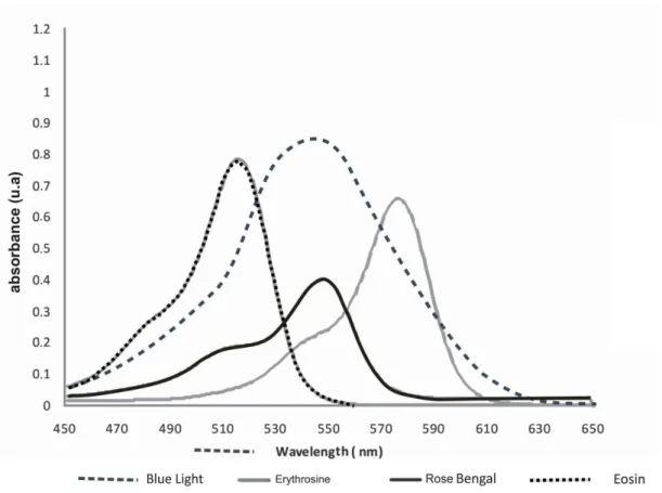
[16]. The scientific literature reports that the interaction between these light sources and the photosensitizers that absorb at this wavelength, such as methylene blue (MB), toluidine blue ortho (TBO) and malachite green (MG), can result in significant microbial killing [17,18,19,20]. Additionally, research has also shown that blue light (380 – 520 nm) is an attractive option for PDT, because blue light sources can be used in combination with other photosensitizers, such as rose bengal (RB), eosin (EOS) and erythrosine (ERI), to photoinactivate oral microorganisms [21,22]. However, no other study has evaluated all these photosensitizers in standardized experimental conditions, by using the same molar concentration and inoculum of S. mutans.
The production of ROS is a molecular reaction dependent on the concentration of the photosentitizer. Thus, the current study used red and blue light sources to activate various photosensitizers with the same molar concentration and analyzed their antimicrobial activities on planktonic suspensions of S. mutans. Furthermore, the singlet oxygen generated by each photosensitizer upon irradiation was quantified using a tryptophan degradation assay.
2. MATERIALS AND METHODS 2.1 Streptococcus mutans
A standard suspension of S. mutans (strain UA159) containing 1 – 2 x 10⁸ viable cells ml-1
was prepared as follows. First, brain heart infusion broth (BHI, Difco, Kansas City, MO, USA) was inoculated with S. mutans and was incubated for 18 h at 37°C in an atmosphere of 10% CO2 (Thermo Fisher Scientific Inc., Waltham, MA, USA). The bacterial culture was then centrifuged at 4,000 g for 5 min (5415R, Eppendorf, São Paulo, SP, Brazil), and the supernatant was discarded. The cell pellet was resuspended in 5 ml of a sterile solution of 0.9% sodium chloride (NaCl).
The number of viable cells in each suspension was determined with a spectrophotometer (Amersham Biosciences Ultrospec 1100 pro, GE Healthcare do Brasil Ltda, São Paulo, SP, Brazil), using a wavelength of 600 nm and an optical density of 0.3120.
Using the standard S. mutans suspension, 150 assays were performed. The photosensitizers methylene blue (MB), toluidine blue ortho (TBO) and malachite green (MG) were tested alone or in combination with irradiation with a red light-emitting diode (LED). Rose bengal (RB), erythrosine (ERI) and eosin (EOS) were tested alone or in combination with irradiation from a blue light source. Ten assays for each of the following conditions were performed: in the absence of both light and photosensitizer; with photosensitizer alone; with light irradiation alone; or in the presence of each photosensitizer and its respective light source.
2.3 Photosensitizer and light
Since PDT is a two-step process in which neither the light source nor the photosensitizer express antimicrobial effect when used alone, we determined in a preliminary study the Minimum Inhibitory Concentration (MIC) for the photosensitizers in order to prevent cytotoxicity of photosensitizers in the absence of light. Difficulty in achieving an agreement among the MIC’s for all studied photosensitizers, led to the establishment of the concentration of 163.5 µM for use in the in vitro S. mutans-experiments. This concentration was coincident with the MICs of TBO and MB. Each photosensitizer solution was prepared in concentrations of 327 µM by dissolving the dye in physiological phosphate-buffered saline (PBS), pH 7.2 with 5% dimethylformamide (DMF) and filtering through a sterile 0.22 μm membrane (MilliporeTM
, São Paulo, SP, Brazil). After filtration, solutions were stored in the dark.
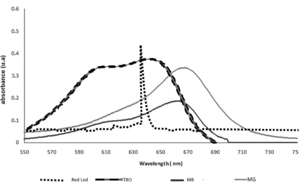
One red LED (80 mW) (MMOptics, São Carlos, SP, Brazil) with a predominant wavelength of 636 nm was used for the photosensitizers MB, TBO and MG, which absorb light of 660, 640 and 675 nm wavelengths, respectively. A blue handheld photopolymerizer (800 mW) (CL-K50, Kondortech São Carlos, SP, Brazil) with a wavelength of 570 nm was used for EOS, ERI and RB, with absorption maxima at 520, 580 and 550 nm, respectively. All devices were set to reach the value of 24 J cm -2 (energy density) according to the following equations:
Fluency = power density × time. Where, power density (PD) is: PD= P (mW)
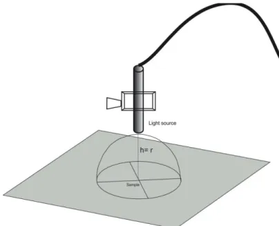
Where, P is the output power of the light source and A is the irradiated area. Since the non-collimate lights may spread as it propagates, the area (A) considered in the present calculation related to the hemisphere area (2 πr2
– in which r is the distance from the end of the light tip to the suspension into the well) (Figure 1). In order to decrease the discrepancy between the applied irradiation times, a 7 mm r distance was chosen for the blue light source, and for the other source, this r distance was set at 3 mm consequently, the applied PDs were respectively 259.9 and 141.5 mW cm-2
.
FIGURE 1. Schematic design of emitting light delivery
FIGURE 2B. Spectrum of the red LED light source system and the absorbance spectra of the TBO, MB and MG in water solution.
2.4 In vitro photosensitization
For each photosensitizer, 0.1 mL of the S. mutans standard suspension was added to each well of sterile, 96-well flat-bottom microtiter plates with lids (TPP, Trasadingen, Switzerland). Next, 0.1 mL of the photosensitizer solution was added to wells in which photosensitizer was tested alone or in combination with light. Control wells received 0.1 mL of 0.9% NaCl solution. Plates were incubated for 5 min on an orbital shaker (TE-145, TECNAL, Piracicaba, SP, Brazil). Wells that were exposed to light, with or without photosensitizer, were irradiated under aseptic conditions in a laminar flow hood in the dark. To prevent light scattering into neighboring wells, plates were covered with a black glass plate with openings corresponding to the diameter of the well [23].
was determined. The results were log-transformed and analyzed by analysis of variance (ANOVA) and the Tukey test. Statistical significance was defined as p ≤
0.05.
2.5 Degradation of Tryptophan by Singlet Oxygen
The same solution of each photosensitizer that was used for experiments was analyzed for photogeneration of singlet oxygen by assessing the reaction of the photosensitizer with tryptophan. One hundred microliters of each photosensitizer was added to 2.5 mL of a 150 µM solution of tryptophan in PBS, pH 7.2, 5% DMF saturated with O2 in a quartz cuvette. Emission by tryptophan at 355 nm after excitation at 280 nm was measured with a fluorescence spectrophotometer (HitachiF4500, Tokyo, Japan), before and during irradiation. The two evaluated light sources expressed different kinetics of tryptophan degradation. Hence, the red light source presented a higher speed of degradation than the blue one. The analysis intervals were 10 sec for the red source and 120 sec for the blue source. Data was collected for up to 2240 sec. The information obtained included the reaction rate between tryptophan and singlet oxygen, assuming first-order kinetics (y = ax + b), and the percent consumption of tryptophan, given the same irradiation potency for each photosensitizer. The observed reaction occurs in two phases, the first one is a photochemical step, which results in the production of singlet oxygen, followed by the reaction of 1
O2 with tryptophan. Thus, the global reaction may be described as follows: Ln[A] – Ln[A]o = -kt, where [A] is the concentration of 3PS in the instant t, [A]o is the concentration of 1PS at the beginning of the reaction, and k is the calculated speed constant and t is the time. Consequently, as k expresses the speed of tryptophan degradation it can be assumed that is similar to 1
O2 production.
3. RESULTS
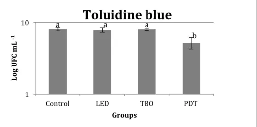
Photodynamic therapy with TBO and MG promoted a significant reduction in the number of CFUmL-1
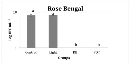
respectively, as shown in figures 5 and 6. PDT performed with ERI had a significant effect on the viability of S. mutans, when compared to control groups, where neither light nor photosensitizer, or light alone was applied. However, when the photosensitizer was utilized alone, a significant reduction in microbial viability was observed, compared to the other control groups (Figure 7). No growth of S. mutans was observed among the groups exposed to RB (Figure 8). These results demonstrate that light sources alone operating with an energy density of 24 J cm-2
have no antimicrobial effect on S. mutans.
FIGURE 3. Effect of toluidine blue ortho (TBO) treatment on the viability of S. mutans. Data (CFU ml-1
) are log-transformed and error bars indicate standard deviation.
FIGURE 4. Effect of malachite green (MG) treatment on the viability of S. mutans. Data (CFU ml-1) are log-transformed and error bars indicate standard deviation.
1 10
Control LED TBO PDT
Lo g U F C m L ‐1 Groups
Toluidine blue
a a a
b
1 10
Control LED MG PDT
Lo g U F C m L ‐1 Groups
Malachite Green
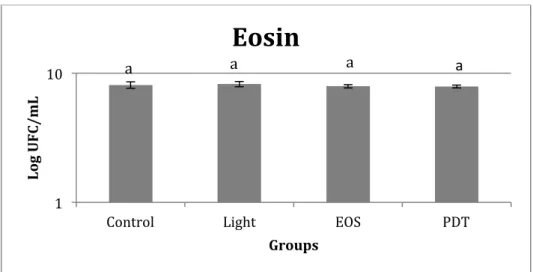
FIGURE 5. Effect of eosin (EOS) treatment on the viability of S. mutans. Data (CFU ml-1
) are log-transformed and error bars indicate standard deviation.
FIGURE 6. Effect of methylene blue (MB) treatment on the viability of S. mutans. Data (CFU ml-1
) are log-transformed and error bars indicate standard deviation. 1
10
Control Light EOS PDT
Lo g U F C /m L Groups
Eosin
a a a a 1 10Control LED MB PDT
Lo g U F C m L ‐ 1 Groups
Methylene Blue
a a a a
1 10
Control Light ERI PDT
Lo g U F C m L ‐1 Groups
Erythrosin
aFIGURE 7. Effect of erythrosin (ERI) treatment on the viability of S. mutans. Data (CFU ml-1
) are log-transformed and error bars indicate standard deviation.
FIGURE 8. Effect of rose bengal (RB) treatment on the viability of S. mutans. Data (CFU ml-1
) are log-transformed and error bars indicate standard deviation.
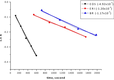
All photosensitizers reduced tryptophan luminescence when irradiated with the convenient wavelength, except for MG. First-order kinetics best describe the reactions, as the relationship between the inverse natural log of Ln[A]vs. irradiation time is linear in periods equal or inferior to 60 sec for red light source, and for blue source in periods equal or inferior to 2000 sec (Figures 9 and 10). The data obtained indicated by the greater reduction in tryptophan concentration by the former.
1 10
Control Light RB PDT
Lo g U F C m L ‐1 Groups
Rose Bengal
a a b b2 0 4 0 6 0
‐0 .8 ‐0 .6 ‐0 .4 ‐0 .2 0 .0 ‐ L n A
tim e, s ec o n d
MB O (‐1 ,4 0 x 1 0‐2) T B O (‐1 ,1 0 x 1 0‐2)
FIGURE 9. Reduction in tryptophan luminescence when irradiated with a red light source at a convenient wavelength and speed of tryptophan degradation.
0 2 0 0 4 0 0 6 0 0 8 0 0 1 0 0 0 1 2 0 0 1 4 0 0 1 6 0 0 1 8 0 0 2 0 0 0 ‐0 .4
‐0 .3 ‐0 .2 ‐0 .1 0 .0
‐L
n
A
tim e, s ec o n d
E O S (‐4 .9 2 x 1 0‐4) E R I (‐1 .2 0 x 1 0‐4) B R (‐1 .1 7 x 1 0‐4)
FIGURE 10. Reduction in tryptophan luminescence when irradiated with a blue light source at a convenient wavelength and speed of tryptophan degradation.
5. DISCUSSION
Management of dental caries involves prescribing therapeutic regimens to individuals according to their risk levels and optimal conservative treatment decisions [24]. Based on this premise, different approaches to control cariogenic biofilms, including the use of antimicrobial agents, have been employed for dental disinfection [25]. The current study focuses on the use of PDT performed with different photosensitizers, since a better understanding of S. mutans photoinactivation is an important issue in Dentistry. In addition, the resulting production of reactive oxygen species (ROS) was evaluated, particularly 1
O2, because many of the bacteria that cause oral infections do not express proteins that neutralize ROS, such as catalase and superoxide dismutase [26].
Our results demonstrate that for MB, TBO, EOS and MG, neither the photosensitizer alone, nor irradiation in the absence of a photosensitizer, had a significant effect on the viability of S. mutans. Thus, our results confirm those of previous studies on oral microorganisms [12,27,28] that conclude that there is no dark toxicity from either component when used separately. However, for RB and ERI,
complete bacterial killing and a decrease in cell viability, respectively, were observed in those groups where the photosensitizer alone was present. One possible explanation for this is that the concentration used (163.5 µM) is high for these photosensitizers, resulting in bacterial toxicity. According to Goulart et al. [29], RB becomes toxic for A. actinomycetemcomitans at concentrations above 0.1 µM and is dose-dependent up
to 50 µM, when cell death is nearly 100%. A recent study demonstrated that PDT performed using ERI was able to obtain a reduction of 5.16, while RB had a reduction of 6.86 log10 CFU/mL on planktonic cultures of S. mutans when compared to the control group. These photosensitizers did not present cytotoxicity in the absence of light [30]. A concentration as low as 2 μM for both photosensitizers and fluence of 92 J cm-2 (4-fold higher than our) were used, explaining the different results obtained in our study. With regard to S. mutans susceptibility to RB, another study demonstrated that the dye shows toxicity in the dark per se in concentrations above 2.5 μM, which is in agreement with our results [31]. Moreover, the latter study found that in these concentrations RB also presented toxicity to fibroblasts, causing damage to human cells [31].
against S. mutans can be attributed exclusively to the PS action. This phenomenon could be observed by the outstanding antimicrobial action expressed in the presence of RB, in spite of the absence of the light source. We recognize that if proper MICs for different PSs had been used, the antimicrobial activity of each PS might have shown different effects, thus, studies must be performed using lower concentrations of RB and ERI to validate their use in PDT against S. mutans. However, it is essential that the photosensitizer is not harmful to the cells alone, because in many cases it will be present in healthy as well as targeted cells. To ensure this, the dark toxicity of the photosensitizer must be very low, assuring the most destructive ability of the drug comes from the production ROS by irradiation. Consequently, to test PSs in the same molar concentration is important to determine the drug, which is capable of ensuring that healthy cells will be not or minimally affected.
No microbial reduction was demonstrated with EOS. To the best of our knowledge, no other study was performed using EOS in PDT against S. mutans. According to our spectral analysis and to the literature, the peak of light absorption for EOS and ERI are at 520 and 580, respectively, and the light source used in the present study emits light predominantly in 570 nm. So, without considering the overlapping area, these data could explain the slightly better results obtained for ERI than for EOS since ERI-mediated PDT statistically reduced S. mutans when compared to its respective control groups.
Despite the fact that MB and TBO are phenothiazine derivatives and have similar chemical structures with physical and chemical properties and hydrophilic features that allow their free passage across the bacterial membrane [32], the results differed greatly between these two photosensitizers in the present study. Blue methylene (MB) and toluidine blue orto (TBO) present a very similar spectrum absorption in the region of the visible red light (≅ λ = 660 nm and 630 nm
overlapping spectral for MG is approximately 2-fold higher than the that found for MB, while the energy absorbed by TBO is about 3.5-fold greater than that absorbed by MB. Therefore, the overlapping spectral of red light emission with TBO absorption was important for obtaining phototoxic effects in vitro against S. mutans. Thus, one can speculate that since this overlap was the highest one for this light source, the other photosensitizers themselves absorbed less photons, and therefore lesser amounts of reactive oxygen species were produced. Nevertheless, this hypothesis seems unlikely, since MB was the photosensitizer that produced the highest amount of 1
O2, which indicates that the observed antimicrobial inefficacy did not result from the total energy absorbed.
A distinguishing characteristic between these dyes is the partition coefficient (P), which is almost 3-fold higher for TBO than for MB [33], indicating the higher ability of TBO molecules to permeate and accumulate in the hydrophobic region of the cellular membrane and therefore present better photobactericidal activities. In addition, TBO is a more effective photobactericide and has a greater ability to dimerize than MB. This pattern was consistently observed in both Gram-positive and Gram-negative bacteria [34].
The penetration of photosensitizers into the cell is not a passive process. Since the cell membrane acts as a selective barrier to free diffusion, penetration of photosensitizer molecules depends on their size, charge and solubility, and they may accumulate at particular subcellular locations or be associated with the more hydrophobic regions of membranous organelles [37]. Differences in intracellular concentration or localization between the photosensitizers may be responsible for the variability in efficacy we observed in this study. The fact that both TBO and MB produced higher levels of singlet oxygen after irradiation, while only TBO demonstrated potent antimicrobial activity, may be due to different interactions with the bacterial cell resulting from slight structural variations between the two photosensitizers. It is important to remember that TBO interacts more easily with the bacterial membrane than does MB, owing to its greater solubility in the hydrophobic region of the membrane and the fact that it has the lowest molecular weight of all of the photosensitizers studied here (305.83 g mol-1
), allowing it to diffuse more easily. Thus, it is not without reason that TBO is one of the most widely used photosensitizers in PDT research [23,33]. It should be emphasized that only TBO and MG caused bacterial reduction compared to their respective control groups. However, MG was not able to show a biologically significant reduction, since it only presented a 1.4 log reduction in S. mutans.
bacteria, as has been previously described. Thus, the bulk in vitro -1O2 production may not be related to the ability of the photosensitizer to get into or closer to the bacterial activity site. Even though singlet O2 production was high for MB samples, if this ERO is not in close contact with bacterial cell DNA it will not have any effect on killing S. mutans.
Although without biological significance, MG was another photosensitizer irradiated with a red light source, which delivered promising results against S. mutans. However, MG did not produce singlet oxygen, indicating that the antimicrobial activity of PDT may also be promoted by other ROS. Indeed, a previous study has demonstrated that MG-induced photodamage occurs via the classical type I photosensitization mechanism, with very little contribution from type II reactions [41].
The photosensitizers irradiated by a blue light source produced a lower amount of singlet oxygen than the other photosensitizers irradiated by red light source. ERI exhibited some toxicity to the bacteria, even without irradiation. However, irradiation increased the antimicrobial activity of ERI, suggesting that the ROS formed after irradiation are responsible for this increase. The fact that all of the red compounds produced singlet oxygen, but only ERI exhibited an antimicrobial effect, emphasizes that other factors influence the effectiveness of PDT. These factors may include the pharmacodynamic properties of the photosensitizer, such as cellular penetration, localization and influx and efflux parameters; the intracellular stability and activity of the photosensitizer; and sufficient targeting of the photosensitizer toward the pathogen [37]. Another consideration is that the blue light used was not derived from an LED, and the lack of collimation and coherence may decrease the efficacy of PDT. It can be speculated that a reduction on the quantity of blue light delivered or absorbed by the photosensitizers occurred due to a higher dissipation of energy, despite the focusing light tip in this light source [42]. Some of the limitations considered in this study include energy density calculation for lights with filter tip performed similarly to that used for calculation in LED sources, and the lack of a suitable method for calculating dissipated energy.
evaluating the antimicrobial activity of PDT against S. mutans. Due to the parameters chosen to perform the present study such as molar concentration, light source characteristics and pre-irradiation time, RB and ERI exhibited cytotoxicity at concentrations as low as 163.5 µM, while TBO and MG were effective in decreasing S. mutans counts after PDT, although only TBO was effective in reducing 3-log of
these microorganisms.
ACKNOWLEDGEMENTS
The authors thank Bruna Melo de Codes for her help during experimental procedures.
REFERENCES
[1]. B.C. WILSON, S.M. PATTERSON, The physics, biophysics and technology of photodynamic therapy, Phy. Med. Biol 53 (2008) 61-109.
[2]. A.C KÜBLER, Photodynamic therapy, Med Laser Appl 20 (2005) 37-45.
[3]. M. OCHSNER, Photophysical and photobiological processes in the photodynamic therapy of tumors, J Photochem Photobiol B: Biol 39(1997) 1–18. [4]. J.J.L. CADET, G.R.RAVANAT, M.H. MARTINEZ, P. MEDEIROS, DI MASCIO, Singlet oxygen oxidation of isolated and cellular DNA: Product formation and mechanistic insights, Photochem. Photobiol 82 (2006) 1219–1225.
[5]. R. SCHMIDT, Photosensitized generation of singlet oxygen, Photochem. Photobiol 82 (2006) 1161–1177.
[6]. Z. MALIK, J. HANANIA, Y. NITZAN, Bactericidal effects of photoactivated porphyrins--an alternative approach to antimicrobial drugs, J. Photochem. Photobiol. B5 (1990) 281-293.
[7]. M. BHATTI, A. MACROBERT, S. MEGHJI, B. HENDERSON, M. WILSON, Effect of dosimetric and physiological factors on the lethal photosensitization of Porphyromonasgingivalis in vitro, Photochem. Photobiol 65 (1997) 1026-31.
[8]. M.R. HAMBLIN, T. HASAN, Photodynamic therapy: a new anti-microbial approach to infectious disease?, Photochem Photobial Sci 3 (2004) 436–450.
[10]. J.S.M. GIUSTI, L. SANTOS-PINTO, A.C PIZZOLITO, K. HELMERSON, E. CARVALHO-FILHO, C. KURACHI, V.S. BAGNATO, Antimicrobial photodynamic action on dentin using a light-emitting diode light source, Photomed. Laser. Surg 26 (2008) 279-285.
[11]. I.C.J. ZANIN, R.B. GONCALVES, A. BRUGNERA-JR, C.K. HOPE, J. PRATTEN, Susceptibility of Streptococcus mutans biofilms to photodynamic therapy: an in vitro study, J. Antimicrob. Chemother 56 (2005) 324-330.
[12]. I.C. ZANIN, M.M. LOBO, L.K. RODRIGUES, L.A. PIMENTA, J.F. HOFLING, R.B. GONCALVES, Photosensitization of in vitro biofilms by toluidine blue O combined with a light-emitting diode, Eur. J. Oral. Sci 114 (2006) 64-69. [13]. M. MERCHAT, G. BERTOLINI, P. GIACOMINI, A. VILLA NUEVA, G. JORI, Meso-substituted cationic porphyrins as efficient photosensitizers of Gram-positive and Gram-negative bacteria, J. Photochem. Photobiol: B 32 (1996) 153–157. [14]. M. WILSON, Photolysis of oral bacteria and its potential use in the treatment of caries and periodontal disease, J. Appl. Bacteriol 75 (1993) 299-306.
[15]. K. PLAETZER, T. KIESSLICH, T. VERWANGER, B. KRAMMER, The modes of cell death induced by PDT: An Overview, Med. Laser. Appl 18 (2003) 7-19.
[16]. B.C. WILSON, The physics of photodynamic therapy, Phys. Med. Biol 31 (1986) 327-360.
[17]. J.P.M. LIMA, M.A. SAMPAIO DE MELO, F.M.BORGES, A.H. TEIXEIRA, C. STEINER-OLIVEIRA, M. NOBRE DOS SANTOS, L.K. RODRIGUES, I.C. ZANIN, Evaluation of the antimicrobial effect of photodynamic antimicrobial therapy in an in situ model of dentine caries, Eur. J. Oral. Sci. 117 (2009) 568-74.
[18]. M.A.S. MELO, D.M. DE-PAULA, J.P.M. LIMA, F.M.C. BORGES, C. STEINER-OLIVEIRA, M. NOBRE-DOS-SANTOS, I.C.J. ZANIN, E.B. BARROS, L. K.A. RODRIGUES, In Vitro Photodynamic Antimicrobial Chemotherapy in Dentine Contaminated by Cariogenic Bacteria, Laser Physics 20 (2010) 1–10.
[19]. L.S. PELOI, R.S.S. SOARES, C.E.G. BIONDO, V.R. SOUZA, N. HIOKA, E. KIMURA, Photodynamic effect of light-emitting diode light on cell growth inhibition induced by methylene blue, J. Biosci. 33 (2008) 231–237
Bactericidal effect of malachite green and red laser on Actinobacillus actinomycetemcomitans, J. Photochem. Photobiol B 86 (2007) 70-6.
[21]. D. METCALF, C. ROBINSON, D. DEVINE, S. WOOD, Enhancement of erythrosine-mediated photodynamic therapy of Streptococcus mutans biofilms by light fractionation, J. Antimicrobial. Chemotherapy 58 (2006) 190–192.
[22]. M. NISNEVITCH, F. NAKONECHNY, Y. NITZAN, K. BIOORG, Photodynamic antimicrobial chemotherapy by liposome-encapsulated water-soluble photosensitizers, Bioorg. Khim 6 (2010) 396-402.
[23]. R.C. SOUZA, J.C. JUNQUEIRA, R.D. ROSSONI, C.A. PEREIRA, E. MUNIN, A.O. JORGE, Comparison of the photodynamic fungicidal efficacy of methylene blue, toluidine blue, malachite green and low-power laser irradiation alone against Candida albicans, Lasers. Med. Sci 25 (2010) 385-9.
[24]. K. ANUSAVICE, Clinical decision-making for coronal caries management in the permanent dentition, J. Dent. Educ 65 (2001) 1143-1146.
[25]. M.S. WOLFF, C. LARSON, The cariogenic dental biofilm: good, bad or just something to control?, Braz. Oral. Res. 23 (2009) 31-38 .
[26]. K.L. RUOFF, Miscellaneous catalase-negative, gram-positive cocci: Emerging opportunists, J. Clin. Microbiol 40 (2002) 1129-1133.
[27]. J.A. WILLIAMS, G.J. PEARSON, M.J. COLLES, M. WILSON, The effect of variable energy input from a novel light source on the photoactivated bactericidal action of toluidine blue O on Streptococcus Mutans, Caries Res. 37 (2003) 190-193. [28]. N.S. SOUKOS, S.S. SOCRANSKY, S.E. MULHOLLAND, S. LEE, A.G. DOUKAS, Photomechanical drug delivery into bacterial biofilms, Pharmaceutical. Res 17 (2003) 405-409.
[29]. R.C. GOULART, M. BOLEAN, T.P. PAULINO, G. THEDEI, S.L.S. SOUZA, A.C. TEDESCO, P.CIANCAGLINI, Photodynamic therapy in Planktonic and biofilm cultures of Aggregatibacter actinomycetemcomitans, Photomed. Laser. Surg 28 (2010) 53-60.
[31]. T. P. PAULINO, K. F. RIBEIRO, G. THEDEI JR., A. C. TEDESCO, P. CIANCAGLINIA, Use of hand held photopolymerizer to photoinactivate Streptococcus mutans, Arch. Oral. Biol 50 (2005) 353 -359.
[32] G. JORI, C. FABRIS, M. SONCIN, S. FERRO, O. COPPELLOTTI, D. DEI, L. FANTETTI, G. CHITI, G. RONCUCCI, Photodynamic Therapy in the Treatment of Microbial Infections: Basic Principles and Perspective Applications, Lasers. Surg. Med 38 (2006) 468–481.
[33]. M.N. USACHEVA, M.C. TEICHERT, M.A. BIEL, Comparison of the methylene blue and toluidine blue photobactericidal efficacy against Gram-positive and Gram-negative microorganisms, Lasers Surg Med29 (2001) 165–173.
[34]. M. N. USACHEVA, M. C. TEICHERT, M.A. BIEL, The role of the methylene blue and toluidine blue monomers and dimers in the photoinactivation of bacteria, J Photochem and Photobiol B: Biol 71 (2003) 87–98.
[35]. S. WOOD, D. METCALF, D. DEVINE, C. ROBINSON, Erythrosine is a potential photosensitizer for the photodynamic therapy of oral plaque biofilms, J Antimicrob Chemother 57 (2006) 680-4.
[36]. M. WAINWRIGHT, Methylene blue derivatives-suitable photoantimicrobials for blood product disinfection?, Int J Antimicrob Agents 16 (2000) 381-94.
[37]. O.E. AKILOV, K. O’RIORDAN, S. KOSAKA, T. HASAN, Photodynamic therapy against intracellular pathogens: Problems and potentials, Med. Laser. Appl 21 (2006) 251-260.
[38]. E.S. NYMAN, P.H. HYNNINEN, Research advances in the use of tetrapyrrolic photosensitizers for photodynamic therapy, J. Photochem. Photobiol. B: 73 (2004) 1– 28.
[39]. D.P. VALENZENO, Photomodification of biological membranes with emphasis on singlet oxygen mechanisms, Photochem. Photobiol 46 (1987) 147–160.
[40]. T. MAISCH, Anti-microbial photodynamic therapy: useful in the future?, Lasers Med Sci 22 (2007) 83–91.
[41]. J.A. BARTLETT, G.L. INDIG, Spectroscopic and photochemical properties of malachite green non covalent bound to bovine serum albumin, Dyes. Pigments (1999) 43 219-226.
3.2. CAPÍTULO 2
Confocal Raman study of photosensitizer penetration in artificially dentin caries lesions
Juliana Paiva Marques Lima ROLIM1
; Mary Anne Sampaio DE-MELO1
;Iriana Carla Junqueira ZANIN1; José Júnior Alves DA-SILVA2; Alexandre Rocha PASCHOAL2; Alejandro Pedro AYALA2; Nádia Accioly Pinto NOGUEIRA1, Lidiany Karla Azevedo RODRIGUES1
1
Post-graduation Program, Faculty of Pharmacy, Dentistry and Nursing, Federal University of Ceará, Brazil, 944 Cap. Francisco Pedro Street - Rodolfo Teófilo, Zip Code: 60430-170
2
Physics Department, Federal University of Ceara, Fortaleza, Ceará, Brazil, Pici Campus – Build # 922 – Fortaleza, Ceara, Brazil Zip code: 60455-760
Address all correspondence to:
Prof. Lidiany Karla Azevedo Rodrigues,DDS, MSc, PhD, Associate Professor
Department of Dentistry, Faculty of Pharmacy, Dentistry and Nursing, Federal University of Ceara, Fortaleza, Ceara, Brazil
Cap. Francisco Pedro Street - Rodolfo Teófilo – Zip Code: 60430-170 Phone- #558533668410, Fax- #558533668232
Email: lidianykarla@yahoo.com
ABSTRACT
INTRODUCTION
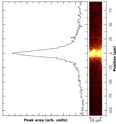
Photodynamic antimicrobial therapy (PACT) is a treatment modality for tissue infection that involves delivering harmless visible light of an appropriate wavelength to promote molecules from non-toxic dyes or photosensitizer (PS) into the excited state. This excited PS then transfers energy to ground-state molecular oxygen to produce excited-state singlet oxygen and free radicals that can oxidise many organisms, such as bacteria, fungi and yeast, and lead to the injury and death of microorganisms (1,2). Thus, PACT has been associated with minimal intervention dentistry as a potential alternative to oral bacteria inactivation in the remaining demineralized dentine tissue, contributing to a minimal removal of carious tissues and a more conservative approach for dealing with deep caries lesions (3). Several studies have suggested PACT is an effective approach to eradicate cariogenic bacteria that infect the tooth tissue (4-7).
Considering the anatomical features of dentin, which is a porous, hard, mineralised connective tissue composed primarily of tubular hydroxyapatite-coated collagen type I fibrils with changes in the relative proportions of dentinal tubules within different areas of the dentin (8), bacteria from carious lesions and their bacterial products can penetrate and diffuse profoundly through the dentinal tubule toward the pulp and promote inflammatory changes in the pulp-dentin complex (9). Thus, a key factor for clinical success is that the photosensitizer should be able to achieve and interact with the target bacteria inside the tissues.