The genome and transcriptome of
Phalaenopsis
yield insights into floral
organ development and flowering
regulation
Jian-Zhi Huang1,
*, Chih-Peng Lin2,4,
*, Ting-Chi Cheng1, Ya-Wen Huang1, Yi-Jung Tsai1, Shu-Yun Cheng1, Yi-Wen Chen1, Chueh-Pai Lee2, Wan-Chia Chung2, Bill Chia-Han Chang2,3, Shih-Wen Chin1, Chen-Yu Lee1and Fure-Chyi Chen1
1Department of Plant Industry, National Pingtung University of Science and Technology,
Pingtung, Taiwan
2Yourgene Bioscience, Shu-Lin District, New Taipei City, Taiwan
3Faculty of Veterinary Science, The University of Melbourne, Parkville, Victoria, Australia 4Department of Biotechnology, School of Health Technology, Ming Chuan University, Gui Shan
District, Taoyuan, Taiwan
*These authors contributed equally to this work.
ABSTRACT
The Phalaenopsisorchid is an important potted flower of high economic value around the world. We report the 3.1 Gb draft genome assembly of an important winter floweringPhalaenopsis‘KHM190’ cultivar. We generated 89.5 Gb RNA-seq and 113 million sRNA-seq reads to use these data to identify 41,153 protein-coding genes and 188 miRNA families. We also generated a draft genome forPhalaenopsis pulcherrima‘B8802,’ a summer flowering species, via resequencing. Comparison of genome data between the two Phalaenopsiscultivars allowed the identification of 691,532 single-nucleotide polymorphisms. In this study, we reveal that the key role ofPhAGL6bin the regulation of labellum organ development involves alternative splicing in the big lip mutant. Petal or sepal overexpressingPhAGL6b leads to the conversion into a lip-like structure. We also discovered that the gibberellin pathway that regulates the expression of flowering time genes during the reproductive phase change is induced by cool temperature. Our work thus depicted a valuable resource for the flowering control, flower architecture development, and breeding of the Phalaenopsisorchids.
Subjects Genomics, Plant Science
Keywords Phalaenopsis, Draft genome,PhAGL6b, Flower organ development, Flowering time
INTRODUCTION
Phalaenopsis is a genus within the family Orchidaceae and comprises approximately 66 species distributed throughout tropical Asia (Christenson, 2002). The predicted Phalaenopsisgenome size is approximately 1.5 gigabases (Gb), which is distributed across 19 chromosomes (Lin et al., 2001).Phalaenopsisflowers have a zygomorphic floral structure, including three sepals (in the first floral whorl), two petals and the third petal develops into a labellum in early stage of development, which is a distinctive feature of a Submitted2 February 2016
Accepted17 April 2016
Published12 May 2016
Corresponding authorsss Bill Chia-Han Chang, [email protected] Shih-Wen Chin, [email protected] Chen-Yu Lee, [email protected] Fure-Chyi Chen, [email protected] Academic editor Sheila McCormick
Additional Information and Declarations can be found on page 13
DOI10.7717/peerj.2017
Copyright 2016 Huang et al.
Distributed under
highly modified floral part in second floral whorl unique to orchids. The gynostemium contains the male and female reproductive organs in the center (Rudall & Bateman, 2002). In the ABCDE model, B-class genes play important role to perianth development in orchid species (Chang et al., 2010;Mondrago´n-Palomino & Theissen, 2011;Tsai et al., 2004). In addition,PhAGL6a andPhAGL6b, expressed specifically in thePhalaenopsis labellum, were implied to play as a positive regulator of labellum formation (Huang et al., 2015;Su et al., 2013). However, the relationship between the function of genes involved in floral-organ development and morphological features remains poorly understood.
Phalaenopsisorchids are produced in large quantity annually and are traded as the most important potted plants worldwide. During greenhouse production of young plants, the high temperature > 28C was routinely used to promote vegetative growth and inhibit
spike initiation (Blanchard & Runkle, 2006). Conversely, a lower ambient temperature (24/18C day/night) is used to induce spiking (Chen et al., 2008) to produce flowering
plants. Spike induction inPhalaenopsisorchid by this cool temperature is the key to precisely controlling its flowering date. Several studies have indicated that cool temperature during the night are necessary forPhalaenopsisorchids to flower (Blanchard & Runkle, 2006; Chen et al., 1994;Chen et al., 2008;Wang, 1995). Despite a number of expressed sequence tags (ESTs), RNA-seqs and sRNA-seqs from several tissues ofPhalaenopsishave been reported and deposited in GenBank or OrchidBase (An & Chan, 2012;An, Hsiao & Chan, 2011;Hsiao et al., 2011;Su et al., 2011), only a few flowering related genes or miRNAs have been identified and characterized. In addition, the clues to the spike initiation during reproductive phase change in the shorten stem, which may produce signals related to flowering during cool temperature induction, have not been dealt with. At this juncture, the molecular mechanisms leading to spiking ofPhalaenopsishas yet to be elucidated.
Here we report a high-quality genome and transcriptomes (mRNAs and small RNAs) of PhalaenopsisBrother Spring Dancer ‘KHM190,’ a winter flowering hybrid with spike formation in response to cool temperature. We also provide resequencing data for summer flowering speciesP. pulcherrima‘B8802.’ Our comprehensive genomic and transcriptome analyses provide valuable insights into the molecular mechanisms of important biological processes such as floral organ development and flowering time regulation.
METHODS SUMMARY
RESULTS AND DISCUSSION
Genome sequencing and assemblyWe sequenced the genome of the Phalaenopsisorchid cultivar ‘KHM190’ (Appendix S1,
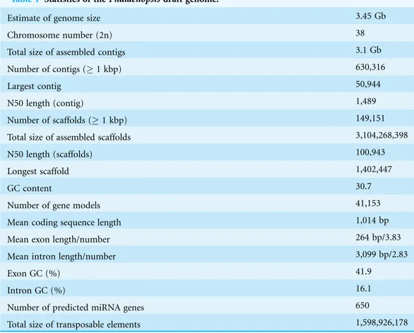
Fig. S1a) using the Illumina HiSeq 2000 platform and assembled the genome with the Velvet assembler, using 300.5 Gb (90-fold coverage) of filtered high-quality sequence data (Appendix S1,Table S1). This cultivar has an estimated genome size of 3.45 Gb on the basis of a 17 m depth distribution analysis of the sequenced reads (Appendix S1,Figs. S2
andS3;Tables S2andS3). De novo assembly of the Illumina reads resulted in a sequence of 3.1 Gb, representing 89.9% of thePhalaenopsisorchid genome. Following gap closure, the assembly consisted of 149,151 scaffolds (1,000 bp), with N50 lengths of 100 and 1.5 kb for the contigs. Approximately 90% of the total sequence was covered by 6,804 scaffolds of > 100 kb, with the largest scaffold spanning 1.4 Mb (Appendix S1,
Tables S3–S5andData S17). The sequencing depth of 92.5% of the assembly was more than 20 reads (Appendix S1,Fig. S3), ensuring high accuracy at the nucleotide level. The GC content distribution in the Phalaenopsisgenome was comparable with that in the genomes ofArabidopsis(The Arabidopsis Genome Initiative, 2000),Oryza (International Rice Genome Sequencing Project, 2005 andVitis(Jaillon et al., 2007) (Appendix S1,Fig. S4).
Gene prediction and annotation
Approximately 59.74% of thePhalaenopsis genome assembly was identified as repetitive elements, including long terminal repeat retrotransposons (33.44%), DNA transposons (2.91%) and unclassified repeats (21.99%) (Appendix S1,Fig. S5andTable S6). To facilitate gene annotation, we identified 41,153 high-confidence and medium-confidence protein-coding regions with complete gene structures in thePhalaenopsisgenome using RNA-Seq (114.1 Gb for a 157.6 Mb transcriptome assembly), based on 15 libraries representing four tissues (young floral organs, leaves, shortened stems and protocorm-like bodies (PLBs)) (Appendix S1,Table S7andData S18), and we used transcript assemblies of these regions in combination with publically available expressed sequence tags (Su et al., 2011;Tsai et al., 2013) for gene model prediction and validation (Data S1–S2). We predicted 41,153 genes with an average mRNA length of 1,014 bp and a mean number of 3.83 exons per gene (Table 1andData S3). In addition to protein coding genes, we identified a total of 562 ribosomal RNAs, 655 transfer RNAs, 290 small nucleolar RNAs and 263 small nuclear RNAs in thePhalaenopsisgenome (Appendix S1,Table S8). We also obtained 92,811,417 small RNA (sRNA) reads (18–27 bp), representing 6,976,375 unique sRNA tags (Appendix S1,Fig. S6andData S6–S7). A total of 650 miRNAs distributed in 188 families were identified (Data S8), and a total of 1,644 miRNA-targeted genes were predicted through the alignment of conserved miRNAs to our gene models (Appendix S1,
Fig. S7andData S9–S10).
The Phalaenopsis gene families were compared with those ofArabidopsis
We identified 41,153 Phalaenopsis genes in 15,855 families, with 8,532 gene families being shared withArabidopsis, Oryzaand Vitis. Another 5,143 families, containing 12,520 genes, were unique toPhalaenopsis (Fig. 1). In comparison with the 29,431 protein-coding genes estimated for thePhalaenopsis equestrisgenome (Cai et al., 2015), our gene set for Phalaenopsis ‘KHM190’ contained 11,722 more members, suggesting a more wider representation of genes in this work. This difference in gene number may be due to different approaches betweenPhalaenopsis ‘KHM190’ andPhalaenopsis equestris. Besides,Phalaenopsis‘KHM190’ is a hybrid while P. equestris species, which may show gene number difference due to different genetic background. To better annotate the Phalaenopsis genome for protein-coding genes, we generated RNA-seq reads obtained from four tissues as well as publically available expressed sequence tags for cross reference. We defined the function of members of these families using (The Gene Ontology Consortium, 2008), the Kyoto Encyclopedia of Genes and Genomes (KEGG) (Kanehisa et al., 2012) and Pfam protein motifs (Finn et al., 2014) (Fig. 2;
Data S3–S5and S19).
The genes in the High confidence (HC) and Medium Confidence (MC) gene sets were functionally annotated based on homology to annotated genes from the NCBI non-redundant database (Data S3). The functional domains ofPhalaenopsis genes were identified by comparing their sequences against protein databases, including (The Gene Ontology Consortium, 2008), KEGG (Kanehisa et al., 2012) and Pfam
Table 1 Statistics of thePhalaenopsisdraft genome.
Estimate of genome size 3.45 Gb
Chromosome number (2n) 38
Total size of assembled contigs 3.1 Gb
Number of contigs (1 kbp) 630,316
Largest contig 50,944
N50 length (contig) 1,489
Number of scaffolds (1 kbp) 149,151
Total size of assembled scaffolds 3,104,268,398
N50 length (scaffolds) 100,943
Longest scaffold 1,402,447
GC content 30.7
Number of gene models 41,153
Mean coding sequence length 1,014 bp
Mean exon length/number 264 bp/3.83
Mean intron length/number 3,099 bp/2.83
Exon GC (%) 41.9
Intron GC (%) 16.1
Number of predicted miRNA genes 650
(Finn et al., 2014;Finn, Clements & Eddy, 2011) databases. GO terms were obtained using the Blast2GO program (Conesa & Gotz, 2008). In the GO annotations, 16,034, 27,294 and 16,360 genes were assigned to the biological process, cellular component, and molecular function categories, respectively (Fig. 2A). Based on KEGG pathway mapping, we were able to assign a significant proportion of thePhalaenopsisgene sets to KEGG functional or biological pathway categories (11,452 sequences; 140 KEGG orthologous terms) (Data S4). To investigate protein families, we compared the Pfam domains ofPhalaenopsisgenome. A total of 1,842 Pfam domains were detected among the Phalaenopsissequences. The most abundant protein domains inPhalaenopsis genome were pentatricopeptide repeats (PPRs, pfam01535), followed by the WD40 (pfam00400), EF hand (pfam00036) and ERM (Ezrin/radixin/moesin, pfam00769) domains (Fig. 2B andData S5). Furthermore, conserved domains could be identified in 50.17% of the predicted protein sequences based on comparison against Pfam databases. In addition, we identified 2,610 transcription factors (TFs) (6.34% of the total genes) and transcriptional regulators in 55 gene families (Appendix S1,
Figs. S8–S10and Datas S11–S12).
Regulation ofPhalaenopsis floral organ development
The relative expression of allPhalaenopsis genes was compared through RNA-Seq analysis of shoot tip tissues from shortened stems, leaf, floral organs and PLB samples, in addition to vegetative tissues, reproductive tissues, and germinating seeds from P. aphrodite (Su et al., 2011; Tsai et al., 2013) (Appendix S1,Fig. S12 andData S1). Phalaenopsisorchids exhibit a unique flower morphology involving outer tepals, lateral inner tepals and a particularly conspicuous labellum (lip) (Rudall & Bateman, 2002). However, our understanding of the regulation of the floral organ development of the genus is still in its infancy. To comprehensively characterize the genes involved in the development ofPhalaenopsis floral organs, we obtained RNA-Seq data for the sepals, petals and labellum of both the wild-type and peloric mutant of Phalaenopsis ‘KHM190’ at the 0.2 cm floral bud stage, at which shows early sign of labellum differentiation. This mutant presented an early peloric fate in its lateral inner tepals. In a peloric flower, the lateral inner tepals are converted into a lip-like morphology at this young bud stage (Appendix S1, Figs. S11BandS12A). We identified 3,743 genes
Figure 1 Venn diagram showing unique and shared gene families between and amongPhalaenopsis,
that were differentially expressed in the floral organs of the wild-type and peloric mutant plants. Gene Ontology analysis of the differentially expressed genes in Phalaenopsis floral organs revealed functions related to biological regulation, developmental processes and nucleotide binding, which were significantly altered A
B
in both genotypes (Huang et al., 2015). TFs seem to play a role in floral organ development. Of the 3,309 putative TF genes identified in the Phalaenopsis
genome showed differences in expression between the wild-type and peloric mutant plants (Data S11).
MADS-box genes are of ancient origin and are found in plants, yeasts and animals (Trobner et al., 1992). This gene family can be divided into two main lineages, referred to as types I and II. Type I genes only share sequence similarity with type II genes in the MADS domain (Alvarez-Buylla et al., 2000). Most of the well-studied plant genes are type II genes and contain three domains that are not present in type I genes: an intervening (I) domain, a keratin-like coiled-coil (K) domain, and a C-terminal (C) domain (Munster et al., 1997). These genes are best known for their roles in the specification of floral organ development, the regulation of flowering time and other aspects of reproductive development (Dornelas et al., 2011). In addition, MADS-box genes are also widely expressed in vegetative tissues (Messenguy & Dubois, 2003;Parenicova et al., 2003). The ABCDE model comprises five major classes of homeotic selector genes: A, B, C, D and E, most of which are MADS-box genes (Theissen, 2001). However, research on the ABCDE model was mainly focused on herbaceous plants and has not fully explained how diverse angiosperms evolved. The function of many other genes expressed during floral development remains obscure.Phalaenopsisexhibits unique flower morphology
involving three types of perianth organs: outer tepals, lateral inner tepals, and a labellum (Rudall & Bateman, 2002). Despite its unique floral morphological features, the
molecular mechanism of floral development inPhalaenopsisorchid remains largely unclear, and further research is needed to identify genes involved in floral differentiation. Recently, several remarkable research studies onPhalaenopsisMADS-box genes have revealed important roles of some of these genes in floral development, such as four B-class DEF-like MADS-box genes that are differentially expressed between wild-type plants and peloric mutants with lip-like petals (Tsai et al., 2004) and aPI-likegene,PeMADS6, that is ubiquitously expressed in petaloid sepals, petals, columns and ovaries (Tsai et al., 2005).
In thePhalaenopsisgenome sequence assembly, a total of 122 genes were predicted to encode MADS-box family proteins (Appendix S1,Fig. S8andData S12). To obtain a more accurate classification, phylogenetic trees were constructed via the neighbour-joining method, with 1000 bootstraps using MEGA5 (Tamura et al., 2011). The differentially expressed genes (DEGs) among 122PhalaenopsisMADS-box genes were obtained from our PhalaenopsisRNA-Seq data (Data S11). The expression profile indicated that most MADS-box genes are widely expressed in diverse tissues. These results will be helpful in elucidating the regulatory roles of these genes in thePhalaenopsis floral organ development.
observed in the wild-type flower (Fig. 3B). Interestingly, we identified four alternatively spliced forms ofPhAGL6b that were specifically expressed only in the petaloid labellum of the big lip mutant (Figs. 3C and 3D; Appendix S1 andFig. S11). To determine whether the alternatively spliced forms of PhAGL6b affect the conversion of the labellum to a petal-like organ in the big lip mutant, we performed RT-PCR of total RNA extracted from the labellum organs of plants with different big lip mutant phenotypes and wild-type plants (Appendix S1, Table S11 andFig. 4A) to amplify the PhAGL6b transcripts. Interestingly, among all of the big lip mutant phenotypes, 500–700 bp bands were detected, corresponding toPhAGL6balternatively spliced forms, which were not found in any of the other orchid plants (Fig. 4A). We further examined the expression of PhAGL6b and its alternatively spliced forms in the labellum organs of Phalaenopsis plants with different big lip phenotypes and wild-type plants via real-time PCR (Appendix S1, Table S11). In the big lip mutants, the expression of native PhAGL6b was reduced by 42–70%, whereas all of the alternatively spliced forms were expressed more strongly compared with the wild-type plants (Fig. 4B). In summary, the RT-PCR and real-time PCR experiments corroborated the specific expression of the alternatively spliced forms of PhAGL6bin the petal-like lip of big lip mutants. Thus, PhAGL6b might play crucial role in the development of the labellum in Phalaenopsis.
The four isoforms of the encodedPhAGL6bproducts differ only in the length of their C-terminus region (Fig. 3D). C-domain is important for the activation of transcription of target genes (Honma & Goto, 2001) and may affect the nature of the interactions with other MADS-box proteins in multimeric complexes (Geuten et al., 2006;Gramzow & Theissen, 2010). In Oncidium, L (lip) complex (OAP3-2/OAGL6-2/OAGL6-2/OPI) is required for lip formation (Hsu et al., 2015). ThePhalaenopsis PhAGL6bis an orthologue of OAGL6-2. In our study, thePhAGL6band its different spliced forms may each other compete thePhalaenopsisL-like complex to affect labellum development as reported in Oncidium(Hsu et al., 2015). This provides a novel clue further supporting the notion that PhAGL6bmay function as a key floral organ regulator inPhalaenopsisorchids, with broad impacts on petal, sepal and labellum development (Fig. 3E).
Control of flowering time in Phalaenopsis
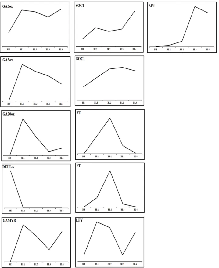
The flowering of Phalaenopsis orchids is a response to cues related to seasonal changes in light (Wang, 1995), temperature (Blanchard & Runkle, 2006) and other external influences (Chen et al., 1994). A cool night temperature of 18–20
C for approximately four weeks will generally induce spiking in most Phalaenopsishybrids, while high temperature inhibits it. To compare gene expression between a constant high-temperature (30/27C; day/night) and inducing cool temperature (22/18C), we collected shoot
condition relative to BH) (Data S13). The identified flowering-related genes correspond to transcription factors and genes involved in signal transduction, development and metabolism (Fig. 3 andData S14). The classification of these genes includes the
following categories: photoperiod, gibberellins (GAs), ambient temperature, light-quality pathways, autonomous pathways and floral pathway integrators (Fornara, de Montaigu & Coupland, 2010;Mouradov, Cremer & Coupland, 2002). However, the genes involved in the photoperiod, ambient temperature, light quality and autonomous pathways did not show significant changes in the floral meristems during the cool temperature treatments (Appendix S1,Fig. S13andData S14). By contrast, the expression patterns of genes involved in pathways that regulate flowering, comprising a total of 22 GA pathway-related genes, were pathway-related to biosynthesis, signal transduction and responsiveness. The GA pathway-related genes and the floral pathway integrator genes have been revealed as representative key players in the link between flowering promotion pathways and the floral transition regulation network in several plant species (Mutasa-Go¨ttgens & Hedden, 2009). In contrast to the expression patterns observed in BL and BH, the GA biosynthetic pathway and positively acting regulator genes showed high expression levels in BL. Furthermore, the expression level of negatively acting regulators, like DELLA genes
Figure 3 Possible evolutionary relationship ofPhAGL6b in the regulation of lip formation and floral symmetry inPhalaenopsisorchid.
(A) Wild-type flower. (B) A big lip mutant ofPhalaenopsisWorld Class ‘Big Foot.’ (C) Representative RT-PCR result showing the mRNA spli-cing pattern ofPhAGL6bin wild-type (W) and big lip mutant (M). (D) Alignment of the amino acid sequences of alternatively spliced forms of
PhAGL6b. (E) Model ofPhAGL6bspatial expression for controllingPhalaenopsisfloral symmetry. Ectopic expression ofPhAGL6bin the distal domain (petal; pink), petal converts into a lip-like structure that leads to radial symmetry. Ectopic expression in proximal domain, (sepal; blue) sepal converts into a lip-like structure that leads to bilateral symmetry. The alternative processing ofPhAGL6btranscripts produced in proximal domain (labellum; pink), labellum converts into a petal-like structure that leads to radial symmetry.PhAGL6bexpression patterns inPhalaenopsis
A
B
identified, was suppressed by the cool temperature which allowing the activation of flowering related genes. The genes included in the flowering promotion pathways and floral pathway integrators were generally upregulated in BL (Figs. 5 and6;Data S11). These findings suggest that the GA pathway may play a crucial role in the regulation of flowering time inPhalaenopsisorchid during cool temperature.
Genetic polymorphisms forPhalaenopsisorchids
The Phalaenopsisgenome assembly also provides the basis for the development of molecular marker-assisted breeding. Analysis of thePhalaenopsisgenome revealed a total of 532,285 simple sequence repeats (SSRs) (Appendix S1,Fig. S14,Table S9andData S15). To enable the identification of single nucleotide polymorphisms (SNPs), we re-sequenced the genome of a summer flowering species,P. pulcherrima ‘B8802,’ with about tenfolds coverage. Comparison of the genome data from the twoPhalaenopsisaccessions (KHM190 and B8802) allowed the discovery of 691,532 SNPs, which should be valuable for future development of SNP markers forPhalaenopsismarker-assisted selection (Appendix S1,Fig. S15,Table S10andData S16).P. pulcherrimais an important parent for small flower and summer-flowering cultivars in breeding program. These SNP markers may contribute valuable tools for varietal identification, genetic linkage map development, genetic diversity analysis, and marker-assisted selection breeding in Phalaenopsis orchid.
Figure 6 Predicted pathway in the regulation of spike induction inPhalaenopsis.Red indicates that the involved genes are more highly expressed in the GA biosynthesis pathway; pink gene names indicate their differential expression in the GA response pathway. Blue gene names represent the activation of flower architecture genes. Red arrows show the steps of the GA signaling stage; Pink arrows direct the steps of inflorescence evocation stage; Blue arrows reveal the steps of flower stalk initiation stage. Inverted T indicates the genes downregulated 2X over.GA20ox,GA3ox,GAMYB,FT,SOC1,LFYandAP1
CONCLUSION
In this study, we sequenced, de novo assembled, and extensively annotated the genome of one of the most importantPhalaenopsis hybrids. We also annotated the genome with a wealth of RNA-seq and sRNA-seq from different tissues, and many genes and miRNAs related to floral organ development, flowering time and protocorm (embryo) development were identified. Importantly, this RNA-Seq and sRNA-seq data allowed us to further improve the genome annotation quality. In addition, mining of SSR and SNP molecular markers from the genome and transcriptomes is currently being adopted in advanced breeding programs and comparative genetic studies, which should contribute to efficientPhalaenopsiscultivar development. Despite that theP. equestrisgenome has been reported recently (Cai et al., 2015), focus on floral organ development and flowering time regulation has not been dealt with. In our study, we obtained transcriptomes from shortened stems (which initiate spikes in response to low ambient temperature) and floral organs, and generated valuable data on potentially regulating flowering time key genes and floral organ development. The genome and transcriptome information of our work should provide a constructive reference resource to upgrade the efficiency of cultivation and the genetic improvement ofPhalaenopsisorchids.
ADDITIONAL INFORMATION AND DECLARATIONS
Funding
This work was supported by grants from the Agriculture and Food Agency, Council of Agriculture, Taiwan (grant numbers 102AS-9.1.1-FD-Z2(1), 103AS-9.1.1-FD-Z2(1), and 104AS-9.1.1-FD-Z2(1)). The funders had no role in study design, data collection and analysis, decision to publish, or preparation of the manuscript.
Grant Disclosures
The following grant information was disclosed by the authors:
Agriculture and Food Agency, Council of Agriculture, Taiwan: 102AS-9.1.1-FD-Z2(1), 103AS-9.1.1-FD-Z2(1), and 104AS-9.1.1-FD-Z2(1).
Competing Interests
The authors declare that they have no competing interests.
Chih-Peng Lin, Chueh-Pai Lee, Wan-Chia Chung and Bill Chia-Han Chang are employees of Yourgene Bioscience, Taiwan.
Author Contributions
Jian-Zhi Huang conceived and designed the experiments, performed the experiments, analyzed the data, contributed reagents/materials/analysis tools, wrote the paper, prepared figures and/or tables, reviewed drafts of the paper.
Ya-Wen Huang performed the experiments.
Yi-Jung Tsai performed the experiments.
Shu-Yun Cheng performed the experiments.
Yi-Wen Chen performed the experiments.
Chueh-Pai Lee performed the experiments, contributed reagents/materials/analysis tools.
Wan-Chia Chung performed the experiments, contributed reagents/materials/analysis tools.
Bill Chia-Han Chang analyzed the data, contributed reagents/materials/analysis tools.
Shih-Wen Chin conceived and designed the experiments, analyzed the data, contributed reagents/materials/analysis tools, wrote the paper, prepared figures and/or tables, reviewed drafts of the paper.
Chen-Yu Lee conceived and designed the experiments, analyzed the data, contributed reagents/materials/analysis tools, wrote the paper, prepared figures and/or tables, reviewed drafts of the paper.
Fure-Chyi Chen conceived and designed the experiments, analyzed the data, contributed reagents/materials/analysis tools, wrote the paper, prepared figures and/or tables, reviewed drafts of the paper.
DNA Deposition
The following information was supplied regarding the deposition of DNA sequences:
SRR1747138,SRR1753943,SRR1753944,SRR1753945,SRR1753946,SRR1753947,
SRR1753948,SRR1753949,SRR1753950,SRR1752971,SRR1753106,SRR1753165,
SRR1753166 SRR1762751,SRR1762752,SRR1762753,SRR1760428,SRR1760429,
SRR1760430,SRR1760432,SRR1760433,SRR1760435,SRR1760436,SRR1760438,
SRR1760439,SRX396172,SRX396784,SRX396785,SRX396786,SRX396787,SRX396788 SRR1760091,SRR1760211,SRR1760212,SRR1760213,SRR1760270,SRR1760271,
SRR1760523,SRR1760524,SRR1760525,SRR1760526,SRR1760527,SRR1760528,
SRR1760530,SRR1760531,SRR1760532.
Data Deposition
The following information was supplied regarding data availability: The research in this article did not generate any raw data.
Supplemental Information
Supplemental information for this article can be found online athttp://dx.doi.org/ 10.7717/peerj.2017#supplemental-information.
REFERENCES
Alvarez-Buylla ER, Pelaz S, Liljegren SJ, Gold SE, Burgeff C, Ditta GS, Ribas de Pouplana L, Martinez-Castilla L, Yanofsky MF. 2000.An ancestral MADS-box gene duplication occurred before the divergence of plants and animals.Proceedings of the National Academy of Sciences of the United States of America97(10):5328–5333DOI 10.1073/pnas.97.10.5328.
temperature in Phalaenopsis aphrodite subsp. formosana.Plant and Cell Physiology 53(10):1737–1750DOI 10.1093/pcp/pcs118.
An FM, Hsiao SR, Chan MT. 2011.Sequencing-based approaches reveal low ambient temperature-responsive and tissue-specific microRNAs in phalaenopsis orchid.PLoS ONE 6(5):e18937DOI 10.1371/journal.pone.0018937.
Blanchard MG, Runkle ES. 2006.Temperature during the day, but not during the night, controls flowering of Phalaenopsis orchids.Journal of Experimental Botany57(15):4043–4049
DOI 10.1093/jxb/erl176.
Cai J, Liu X, Vanneste K, Proost S, Tsai WC, Liu KW, Chen LJ, He Y, Xu Q, Bian C, Zheng Z, Sun F, Liu W, Hsiao YY, Pan ZJ, Hsu CC, Yang YP, Hsu YC, Chuang YC, Dievart A, Dufayard JF, Xu X, Wang JY, Wang J, Xiao XJ, Zhao XM, Du R, Zhang GQ, Wang M, Su YY, Xie GC, Liu GH, Li LQ, Huang LQ, Luo YB, Chen HH, Van de Peer Y, Liu ZJ. 2015.The genome sequence of the orchid Phalaenopsis equestris.Nature Genetics47(1):65–72 DOI 10.1038/ng.3149.
Chang YY, Kao NH, Li JY, Hsu WH, Liang YL, Wu JW, Yang CH. 2010.Characterization of the possible roles for B class MADS box genes in regulation of perianth formation in orchid.Plant Physiology152(2):837–853DOI 10.1104/pp.109.147116.
Chen W-S, Liu H-Y, Liu Z-H, Yang L, Chen W-H. 1994.Geibberllin and temperature influence carbohydrate content and flowering in Phalaenopsis.Physiologia Plantarum90(2):391–395 DOI 10.1111/j.1399-3054.1994.tb00404.x.
Chen WH, Tseng YC, Liu YC, Chuo CM, Chen PT, Tseng KM, Yeh YC, Ger MJ, Wang HL. 2008.
Cool-night temperature induces spike emergence and affects photosynthetic efficiency and metabolizable carbohydrate and organic acid pools in Phalaenopsis aphrodite.Plant Cell Reports27(10):1667–1675DOI 10.1007/s00299-008-0591-0.
Christenson EA. 2002.Phalaenopsis: A Monograph. Portland: Timber Press.
Conesa A, Gotz S. 2008.Blast2GO: a comprehensive suite for functional analysis in plant genomics.International Journal of Plant Genomics2008:619832DOI 10.1155/2008/619832.
Dornelas MC, Patreze CM, Angenent GC, Immink RG. 2011.MADS: the missing link between identity and growth?Trends in Plant Science16(2):89–97DOI 10.1016/j.tplants.2010.11.003.
Finn RD, Bateman A, Clements J, Coggill P, Eberhardt RY, Eddy SR, Heger A, Hetherington K, Holm L, Mistry J, Sonnhammer EL, Tate J, Punta M. 2014.Pfam: the protein families database.Nucleic Acids Research42(D1):D222–D230DOI 10.1093/nar/gkt1223.
Finn RD, Clements J, Eddy SR. 2011.HMMER web server: interactive sequence similarity searching.Nucleic Acids Research39:W29–W37DOI 10.1093/nar/gkr367.
Fornara F, de Montaigu A, Coupland G. 2010.SnapShot: control of flowering in Arabidopsis.Cell 141(3):e551–e552DOI 10.1016/j.cell.2010.04.024.
Geuten K, Becker A, Kaufmann K, Caris P, Janssens S, Viaene T, Theißen G, Smets E. 2006.
Petaloidy and petal identity MADS-box genes in the balsaminoid genera Impatiens and Marcgravia.The Plant Journal47(4):501–518DOI 10.1111/j.1365-313X.2006.02800.x.
Gramzow L, Theissen G. 2010.A hitchhiker’s guide to the MADS world of plants.Genome Biology 11(6):214DOI 10.1186/gb-2010-11-6-214.
Honma T, Goto K. 2001.Complexes of MADS-box proteins are sufficient to convert leaves into floral organs.Nature409(6819):525–529DOI 10.1038/35054083.
Hsiao YY, Chen YW, Huang SC, Pan ZJ, Fu CH, Chen WH, Tsai WC, Chen HH. 2011.Gene discovery using next-generation pyrosequencing to develop ESTs for Phalaenopsis orchids.
Hsu H-F, Hsu W-H, Lee Y-I, Mao W-T, Yang J-Y, Li J-Y, Yang C-H. 2015.Model for perianth formation in orchids.Nature Plants1(5):15046DOI 10.1038/nplants.2015.46.
Huang JZ, Lin CP, Cheng TC, Chang BC, Cheng SY, Chen YW, Lee CY, Chin SW, Chen FC. 2015.
A de novo floral transcriptome reveals clues into Phalaenopsis orchid flower development.
PLoS ONE10(5):e123474DOI 10.1371/journal.pone.0123474.
International Rice Genome Sequencing Project. 2005.The map-based sequence of the rice genome.Nature436:793–800DOI 10.1038/nature03895.
Jaillon O, Aury JM, Noel B, Policriti A, Clepet C, Casagrande A, Choisne N, Aubourg S, Vitulo N, Jubin C, Vezzi A, Legeai F, Hugueney P, Dasilva C, Horner D, Mica E, Jublot D, Poulain J, Bruyere C, Billault A, Segurens B, Gouyvenoux M, Ugarte E, Cattonaro F, Anthouard V, Vico V, Del Fabbro C, Alaux M, Di Gaspero G, Dumas V, Felice N, Paillard S, Juman I, Moroldo M, Scalabrin S, Canaguier A, Le Clainche I, Malacrida G, Durand E, Pesole G, Laucou V, Chatelet P, Merdinoglu D, Delledonne M, Pezzotti M, Lecharny A, Scarpelli C, Artiguenave F, Pe ME, Valle G, Morgante M, Caboche M, Adam-Blondon AF, Weissenbach J, Quetier F, Wincker P. 2007.The grapevine genome sequence suggests ancestral hexaploidization in major angiosperm phyla.Nature449(7161):463–467
DOI 10.1038/nature06148.
Kanehisa M, Goto S, Sato Y, Furumichi M, Tanabe M. 2012.KEGG for integration and interpretation of large-scale molecular data sets.Nucleic Acids Research40(D1):D109–D114 DOI 10.1093/nar/gkr988.
Li L, Stoeckert CJ Jr, Roos DS. 2003.OrthoMCL: identification of ortholog groups for eukaryotic genomes.Genome Research13(9):2178–2189DOI 10.1101/gr.1224503.
Lin S, Lee HC, Chen WH, Chen CC, Kao YY, Fu YM, Chen YH, Lin TY. 2001.Nuclear DNA contents ofPhalaenopsissp. andDoritis pulcherrima.Journal of the American Society for Horticultural Science126(2):195–199.
Messenguy F, Dubois E. 2003.Role of MADS box proteins and their cofactors in combinatorial control of gene expression and cell development.Gene 316:1–21DOI 10.1016/S0378-1119(03)00747-9.
Mondrago´n-Palomino M, Theissen G. 2011.Conserved differential expression of paralogous DEFICIENS- and GLOBOSA-like MADS-box genes in the flowers of Orchidaceae: refining the ‘orchid code.’The Plant Journal66(6):1008–1019DOI 10.1111/j.1365-313X.2011.04560.x.
Mouradov A, Cremer F, Coupland G. 2002.Control of flowering time: interacting pathways as a basis for diversity.The Plant Cell14(Suppl):S111–S130DOI 10.1105/tpc.001362.
Munster T, Pahnke J, Di Rosa A, Kim JT, Martin W, Saedler H, Theissen G. 1997.Floral homeotic genes were recruited from homologous MADS-box genes preexisting in the common ancestor of ferns and seed plants.Proceedings of the National Academy of Sciences of the United States of America94(6):2415–2420DOI 10.1073/pnas.94.6.2415.
Mutasa-Go¨ttgens E, Hedden P. 2009.Gibberellin as a factor in floral regulatory networks.Journal of Experimental Botany60(7):1979–1989DOI 10.1093/jxb/erp040.
Parenicova L, de Folter S, Kieffer M, Horner DS, Favalli C, Busscher J, Cook HE, Ingram RM, Kater MM, Davies B, Angenent GC, Colombo L. 2003.Molecular and phylogenetic analyses of the complete MADS-box transcription factor family in Arabidopsis: new openings to the MADS world.The Plant Cell15(7):1538–1551 DOI 10.1105/tpc.011544.
monocots.Biological Reviews of the Cambridge Philosophical Society77(3):403–441 DOI 10.1017/S1464793102005936.
Su CL, Chao YT, Alex Chang YC, Chen WC, Chen CY, Lee AY, Hwa KT, Shih MC. 2011.De novo assembly of expressed transcripts and global analysis of the Phalaenopsis aphrodite
transcriptome.Plant and Cell Physiology52(9):1501–1514DOI 10.1093/pcp/pcr097.
Su CL, Chen WC, Lee AY, Chen CY, Chang YC, Chao YT, Shih MC. 2013.A modified ABCDE model of flowering in orchids based on gene expression profiling studies of the moth orchid Phalaenopsis aphrodite.PLoS ONE8(11):e80462DOI 10.1371/journal.pone.0080462.
Tamura K, Peterson D, Peterson N, Stecher G, Nei M, Kumar S. 2011.MEGA5: molecular evolutionary genetics analysis using maximum likelihood, evolutionary distance, and maximum parsimony methods.Molecular Biology and Evolution28(10):2731–2739 DOI 10.1093/molbev/msr121.
The Arabidopsis Genome Initiative. 2000.Analysis of the genome sequence of the flowering plant Arabidopsis thaliana.Nature408(6814):796–815DOI 10.1038/35048692.
The Gene Ontology Consortium. 2008.The Gene Ontology project in 2008.Nucleic Acids Research36(Suppl 1):D440–D444DOI 10.1093/nar/gkm883.
Theissen G. 2001.Development of floral organ identity: stories from the MADS house.Current Opinion in Plant Biology4(1):75–85DOI 10.1016/S1369-5266(00)00139-4.
Trapnell C, Pachter L, Salzberg SL. 2009.TopHat: discovering splice junctions with RNA-Seq.
Bioinformatics25(9):1105–1111DOI 10.1093/bioinformatics/btp120.
Trapnell C, Roberts A, Goff L, Pertea G, Kim D, Kelley DR, Pimentel H, Salzberg SL, Rinn JL, Pachter L. 2012.Differential gene and transcript expression analysis of RNA-seq experiments with TopHat and Cufflinks.Nature Protocols7(3):562–578DOI 10.1038/nprot.2012.016.
Trobner W, Ramirez L, Motte P, Hue I, Huijser P, Lonnig WE, Saedler H, Sommer H, Schwarz-Sommer Z. 1992.GLOBOSA: a homeotic gene which interacts with DEFICIENS in the control of Antirrhinum floral organogenesis.The EMBO Journal11(13):4693–4704.
Tsai WC, Fu CH, Hsiao YY, Huang YM, Chen LJ, Wang M, Liu ZJ, Chen HH. 2013.OrchidBase 2.0: comprehensive collection of Orchidaceae floral transcriptomes.Plant and Cell Physiology 54(2):e7DOI 10.1093/pcp/pcs187.
Tsai WC, Kuoh CS, Chuang MH, Chen WH, Chen HH. 2004.Four DEF-like MADS box genes displayed distinct floral morphogenetic roles in Phalaenopsis orchid.Plant and Cell Physiology 45(7):831–844DOI 10.1093/pcp/pch095.
Tsai WC, Lee PF, Chen HI, Hsiao YY, Wei WJ, Pan ZJ, Chuang MH, Kuoh CS, Chen WH, Chen HH. 2005.PeMADS6, a GLOBOSA/PISTILLATA-like gene in Phalaenopsis equestris involved in petaloid formation, and correlated with flower longevity and ovary development.Plant and Cell Physiology46(7):1125–1139DOI 10.1093/pcp/pci125.
Wang Y-T. 1995.Phalaenopsis orchid light requirement during the induction of spiking.
HortScience30(1):59–61.