ISSN 1553-3468
© 2010 Science Publications
Corresponding Author: Abdel E Ghaly, Department of Process Engineering and Applied Science, Dalhousie University, Halifax, Nova Scotia, Canada Tel: (902) 494-6014; email: [email protected]
Extraction and Purification of Collagenase Enzymes: A Critical Review
Said M Daboor, Suzanne M Budge, Abdel E Ghaly, Su-Ling Brooksand Deepika Dave Department of Process Engineering and Applied Science, Faculty of Engineering,
Dalhousie University, Halifax, Nova Scotia, Canada
Abstract: Problem statement: Enzymes have vital roles in several industrial processes (foods, cosmetics, nutraceuticals and pharmaceuticals) due to their highly selective nature and high activity at very low concentrations. Recent efforts to identify new sources of useful enzymes have been concentrated on the marine environment because of the potential to make use of processing wastes. About 35-50% of the mass of the fish caught is a waste that is disposed off at sea or in landfills. The extraction of enzymes from fish processing waste can reduce environment problems and improve the economics of the fish industry. Collagenases are a group of enzymes that can be extracted from fish waste. Approach: Comprehensive reviews of the literature on the extraction, purification, characterization and use of collagenases was carried out. Results: Collagenases have different molecular weights based on their types and sources. They have the ability to break down the peptide bonds in collagen at physiological pH. They are classified into two types: serine and metallocollagenase. Collagenolytic activities have been shown at a wide range of temperatures (20-40°C) and pH (6-8). Many activators can be used to achive collagenase activity including 4-Aminophenylmercuric Acetate (APMA), trypsin, potassium or sodium thiocyanate, iodoacetamide and potassium iodide. Dithiothreitol (DTT), mercaptoethanol, ethylendiaminetetracetic acid, o-phenanthroline and cysteine inactivate the enzyme. Collagenases enzymes can be extracted with a variety of techniques using different buffering systems (tris-HCl, sodium bicarbonate, calcium chloride and cacodylate). All techniques involve the use of ammonium sulphate fractionation and centrifugation to precipitate the enzyme. Collagenases are normally purified using chromatographic techniques such as gel-filtration, ion-exchange and affinity column chromatography. Collagenase can be assayed with a number of methods, including: colorimetric absorbance, viscometry, radioactivity and fluorescence spectroscopy. Collagenases are partly responsible for toughness in red meats and are used as tenderizers in food industry, have application in the fur and hide tanning to ensure uniform dying of leather, used in medicine to treat burns and ulcers, eliminate scar tissues, transplantation of organs.
Conclusion: Understanding of the nature of the enzymes and identifying the most suitable resources and the methods for their extraction and purification will have significant impact on the fish processing, food and medical industries.
Key words: Fish waste, collagenase enzymes, metallocollagenase, collagenolytic, Dithiothreitol (DTT), Aminophenylmercuric Acetate (APMA), phenanthroline and cysteine, extraction and purification, Collagen Binding Domain (CBD), Matrix Metalloproteinase (MMP)
INTRODUCTION
Enzymes are used in a variety of industrial processes to create an array of foods (Foegeding and Larick, 1986; Cronlund and Woychik, 1987; Christensen, 1989; Ashie and Lanier, 2000; Dĩaz-López and García-Carreńo, 2000; Shahidi and JanakKamil, 2001), cosmetics (Griffith et al., 1969; Lods et al., 2000; Sim et al., 2000; Spök, 2006; Mohorcic et al.,
(a)
(b)
Fig. 1: Triple helix structure of collagen: A (Adapted from Morris and Gonsalves, 2010) and B (Adapted from TQS, 2004)
Recent efforts to identify new sources of useful enzymes have been concentrated on the marine environment because of the potential to make use of processing wastes (Shahidi and JanakKamil, 2001). It is estimated that approximately 35% of the mass of all fish caught results in a waste (Park et al., 2002) that is typically disposed of at sea or in landfills (Shahidi, 1994). Thus, the extraction of enzymes from fish processing waste represents a solution to the costly
disposal of waste and serves as a value-added processing step that improves the economics of the fish industry while minimizing environmental problems (Haard et al., 1994; Shahidi, 1994; Venugopal and Shahidi, 1995).
As proteolytic enzymes, collagenases have a number of industrial applications. Collagen is partly responsible for toughness in red meats and used as tenderizers in the food industry (Foegeding and Larick, 1986; Cronlund and Woychik, 1987). Collaganases have applications in fur and hide tanning to help ensure a uniform dying of leathers (Goshev et al., 2005; Kanth et al., 2008). However, the most common uses of these enzymes appear to be in medicine. They are used to treat burns and ulcers (Agren et al., 1992; Püllen et al., 2002), to eliminate scar tissue (Shmoilov et al., 2006) and play an important role in the successful transplantation of specific organs (Klöck et al., 1996; Kin et al., 2007). As more cost-effective methods are developed for the isolation and purification of collagenases, the range of biotechnological applications will surely expand. Therefore, understanding the nature of these enzymes and identifying the most suitable methods for their recovery and purification are paramount.
Table 1: Collagen α chains, number of amino acids and National Center for Biotechnology Information (NCBI) reference numbers (Adapted from Gordonand Hahn, 2010)
α chain Number of amino acids NCBI reference number
α1 (I) 1464 (includes 22 aa SP) NP_000079
α2(I) 1366 aa (includes SP) NP_000080
α1(II)A 1487 aa (includes 25 aa SP) NP_001835
α1(II)B Same as a1(II)A but lacks vWC domain NP_149162
α1(III) 1466 aa (includes 23 aa SP) NP_000081
α1(IV) 1669 aa (includes 27 aa SP) NP_001836
α2(IV) 1712 aa (includes 25 aa SP) NP_001837
α3(IV) 1670 aa (includes 28 aa SP) NP_000082
α4(IV) 1690 aa (includes 38 aa SP) NP_000083
α5(IV) 1685 aa (includes 26 aa SP) NP_000486
α6(IV) 1691 aa (includes 21 aa SP) NP_001838
(B, so form P_378667)
α1(V) 1838 aa (includes SP) NP_000084
α2(V) 1499 aa (includes SP) NP_000384
α3(V) 1745 aa (includes 29 aa SP) NP_056534
α1(VI) 1028 aa (includes 19 aa SP) NP_001839
α2(VI) 1019 aa (includes 20 aa SP) NP_001840 NP_001840
2C2a and 2C2a iso forms
α3(VI) (with 25 aa SP) 3177 NP_004360 –
multiple splicings
mu α4(VI) Not in human; mouse = 2309 aa Swiss-Prot A2AX52
α5(VI) 2611 (includes SP) Iso forms 2 and 3 NP_694996 (partial- shows 2526 aa)
α6(VI) 2263 aa (includes SP) NP_001096078
α1(VII) 2944 aa (includes 16 aa SP) NP_000085
α1(VIII) 744 aa (includes 28 aa SP) NP_065084;
cmkmNP_001841
α2(VIII) 703 aa (includes SP) NP_005193
α1(IX) 921 aa (includes 23 aa SP), Short form NP_001842
678 aa (includes 23 aa SP)
α2(IX) 689 aa (includes SP) NP_001843
α3(IX) 684 aa (includes SP) NP_001844
α1(X) 680 aa (includes 18 aa SP) NP_000484
α1(XI)A 1806 aa (includes 36 aa SP) NP_001845
α1(XI)B 1818 aa (includes 36 aa SP) NP_542196
α1(XI)C 1767 aa (includes 36 aa SP) NP_542197
α2(XI)) 1736 aa (includes 22 aa SP Iso forms 2 and 3 NP_542411
α3(XI) Same as α1(II)A NP_001835
α1(XII) 3063 aa (includes 23 aa SP) Short form 1899 NP_004361 aa, includes same SP as long form (has NC1 variants)
α1(XIII) 717 aa (transmembranous) NP_005194
(20+ splice variants) α1(XIV) 1796 aa (includes SP) Short form without NP_066933
N-terminal FNIII domain; NC1 variants) (has NC1 variants)
α1(XV) 1388 aa (includes 25 aa SP) NP_001846
α1(XVI) 1604 aa (includes SP) NP_001847
α1(XVII) 1497 aa (transmembranous) NP_000485
α1(XVIII) 1516 aa (includes 23 aa SP) Short form NP_085059 NP_569712 1336 aa (includes 33 aa SP)
α1(XIX) 1142 aa (includes 23 aa SP) NP_001849
α1(XX) Not in human; ch=1472 aa (without SP) NP_001004392
α1(XXI) 957 aa (includes 22 aa SP) NP_110447
α1(XXII) 1626 aa (includes SP) NP_690848
α1(XXIII) 540 aa (transmembranous) NP_775736
α1(XXIV) 1714 aa (includes SP) NP_690850
α1(XXV) 654 aa (transmembranous) Iso form 2 is 642 aa NP_942014
α1(XXVI) 439 aa (includes SP) NP_597714
α1(XXVII) 1860 aa (includes 41 aa SP) NP_116277
α1(XXVIII) 1125 aa (includes SP) NP_001032852
Fig. 2: Assembly of trimeric molecules from α chain for collagen Types I and V (Adapted form Gordon and Hahn, 2010)
(a)
(b)
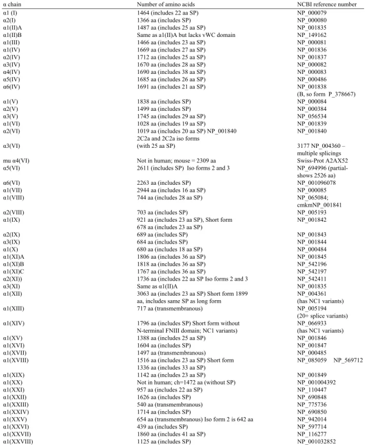
Fig. 3: Collagen triple-helical peptides: (A) manually aligned into the active site of the catalytic domain of porcine MMP-1, (B) rotated 90° to the left, the active site shown as a cleft is unoccupied by the triple-helical peptide substrate, pink: catalytic domain, blue: Hpx, purple: zinc ion (Adapted from Kramer et al., 2001)
Collagens have been classified based on the expression of different genes during tissue construction (Lozano et al., 1985). Collagen type I is the most common type that is found in bone, tendon, skin and ligaments, while collagen type III is the second most common and is found in elastic tissues such as blood vessels and various internal organs (Miller et al., 1971; Kielty and Grant, 2002). The abundance of types V and XI are low but they are found associated with the types I and II in bone and cartilage as well as in other tissues (Prockop and Kivirikko, 1995; Myllyharju and Kivirikko, 2001; Kielty and Grant, 2002). Figure 2 shows the assembly of trimeric molecules for types I and V collagenases while Fig. 3 shows the collagen triple-helical peptides.
Collagenase and collagenolytic enzymes:
Collagenolytic enzymes: Very few enzymes are capable of breaking down the complex triple helix structure of collagen (Hayashi et al., 1980; Hulboy et al., 1997; Visse and Nagase, 2003). The enzymes that are capable of degrading collagen (including cathepsin and elastase) are known generally as collagenolytic enzymes. Cathepsin K cleaves collagen type I in an acidic medium (Garnero et al., 1998). Elastase is the most well-studied collagenolytic enzyme and is considered a serine protease enzyme (Brown and Wold, 1973). It is principally responsible for the breakdown of elastin (a highly viscous insoluble protein found in connective tissue). Together with collagenase, they determine the mechanical properties of connective tissue by cleaving particular peptide bonds (Asgeirsson and Bjarnason, 1993; De-Vecchi and Coppes, 1996). Kafienah et al. (1998) isolated and purified human neutrophil elastase with the ability to cleave collagen type I which is resistant to attack by most proteolytic enzymes. Elastase has been isolated from marine and fresh water fish species (Cohen et al., 1981; Clark et al., 1985; Asgeirsson and Bjarnason, 1993; Gildberg and Øverbø, 1990; Raa and Walther, 1989). In some situations, collagen is considered to be a poor substrate for collagenase, so that the initiation of collagen breakdown is inhibited. However, if the substrate is initially attacked by elastase, proteoglycans are removed from the collagen fibers which make it more susceptible to collagenase attack (Baici et al., 1982; Zeydel et al., 1986).
(a)
(b)
(c)
Fig. 4: Model of different collagen binding sites and triple helicase mechanisms of membrane type (MT1-MMP), MMP-2, gelatinase A (MMP-2), MMP -1 and MMP -8, (A) sMT1-MMP, MMP-2, MMP-1, MMP-8 and type I collagen. (B) native collagen binding and unwinding by MMPs. MT1-MMP, MMP-1 and MMP-8 utilize the hemopexin C domain to bind collagen in the vicinity of the cleavage site and to induce localized helix unwinding, MMP-2 utilizes the Collagen Binding Domain (CBD) for this function, while MMP-1 and MMP-8 bind at a different site than MT1-MMP and MMP-2, (C) binding and localized unwinding in the vicinity of the collagenase cleavage site by recombinant MT1-LCD (membrane type- linker/hemopexin C domain) and MMP-2 CBD; by competitive inhibition this interaction blocks collagen cleavage by MT1-MMP and MMP-2, respectively, but promotes enhanced cleavage by MMP-1 and MMP-8 which to bind at a different site (Adapted from Tam et al., 2004)
MMP-18. MMP-2 is known as gelatinase A (Aimes and Quigley, 1995; Patterson et al., 2001). Figure 4 shows collagen binding sites and triple helicase mechanisms of MMP types.
Elastase and collagenase display about the same collagenolytic potential on human cartilage on a weight
basis and the elastase/collagenase system from human polymorphonuclear leukocytes may represent a cooperative proteolytic complex in the destruction of cartilage in rheumatoid arthritis (Baici et al., 1982).
Collagenase enzymes: Collagenase enzymes, as specific enzymes for the collagen substrate, have been isolated and characterized from both microbial cells and animal tissues. Microbial collagenases have been recovered from pathogenic microorganisms, principally Clostridium histolyticum. These collagenases split each polypeptide chain of collagen at multiple sites (Goldberg et al., 1986). They are thought to function as an exotoxin, causing hydrolysis of collagen in the host cells and disrupting metabolism in connective tissues (Lecroisey and Keil, 1979). Bacterial collagenases are quite versatile, being capable of hydrolyzing both water-insoluble native collagens and water-soluble denatured collagens (Mookhtiar et al., 1985).
While much of the research with microbial collagenases has focused on a single species, tissue collagenases have been isolated and characterized from a number of different tissues in many animals. Since tissue collagenases are digestive enzymes, they are commonly isolated from the digestive tracts of various fish and invertebrates including: tadpole tailfin (Gross and Nagai, 1965; Nagai et al., 1966), rabbit skin (Fullmer and Gibson, 1966), rat uterus (Jeffrey and Gross, 1967), rheumatoid synovial tissue (Evanson et al., 1968), mouse bones (Sakamoto et al., 1972), crabs (Eizen and Jeffrey, 1969; Grant et al., 1983; Klimova et al., 1990; Sellos and Van Wormhoudt, 1992; Gerasimova and Kupina, 1996; Zefirova et al., 1996), fresh water prawns (Baranowski et al., 1984), crayfish (Garcia-Carreno et al., 1994), Atlantic cod (Kristjánsson, et al., 1995), tropical shrimp (Penaeus vannamei) (Sellos and Van Wormhoudt, 1992; Van Wormhoudt et al., 1992) and catfish (Parasilurus asotus) (Klimova et al.,1990; Sellos and Van Wormhoudt, 1992).
molecular weights ranging from 80-120 kDa. It seems that bacterial collagenases typically have molecular weight >55 kDa while molecular weights of collagenases obtained from animal tissues tend to be lower. For instance, Sakamoto et al. (1972) isolated collagenase with molecular weight of 41 kDa from mouse bones.
McCroskery et al. (1975) reported molecular weights of collagenase from rabbit muscle between 33 kDa and 35 kDa. A number of researchers (Kristjánsson
et al., 1995; Roy et al., 1996; Sivakumar et al., 1999) isolated serine collagenses from digestive glands of marine organism with molecular weights <60 kDa.
The wide range of molecular weight is to be expected for an enzyme such as collagenase that does not have a single structure. Bond and Van Wart (1984) suggested that some of the variation in reported molecular weights of collagenases may be simply due to proteolysis of a larger collagenase precursor. For use in industry, such variation will be less important than overall collagenolytic activity but the potential for variation should be noted.
The production of tissue collagenases is thought to be stimulated in the presence of microbial collagenases which seem to serve as a key factor, similar to the action of exogenous enzymes produced by some microorganisms when added to food or feed (Taoka et al., 2007). They are typically classified as either serine collagenase or metallocollagenase, based on their different physiological functions.
Serine collagenases: Serine collagenases, like all serine proteinases, contain a serine residue in their catalytic sites. They typically have molecular weights in the range of 24,000-36,000Da (Roy et al., 1996). They are normally associated with the digestive organ (Zefirova
et al., 1996), are able to cleave the triple helix structure of collagen types I, II and III and are often involved with hormone production, protein degradation, blood-clotting and fibrinolysis (Neurath, 1984).
Collagenolytic serine proteases (EC 3.4.21.32) were first isolated from the fiddler crab (Uca pugzlator) hepatopancreas (Eisen et al., 1973) but have now been extracted and characterized from the digestive tracts of a variety of fish and aquatic invertebrates (Eizen and Jeffrey, 1969; Grant et al., 1983; Baranowski et al., 1984; Garcia-Carreno et al., 1994; Kristjánsson et al., 1995; Gerasimova and Kupina, 1996; Zefirova et al., 1996). Serine collagenases are much less abundant than metallo collagenases (Gonzales and Robert-Baudouy,
1996). Tsu and Craik (1996) described the serine protease mechanism by the following reactions:
(1)
• Under conditions where acylation is rate-limiting
kcat = k2 (2)
Km = KS (3)
• Under conditions where deacylation is rate-limiting
kcat = k3 (4)
Km = KS[k3/(k2+k3)] (5)
Metallocollagenase: Metallocollagenases are members of the Matrix Metalloproteinase (MMP) family with molecular weights between 30,000 and 150,000 Da (Harris and Vatar, 1982). Like all MMP, metallocollagenases are zinc-dependent enzymes and are inhibited by any chelator that binds those ions. Divalent calcium is required for stability (Stricklin et al., 1977). Only MMP 1, 8, 13, 14 and 18 have activity against native triple-stranded collagen types I, II, III, VII and X (Freije et al., 1994). Metallocollagenases are commonly recovered from animal and fish tissues such as bones, fins, skins and from marine crab hepatopancreas (Sivakumar et al., 1999).
Digestive organs can serve as a source of both serine collagenases and metallocollagenases but most studies have concentrated on digestive glands as a source of serine collagenase. Thus, waste tissues, in addition to digestive glands, can serve as a valuable source of metallocollagenase.
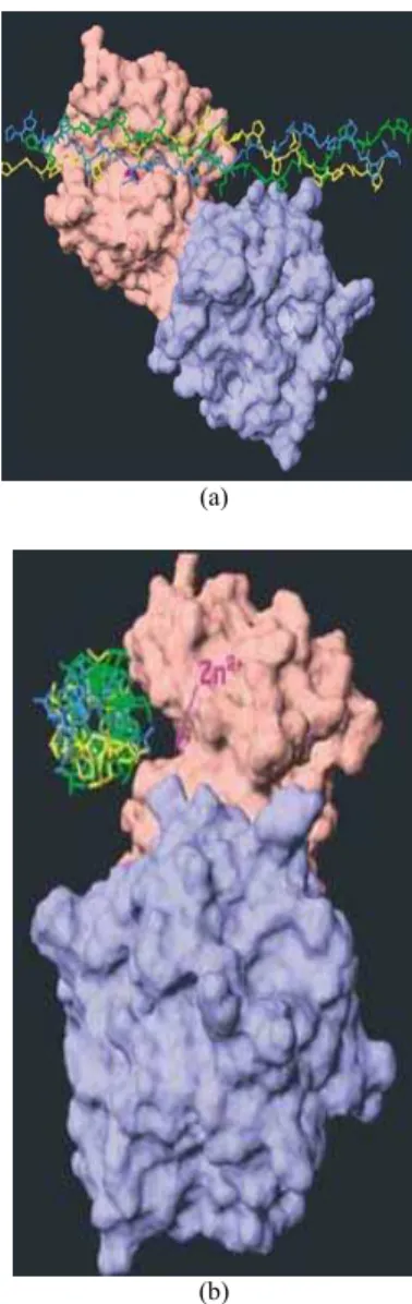
Tris-HCl buffer extraction: A number of very similar procedures have been used to recover collagenase from cells using tris-HCl buffers in a narrow pH range of 7.4-7.6 with low concentrations of CaCl2 (5-20 mM) added to the buffer at temperatures < 4°C (Sakamoto et al.,1972; McCroskery et al., 1975; Ohyama and Hashimoto, 1977). All methods incorporate an ultracentrifugation step (>20 000g) followed by ammonium sulphate fractionation (with concentrations ranging from 25-80%) and precipitate recovery. Sakamoto et al. (1972) presented a simple procedure for extraction from mouse bone, essentially consisting of dissolution of the lyophilized tissue culture medium in tris-HCl buffer containing CaCl2 at a pH of 7.6, followed by ammonium sulphate fractionation and centrifugation (Fig. 5). Several other authors (Baranowski et al., 1984; Teruel and Simpson, 1995; Hernández-Herrero et al., 2003; Burgos-Hernández et al., 2005) employed a very similar technique to extract collagenase from a variety of fish tissues including cod and winter flounder (Pseudopleuronectes americanus) muscles and shrimp hepatopancreas, with the tris-HCl buffer containing CaCl2 but at a pH of 7.4, fractionation with ammonium sulfate (40-80%) and incorporating a filtration step prior to the initial centrifugation (Fig. 6).
Others researchers modified this basic procedure for a number of reasons. Ohyama and Hashimoto (1977) added sodium thiocyanate to the crude extract after centrifugation to serve as a collagenase activator (Fig. 7). McCroskery et al. (1975) and Iijima et al. (1981) added sodium azide to suppress the microbial growth (Fig. 8).
Delaissè et al. (1985) and Gillet et al. (1977) added high sodium chloride concentrations (up to one M) during extraction to encourage the dissociation of collagenase from insoluble collagen. Ohyama and Hashimoto (1977) incorporated repetitive freeze-thaw cycles to disrupt cell walls and make the enzyme more accessible to the buffer. However, very few reports describe yield with each step so it is difficult to access the efficiency of extraction with the variation in techniques.
Sodium bicarbonate buffer extraction: Sivakumar et al. (1999) extracted collaganase from green crab hepatopancreas by homogenizing the tissue in sodium bicarbonate buffer (pH 8.3) containing calcium chloride and then stirring for 36 hours to extract the enzyme.
The collagenase was then isolated by precipitation in cold acetone. The same method was used by Indra et al. (2005) to isolate collagenase from the hepatopancreas of a land snail (Achatina fulica). On an industrial scale, the use of an organic solvent is unlikely to be practical because of risks of fires and explosions. The prolonged period of stirring would, lead to increased chances of microbial growth and the addition of an antimicrobial would increase costs.
Unbuffered water extraction: Unbuffered water containing CaCl2 has been used to extract collagenase from Atlantic cod intestines by Kristjánsson et al. (1995). The pH was adjusted to 7.5 with NaOH after homogenization. The mixture was stirred for 22 h, left standing for 30 h and then centrifuged. The supernatant was concentrated and fractionated by ammonium sulphate (20-50%). The use of calcium ions increases the enzyme thermal stability by about 7-8°C, where calcium bind with the enzyme at a single site (Sipos and Merkel, 1970; Bode and Schwager 1975; Chiancone et al., 1985; Klimova et al., 1990). Maintaining a constant pH without using buffer would be very difficult and is very unlikely that the extraction was actually carried out at a constant pH of 7.5. It is time consuming (two days standing), making it unacceptable for large scale processing and possibly leading to undesirable changes in the homogenate.
Fig. 5: The buffer extraction and fractionation system of mouse bone collagenase enzyme used by Sakamoto et al. (1972)
Fig. 6: The Tris-buffer extraction and fractionation system of collagenase enzyme from freshwater prawn (Baranowski et al., 1984) and winter flounder fish skeletal muscle (Teruel and Simpson, 1995)
°
Fig. 7: The modified Tris-buffer extraction and fractionation procedure of collagenase enzyme from human skin basal cell Epithelioma used by Ohyama and Hashimoto (1977)
Fig. 8: The modified Tris-buffer extraction and fractionation procedure of collagenase enzyme from rabbit tumour tissue used by McCroskery et al. (1975)
° °
°
°
°
°
Fig. 9: Separation principles in chromatographic purification (Adapted from Amersham Biosciences, 2010)
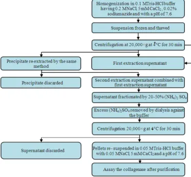
Fig. 10: The Gel filtration procedure used to purify collagenase enzyme by Sivakumar et al. (1999) and Indra et al. (2005)
Collagenase purification: Once a crude collagenase extract is recovered, it must be purified using one of several chromatographic methods that can be classified as: gel filtration, ion exchange, hydrophobic interaction or affinity Figure 9 shows the principle of these purification processes. It should be noted that these chromatography techniques are commonly coupled together so that most purification schemes will begin with gel filtration, followed by either ion
exchange or affinity chromatography or both, depending on the application.
Gel filtration: Gel filtration is known as size exclusion or molecular sieve chromatography. It separates molecules based on size. It is a physical separation where the column packing material contains pores that only molecules within a particular size or mass range can enter and be retained. Many commercial gel matrixes are used such as Sephadex (10, 25, 50, 75, 100 and G-200), Sepharose, Sephacryl, Sepharose CL and Bio-Gel. These different materials have different protein size-exclusion ranges. Proteins can be separated by running them through the appropriate gel resin column. All the procedure should be carried at 4°C and the selection of the gel-powder type is based on the particular protein(s) of interest (Kaufman et al., 1995). Several researchers performed collagenase purification using gel-filtration chromatography. Ohyama and Hashimoto (1977) used Sephadex G-150 to purify collagenase of human skin. Kristjánsson et al. (1995) isolated the purified collagenolytic serine proteinase from the Atlantic cod using phenyl-Sepharose CL-4B. Sivakumar et al. (1999) and Indra et al. (2005) used gel-filtration chromatography on Sephadex G-100 to obtain purified collagenase from hepatopancreas of the marine crab and land snail, respectively (Fig. 10). Several gel-powders have been used including: Polyacrylamide (Sakamoto et al., 1972) and mixtures of polyacrylamide and dextran (Callaway et al., 1986; Sakurai et al., 2009). Regardless of support material, all groups seem to consistently employ a maximum pore size such that 200 kDa is the upper molecular mass limit for solute retention (i.e., all molecules with large molecular masses pass through the column with the solvent). With the uncertainty in molecular mass of collagenase, such a high mass limit seems prudent.
Fig. 11: Chromatographic procedure used to purify collagenase enzyme by Sakamoto et al. (1972)
Diethylaminoethyl (DEAE) cellulose for binding to net negatively charged proteins and (b) carboxymethyl for binding to net positively charged proteins (Kaufman et al., 1995).
At optimal pH levels (near 7.0), collagenase will bear an overall negative charge and anionic exchange packings must be employed. Anion exchange chromatography based on Diethylaminoethyl (DEAE) cellulose or agarose is by far the most common ion exchange technique that has been used by a number of researchers to partially purify collagenase (Keller and
Mandl, 1963; Gross and Nagai, 1965; Hook et al., 1972; Sakamoto et al., 1972; Ijima et al., 1981; Lim et al., 1993; Kristjánsson et al., 1995; Kim et al., 2002) as shown in Fig. 11-13.
Hydrophobic Interaction Chromatography (HIC):
Fig. 12: Chromatographic procedure used to purify collagenase enzyme by Iijima et al. (1981)
This technique is similar to Reverse Phase (RP) chromatography where non-polar structures, such as alkyl ligands, are attached to a support and form reversible interactions with the solutes. With HIC, however, the interactions are much weaker so that proteins are not strongly retained and can be recovered with polar solvents with varying salt concentrations (Queiroz et al., 2001). Kristjánsson et al. (1995) used a column of phenyl-substituted agarose to partially purify collagenase. Sakurai et al. (2009) used this technique but with a more hydrophobic butyl substitute. Generally, the use of this technique for purification of collagenase is not common.
Affinity chromatography: Affinity chromatography uses a specific interaction between a substrate and a biologically active substance and is the most powerful method for protein purification. This method is based on the high affinity of some proteins to specific chemical groups (ligands) covalenty attached to a chromatographic bed material (Kaufman et al., 1995).
Fig. 13: Chromatographic procedure used to purify collagenase enzyme by Kristjánsson et al. (1995)
The interaction may be very specific, with a substrate bonded to the stationary phase that only the enzyme of interest can interact with such as a collagen-collagenase pairing. Stationary phases with collagen as ligand are not commercially available but a number of research groups have prepared their own packing materials to make use of this very specific interaction (Ohyama and Hashimoto, 1977; Ijima et al., 1981; Evans, 1985; Tyagi and Cleutjens, 1996).
Factors affecting collagenase activity: Collagenases are secreted as zymogens or inactive enzyme precursors. These latent enzymes require a change in structure (or activation) to achieve collagenolytic activity. Inactive forms of collagenase may, also, be present due to interaction with other molecules or inhibitors and cleaving the linkages between inhibitors and collagenases may be necessary to generate collagenolytic activity (Sellers et al., 1977). Thus, addition of activators and control of inhibitors during extraction and purification of collagenase becomes critical. In any process used to isolate collagenases, it is crucial to avoid denaturation of the enzyme and maintain its activity. While a number of structures serve as activators or inhibitors, temperature and pH are considered to be the most important factors in retaining collagenolytic activity.
Temperature: Collagenases isolated from marine fish have shown activity at a variety of temperatures, depending on the tissue and species from which they were isolated. Sovik and Rustad (2006) reported that a decrease in enzyme activity seems to become an issue at temperatures >35°C in viscera from tusk (Brosme brosme) and ling (Molva molva). Teruel and Simpson (1995) reported that tissues isolated from other fish such as muscle in winter flounder yielded collagenases that began to denature at temperatures >40°C. Park et al. (2002) reported that some species (mackerel viscera) showed maximal activity at temperatures as high as 55°C. This temperature range seems to be strongly influenced by the type of collagenase examined. Several researchers (Eizen and John, 1969; Zefirova et al., 1996; Sivakumar et al., 1999) showed that the temperature for optimal activity is tissue and species specific. Sovik and Rustad (2006) showed that metallocollagenases isolated from the muscle of cod had maximum activity at 20°C, while maximum activity of serine collagenases isolated from the viscera of the same species was at 50°C. Despite this high degree of variation, it would seem that temperatures <20°C should be sufficient to preserve collagenolytic activity in most tissues.
pH: Collagenase enzymes recovered from fish and aquatic invertebrates seem to exhibit optimal activity at physiological pH (Eisen et al., 1970). For most species and tissues, this physiological pH is within the range of 6.0-8.0. Teruel and Simpson (1995) isolated a collagenolytic enzyme from the skeletal muscle of winter flounder with greatest activity at a pH of 7.5
while the most enzyme activity of collagenolytic serine proteases isolated from digestive tracts of various fish and aquatic invertebrates (including crabs, prawns, crayfish and cod) is within the pH range of 6.5-8.0 (Haard and Simpson, 1994). Other studies found slightly higher pH values (7.0-8.0) to be optimal for collagenolytic activity in the digestive tissues of crabs, mackerel and file fish (Sivakumar, et al., 1999; Kim et al., 2002; Park et al., 2002). Extremes of pH are to be avoided. Kristjánsson et al. (1995) showed that collagenases isolated from Atlantic cod intestine were unstable at pH<7.0 and were denatured at pH 5.0. Haard and Simpson (1994) reported that metallocollagenases were inactivated at pH<6.0. Little information is available at pH>8.0, so it seems prudent to maintain pH between 7.0 and 8.0 to ensure the retention of collagenolytic activity.
Inhibitors: Enzyme inhibitors are molecules that interact with the enzyme or compounds that chelate metal ions required by the enzyme to maintain conformation. Compounds containing sulphydryl groups, such as Dithiothreitol (DTT) and mercaptoethanol, will irreversibly inactivate collagenase through reduction of thiol functionalities (Hook et al., 1971; Woessner, 1991). Metallocollagenases are zinc-dependant and both metallocollagenases and serine collagenases require calcium. The Ethylenediaminetetraacetic Acid (EDTA), a well-known metal chelator, is an effective collagenase inhibitor (Hook et al., 1971; Fullmer et al., 1972; Vaes, 1972; Ohyama and Hashimoto, 1977; Woessner, 1991; Sivakumar et al., 1999; Indra et al., 2005). Interestingly, while required by metallocollagenases, zinc is an effective inhibitor of serine collagenases (Park et al., 2002). O-phenanthroline and cysteine have similar mechanisms of inhibition with both types of collagenase (Seifter et al., 1959; Endo et al., 1987).
Fig. 14: Location of receptor binding domains (RBP) in α2M-N (α2-macroglobulin native) and α 2M-MA (α2-macroglobulin methylamine). The chisel-shaped features at the two ends of the native molecule sequester the RBDs (hatched oval). After thiol ester cleavage, the chisels split and rotate, exposing the RBDs near the tops of the arm-like features of α2M-MA. For clarity, the rotation and translation are depicted with arrows only at the top of the molecule (Adapted from Qazi et al., 1999)
Activators: Typical collagenase is synthesized as a pre-proenzyme and secreted as an inactive pre-proenzyme consisting of: (a) a propeptide catalytic domain, a short linker region rich in proline and (b) a C-terminal Hemopexin (Hpx) domain (Birkedal-Hansen et al., 1975; Sellers et al., 1977; Armour et al., 1984; He et al., 1989). Activation of collagenase by changing the proenzyme to the active form or by removal of inhibition could be useful for controlling collagenase mechanism activity in vivo. The breakdown of the collagen triple helical bond by MMP-1 requires the C- terminal Hpx domain. Clark and Cawston (1989) were the first to report to the role of C-terminal hemopexin (Hpx) in collagen breakdown by MMP-1 (collagenase I). Although the catalytic domain alone has proteolytic activities on noncollageneous proteins and peptides, it did not cleave collagen (Clark and Cawston, 1989; Murphy et al., 1992). Also, it is not easy to determine if the activation process is due to the destruction of a bound endogenous collagenase inhibitor or to the cleavage of a zymogen form to yield an active enzyme. Although, most authors supported the latter
explanation, the structural basis for collagen-degrading specificity is not clearly understood (Woessner, 1977).
The most commonly used activator of collagaenase is arguably 4-Aminophenylmercuric Acetate (APMA). Sellers et al. (1977) were among the first groups to suggest that this reagent causes dissociation of a collagenase-inhibitor complex, resulting in free collagaenase. Sakamoto et al. (1972) suggested that trypsin (another common activator) might be used to bind trypsin inhibitors such as α2-macroglobulin, thereby preventing these from inhibiting collagenase. Woessner (1977) found that trypsin enhanced collagenase activity by almost 30%. α2-macroglobulin is able to inactivate an enormous variety of proteinases (including serine-, cysteine-, aspartic- and metalloproteinases). Sellers et al. (1977) showed that trypsin acts by preferential degradation of the inhibitor portion of the collagenase- α2-macroglobulin complex (Fig. 14). α2-macroglobulin is a major serum protein with diverse functions, including inhibition of protease activity and binding of growth factors, cytokinesand disease factors. It is also a panproteinase inhibitor that is found immunohistochemically in neuritic plaques.
Other activators (potassium or sodium thiocyanate) have been used to denature α2-macroglobulin (Abe and Nagai, 1972; Nagai, 1973). DTT, iodoacetamide and potassium iodide seem to follow a similar mechanism involving the removal, degradation or denaturation of the inhibitor (Abe and Nagai, 1973; Shinkai et al., 1977; Rajabi et al., 1988). Reagents such as DTT require care when used as activators because their thiol-reducing nature that results in degradation of the inhibitor is, also, capable of inhibiting collagenase.
Collagenase enzyme assays: The principles of enzymatic activity are shown in Fig. 15 and 16 and can be described by the following equation:
2
Collagen+H O Collagenas PetidesJJJJJJJJJJJJG (6)
Assays for collagenase can loosely be grouped into four different types: (a) colorimetric, (b) fluorescent, (c) viscometry and (d) radio activity.
Fig. 15: Degradation of interstitial collagen by matrix metalloproteinase (MMP) collagenase: (a) A monomer of types I, II or III interstitial collagen, which shows the position of the glycine 775– leucine/isoleucine 776 bond that is cleaved by MMP-1 (b) Cleavage of collagen by frog collagenase into 3/4:1/4-length fragments, reconstituted into segment long spacing structures that are viewed with electron microscope and (C) Digestion scheme of collagen by collagenolytic protease and Pz-peptidase (Adapted from Watanabe, 2004 and Brinckerhoff and Matrisian, 2002).
In this method, ninhydrin reacts with free amino acids generated by the action of collagenase with the collagen and other substrate (azocoll and casein). Because ninhydrin reacts with all amino acids regardless of source, an obvious problem arises when this method is used with crude enzyme preparations where the background signal may be much larger than that from cleavage of collagenase. Thus, particular care should be taken when using this method on semi-purified extracts, with particular emphasis placed on blank measurements (Lim et al., 1993). Despite this limitation, this assay remains a relatively simple method to perform and a number of studies have successfully used it with minor modifications of the incubation time and/or the amount of the substrate to determine collagenases isolated from a variety of sources (Rosen, 1957; Yoshida and Noda, 1965; Endo
et al., 1987; Sivakumar et al., 1999; Yin et al., 2002; Park et al., 2002; Wu et al., 2010).
(a)
(b)
Fig. 17: Sequence of internally quenched fluorescent collagenase substrate (Adapted from Saikumari and Balaram, 2008)
Fig. 18: Sketched for Ubbelohde viscometer (Adapted from KFDA, 2010)
Synthetic peptides and fluorescent assay:
Collagenase activity can be determined using spectroscopic methods that measure the cleavage of synthetic peptides by collagenase. Pz-PLGPR (p- phenylazobenzyloxycarbonyl- L- prolyl-L-leucyl-glycyl-L-prolyl-D-arginine), Pz-PLGPR (Pz-Pro-Leu-Gly-Pro-R) and PLGPA (Pz-Pro-Leu-Gly-Pro-D-Arg) can simply be incubated for several hours with collagenase at 37°C and the enzyme activity monitored through the ninhydrin assay (Wiinsch and Heidrich, 1963; Nagai et al., 1976; Lecroisey and Keil, 1979; Endo et al.,1987; Matsushita et al., 1994; Hernández-Herrero et al., 2003). However, the advantage over regular ninhydrin-based assays is that the hydrolyzed Pz-product can be separated by extraction with organic solvent from both the
unhydrolyzed reagent and any amino acids that may be present in the sample, thus mimimizing the problem of high background signal normally encountered with regular ninhydrin assays.
Synthetic peptides incorportating a fluorescent label have also been used (Fig. 17). Kojima et al. (1979) employed a fluorescence assay for collagenase- like peptidase using succinyl-Gly-Pro-Leu-Gly-Pro-4-methylcoumaryl-7-amide (Suc-GPLGP-MCA) as a synthetic substrate. Barrett et al. (1989) assayed a clostridial collagenase using N-(2,4-dinitrophenyl)-Pro-Leu-Gly-Pro-Trp-Lys substrate. Bickett et al. (1993); Gould et al. (1999) and Saikumari and Balaram (2008) used similar fluorescently-labeled substrates to achieve high sensitivity in collagenase activity measurements. Variation in these fluorescent techniques include: (a) labeling the products of collagenase activity rather than employing a synthetic substrate, (b) employing a reagent that forms a fluorescent complex with amino acids, (c) adding fluoresamine after incubating the enzyme and (d) using fluorescent substrate (Evans and Ridella, 1984). Similar to the ninhydrin method, it is detecting the amino acids released after collagen hydrolysis. The primary advantage of this technique is the low detection limit. However, caution needs to be employed because any substance that quenches the fluorescence (such as APMA or high protein concentrations) will interfere with this fluorescence assay.
Unfortunately, whether measuring absorbance or fluorescence, assays using synthetic substrates or fluoresamine-labeled product are rarely specific. Thus, one of the greatest obstacles with the use of these techniques is the confusion that may arise due to the action of proteolytic enzymes other than collagenase. Like the ninhydrin method, careful use of blanks is necessary.
Viscometry: Collagen dissolved in solution is very viscous. With collagenase action and degradation of collagen, the viscosity of the solution decreases. This change in viscosity at constant temperature and time correlates with collagenolytic activity and can be easily determined through viscometry (Fig. 18). With the widespread availability of synthetic peptides, this method now has limited use but was popular in the past (Gallop et al., 1957; Lazarus et al., 1968; Eizen and Jeffrey, 1969; Vaes, 1972; McCroskery et al., 1975; Ohyama and Hashimoto, 1977; Sivakumar et al., 1999).
1986) or 14C (Ohyama andHashimoto, 1977; Murphy et al., 1982; Birkedal-Hansen et al., 1985). In contrast to methods using synthetic peptides, these methods are specific for collagen but the only great deterrent to the use of this technique is the cost of labeled substrate. Also, detection limits with techniques based on radioactively-labeled collagen are quite low at the nanogram level (Evans and Ridella, 1984).
CONCLUSION
Enzymes are used in a variety of industrial processes to create an array of foods, cosmetics, nutraceuticals and pharmaceuticals. They offer advantages over chemical techniques including substrate specificity and elevated activity that allow better control of the production processes. However, the use of enzymes in industrial applications requires their large scale production.
There are a few enzymes that breakdown collagen other than collagenase (cathepsin K and elastase), but collagenase enzymes (serine collagenase and metallocollagenase) are specific enzymes for collagen. They are particularly attractive because they do not require special conditions to break down the substrate. Collagenase enzyme can be isolated from digestive organs of different fish and invertebrates. They are secreted as latent form that can be activated with a member of different material that convert it to the active form. 4-Aminophenylmercuric Acetate (APMA) is the most commonly used but trypsin, Dithiothreitol (DTT) and other activators (potassium or sodium thiocyanate) have been used. On the other hand, Ethylenediaminetetraacetic Acid (EDTA), mercaptoethanol, O-phenanthroline and cysteine have similar mechanisms to inactivate collagenases. Collagenase enzymes are effective at physiological pH (6-8) and a wide range of temperature (20-40°C).
Electrophoresis is used to characterize collagenase molecular weights. Collagenase enzymes molecular weights vary significantly based on the type (serine or metallocollagenase) and the source (microbial or animal tissue). Different extraction methods use different buffering systems including: tris-HCl, sodium bicarbonate, calcium chloride and cacodylate buffer but all involve the use of precipitation and centrifugation to isolate the active protein. Most methods are carried out at physiological pH (7.4-7.6) and a temperature ≤ 4°C. The extraction methods used for this enzyme must be free of any organic solvent (for safety) and have low coast. Therefore, the use of buffer system is the most common method. Tris-HCl with a low concentration of CaCl2 (5-20 mM) and some times with NaCl (0.2 mM)
at a pH value within the range of 7.4-7.6 and a temperature below 4°C is the most reported method. To avoid the use of ultra speed centrifugation, glass wool or other simple filters can be used to remove most of the undesirable material, followed by centrifugation at low speed and then ultrafiltration. The enzyme is fractionated by 40-80% of (NH4)2SO4. Once a crude collagenase extract is recovered from its tissues, it must be purified using one of several chromatographic methods: gel filtration, ion exchange, hydrophobic interaction or affinity chromatography. The most effective method is that which uses a combination of several of those techniques.
Collagenase enzymes can be assayed one of four methods: (a) colorimetric, (b) fluorescent, (c) viscometry and (d) radio activity. The most common assay method is that colorimetric ninhydrin method. However, the problem with this method is that the background signal may be much larger than that from collagenase cleavage. Thus, particular care should be taken when using this method. The advantage of fluorescent technique over regular ninhydrin-based assays is that the hydrolyzed Pz-product can be separated by extraction with organic solvent from both the unhydrolyzed reagent and any amino acids that may be present in the sample, thus eliminating the problem of high background signal encountered with regular ninhydrin assays. However, measurements of absorbance or fluorescence (using synthetic substrates or fluoresamine-lablled product) are rarely specific. Thus, one of the greatest obstacles with the use of these techniques is the confusion that may arise due to the action of proteolytic enzymes other than collagenase. Like the ninhydrin method, careful use of blanks is necessary. Efficient production of collagenase requires low-cost procedures and thus must avoid sophisticated methods that increase the production costs.
ACKNOWLEDGMENT
This research was supported by the National Science and Engineering Research Council (NSERC) of Canada.
REFERENCES
Abe, S. and Y. Nagai, 1972. Interaction between tadpole collagenase and human α2-macroglobluin. Biochimica Biophys. Acta, 278: 125-132. DOI: 10.1016/0005-2795(72)90113-4
Agren, M.S., C.J. Taplin, J.F. Woessner Jr, W.H. Eagistein and P.M. Mertz, 1992. Collagenase in wound healing: Effect of wound age and type. J. Investigative Dermatol., 99: 709-714. DOI:10.1111/1523-1747.ep12614202
Aimes, R.T. and J.P. Quigley, 1995. Matrix metalloproteinase-2 is an interstitial collagenase. Inhibitor-free enzyme catalyzes the cleavage of collagen fibrils and soluble native type I collagen generating the specific 3/4- and 1/4-length fragments. J. Biol. Chem., 270: 5872-5876. PMID: 7890717
Armour, P.C., S. Levi, E.E. Golds, A.R. Poole and J.S. Mort et al., 1984. Activation of latent collagenase by serum proteinases that interact with immobilized immunoglobulin G. Rheumatol. Int., 4: 151-155. DOI: 10.1007/BF00541205
Asgeirsson, B. and J.B. Bjarnason, 1993. Properties of elastase from Atlantic cod, a cold-adapted proteinase. Biochimica Biophys. Acta, 1164: 91-100. DOI: 10.1016/0167-4838(93)90116-9
Ashie, I.N.A. and T.C. Lanier, 2000. Transglutaminases in Seafood Processing. In: Seafood Enzymes Utilization and Influence on Postharvest Seafood Quality, Haard, N.F. and B.K. Simpson (Eds.) Marcel Dekker, Inc. New York, ISBN: 0-8247-0326-X
Baici, A., P. Salgam, G. Cohen, K. Fehr and A. Boni, 1982. Action of collagenase and elastase from human polymorphonuclear leukocytes on human articular cartilage. Rheumatol. Int., 2: 11-16. DOI: 10.1007/BF00541264
Baranowski, E.S., W.K. Nip and J.H. Moy, 1984. Partial characterization of a crude enzyme extract from the freshwater prawn, Macrobrachium rosenbergii. J. Food Sci., 49: 1494-1495, 1505. DOI: 10.1111/j.1365-2621.1984.tb12829
Barrett, A.J., K.C. Graham, A.B. Molly and T. Ursula, 1989. A continuous fluorimetric assay for clostridial collagenase and Pz-peptidase activity. Biochem. J., 260: 259-263. PMCID: PMC1138654 Bickett, D.M., M.D. Green, J. Berman, M. Dezube and
A. Howe, et al., 1993. A high throughput luorogenic substrate for interstitial coBagenase (MMP-1) and gelatinase (MMP-9). Anal.
Biochem., 212: 58-64. DOI:
10.1006/ABIO.1993.1291
Biosciences, A., 2010. Affinity chromatography: Principles methods, 18-1022-29. Handbooks from Amersham Biosciences Retrieved on August 6th 2010. http://fachschaft.biochemtech.uni-halle.de/downloads/chromato graphy/affchr.pdf
Birkedal-Hansen, H., C.M. Cobb, R.E. Taylor and H.M. Fullmer, 1975. Trypsin activation of latent collagenase from several mammalian sources. Eur. J. Oral Sci., 83: 302-305. DOI: 10.1111/j.1600-0722.1975.tb00442.x
Birkedal-Hansen, H., R.E. Taylor, A.S. Bhown, J. Katz and H.Y. Lin, et al., 1985. Cleavage of bovine skin type III collagen by proteolytic enzymes. Relative resistance of the fibillar form. J. Biol. Chem., 260: 16411-16417. PMID: 3905816
Bode, W. and P. Schwager, 1975. The refined crystal structure of bovine β-trypsin at 1.8 A resolution. II. Crystallographic refinement, calcium binding site, benzamidine binding site and active site at pH 7.0. J. Molecular Biol., 98: 693-717. DOI: 10.1016/S0022-2836(75)80005-2
Bond, M.D. and H.E. Van Wart, 1984. Characterization of the individual collagenases from Clostridium histolyticum. Biochemistry, 19: 3085-3091. DOI: 10.1021/bi00308 a036
Brinckerhoff, E.C. and L.M. Matrisian, 2002. Matrix metalloproteinases: A tale of a frog that became a prince. Molecular Cell Biol., 3: 207-214. DOI: 10.1038/nrm763
Brown, W.E. and F. Wold, 1973. Alkyl isocyanates as active-site-specific reagents for serine proteases. Identification of the active-site serine as the site of reaction. Biochemistry, 12: 835-840. DOI: 10.1021/bi00729a008
Callaway, J.E., J.A. Jr. Garcia, C.L. Hersh, R.K. Yeh and M.Gilmore-Hebert, 1986. Use of lectin affinity chromatography for the purification of collagenase from human polymorphonuclear leukocytes. Biochemistry, 25: 4757-4762. DOI: 10.1021/bi00365a006
Chiancone E., T. Drakenberg, O. Teleman and S. Forsèn, 1985. Dynamic and structural properties of the calcium binding site of bovine serine proteases and their zymogens: A multinuclear nuclear magnetic resonance and stopped-flow study. J. Molecular Biol., 185: 201-207. DOI: 10.1016/0022-2836(85)90191-3
Christensen, F.M., 1989. Enzyme technology versus engineering technology in the food industry. Biotechnol. Applied Biochem., 11: 249-265. ISSN: 1470-8744
Clark, I.M. and T.E. Cawston, 1989. Fragments of human fibroblast collagenase. Purification and characterization. Biochem. J., 263: 201-206. PMCID: PMC1133409
Clark, J., N.L. Macdonald and J.R. Stark, 1985. Metabolism in marine flatfish-III. Measurement of elastase activity in the digestive tract of dover sole (Solea solea L.). Comparative Biochem. Phys., 81: 695-700. DOI: 10.1016/0305-0491(85)90389-X Cohen, T., A. Gertler and Y. Birk, 1981. Pancreatic
proteolytic enzymes from carp Cyprinus carpio I. Purification and physical properties of trypsin, chymosin, elastase and carboxypeptidase B. Comparative Biochem. Phys., 69: 639-646. DOI: 10.1016/0305-0491(81)90364-3
Cronlund, A.L. and J.H. Woychik, 1987. Solubilization of collagen in restructured beef with collagenases and α-Amylase. J. Food Sci., 52: 857-860. DOI: 10.1111/j.1365-2621.1987.tb14227.x
Delaissè, J.M., Y. Eeckhout, C. Sear, A. Galloway and K. McCullagh, et al., 1985. A new synthetic inhibitor of mammalian tissue collagenase inhibits bone resorption in culture. Biochem. Biophys. Res. Commun., 133: 483-490. DOI: 10.1016/0006-291x(85)90932-5
De-Vecchi, S.D. and Z. Coppes, 1996. Marine fish digestive proteases- relevance to food industry and south-west Atlantic region- a review. J. Food Biochem., 20: 193-214. DOI 10.1111/j.1745-4514.1996.tb00551.x
Di Lullo, G.A., S.M. Sweeney, J. Körkkö, L. Ala-Kokko and J.D. San Antonio, 2002. "Mapping the ligand-binding sites and disease-associated mutations on the most abundant protein in the human, Type I collagen. J. Biol. Chem., 277: 4223-4231. DOI: 10.1074/jbc.M110709200
Dĩaz-López, M. and F.L. García-Carreńo, 2000. Applications of Fish and Shellfish Enzymes in Food and Feed Products. In: Seafood Enzymes, Utilization and Influence on Postharvest Seafood Quality. Haard, N.F. and B.K. Simpson, (Eds.). Marcel Dekker, New York, ISBN: 0-8247-326-x Doi, E., D. Shibata and T. Matoba, 1981. Modification
colorimetric ninhydrin methods for peptidase assay. Anal. Biochem., 118: 173-184. DIO: 10.1016/0003-2697(81)90175-5
Eeckhout, Y., J.M. Delaisse and G. Vaes, 1986. Direct extraction and assay of bone tissue collagenase and its relation to parathyroid-hormone-induced bone resorption. Biochem. J., 239: 793-796. PMCID: PMC1147359
Eisen, A.Z., A.E.A. Bauer and J.J. Jeffrey, 1970. Animal and human collagenases. J. Invest. Dermatol., 55: 359-373. DOI: 10.1111/1523-1747.ep12260483
Eisen, A.Z., K.O. Henderson, J.J. Jeffrey and R.A. Bradshaw, 1973. A collagenolytic protease from the hepatopancreas of the fiddler crab, Uca pugilator. Purification and properties. Biochemistry, 12: 1814-1822.PMID: 4633482
Eizen, A.Z. and J.J. Jeffrey, 1969. An extractable collagenase from crustacean hepatopancreas. Biochimica et Biophys. Acta, 191: 517-526. DOI: 10.1016/0005-2744(69)90345-3
Eizen, A.Z. and J.J. John, 1969. An extractable collagenase from crustacean hepatopancreas. Biochimica et Biophys. Acta, 191: 517-526. DOI: 10.1016/0005-2744(69)90345-3
Endo, A., S. Murakawa, H. Shimizu and Y. Shiraishi, 1987. Purification and properties of collagenase from a Streptomyces Species. J. Biochem., 102: 163-170. ISSN: 1756-2651
Evans, C.H. and J.D. Ridella, 1984. An evaluation of fluorometric proteinase assays which employ fluorescamine. Anal. Biol., 142: 411-420. DOI: 10.1016/0003-2697(84)90485-8
Evans, C.H., 1985. The lanthanide-enhanced affinity chromatography of clostridial collagenase. Biochem. J., 225: 553-356.PMCID: PMC1144624 Evanson, J.M., J.J. Jeffrey and S.M. Krane, 1968.
Studies on collagenase from rheumatoid synovium in tissue culture. J. Clin. Invest., 47: 2639-2651. DOI: 10.1172/JCI105947
Foegeding, E.A. and D.K. Larick, 1986. Tenderization of beef with bacterial collagenase. Meat Sci., 18: 201-214. DOI: 10.1016/0309-1740(86)90034-3 Freije, J.P., I. Diez-Itza, M. Balbin, L.M. Sanchez and
R. Blasco, et al., 1994. Molecular cloning and expression of collagenase-3, a novel human matrix metalloproteinase produced by breast carcinomas. J. Biol. Chem., 269: 16766-16773. PMID: 8207000 Fullmer, H.M. and W. Gibson, 1966. Collagenolytic activity in the Gingivae of man. Nature, 209: 728-729. DOI: 10.1038/209728a0
Fullmer, H.M., R.E. Taylor and R.W. Guthrie, 1972. Human Gingival collagenase: purification, molecular weightand inhibitor studies. J. Dental Res. Suppl., 51: 349-355. DOI: 10.1177/00220345720510022101
Garcia-Carreno, F.L., M.P. Hernandez-Cortes and N.F. Haard, 1994. Enzymes with peptidase and proteinase activity from the digestive system of a freshwater and marine decapod. J. Agric. Food
Chem., 4297: 1456-1461. DOI:
10.1021/jf00043a013
Garnero, P., O. Borel, I. Byrjalsen, M. Ferreras and F.H. Drake, et al., 1998. The collagenolytic activity of Cathepsin K is unique among mammalian proteinases. J. Biol. Chem., 273: 32347-32352. DOI: 10.1074/jbc.273.48.32347
Gerasimova, N.A. and N.M. Kupina, 1996. Properties of proteases from internal organs of the king crab. Applied Biochem. Microbiol., 32: 375-378. ISSN: 0003-6838
Gildberg, A. and K. Øverbø, 1990. Purification and characterization of pancreatic elastase from Atlantic cod (Gadus morhua). Comparative Biochem. Physiol., 97: 775-782. DOI: 10.1016/0305-0491(90)90122-A
Gillet, C., Y. Eeckhout and G. Vaes, 1977. Purification of procollagenase and collagenase by affinity chromatography on Sepharose-collagen. FEBS Lett., 74: 126-128. DOI: 10.1016/0014-5793(77)80768-0
Goldberg, G.I., S.M. Wilhelm, A. Kronberger, E.A. Bauer and G.A. Grant, et al., 1986. Human fibroblast collagenase. Complete primary structure and homology to an oncogene transformation-induced rat protein. The J. Biol. Chem., 261: 6600-6605.PMID: 3009463
Gonzales, T. and J. Robert-Baudouy, 1996. Bacterial aminopeptidases: Properties and functions. FEMS Microbiol. Rev., 18: 319-344. DOI: 10.1016/0168-6445(96)00020-4
Gordon, M.K. and R.A. Hahn, 2010. Collagens. Cell Tissue Res., 33991: 247-257. DOI: 10.1007/s00441-009-0844-4
Goshev, I., A. Gousterova, E. Vasileva-Tonkova and P. Nedkov, 2005. Characterization of the enzyme complexes produced by two newly isolated thermophylic actinomycete strains during growth on collagen-rich materials. Proc. Biochem., 40:
1627-1631. DOI: 10.1016/J.PROCBIO.2004.06.016
Gould, L.J., D.R. Yager, G.M. Mcgeehan and R.E. Diegelmann,1999. Method to analyze collagenase and gelatinase activity by fibroblasts in culture. Vitro Cellular Develop. Biol. Animal, 35: 75-79. DOI: 10.1007/s11626-999-0004-x
Grant, G.A., J.C. Sacchettini and H.G. Welgus, 1983. A collagenolytic serine protease with trypsin-like specificity from the fiddler crab, Uca pugilator. Biochemistry, 22: 354-358. DOI: 10.1021/bi00271a019
Griffith, J.F., J.E. Weaver, H.S. Whitehouse, R.L. Poole and E.A. Newmann, et al., 1969. Safety evaluation of enzyme detergents. Oral and cutaneous toxicity, irritancy and skin sensitization studies. Food Cosmet. Toxicol., 7: 581-593. DOI: 10.1016/S0015-6264(69)80461-X
Gross, J. and Y. Nagai, 1965. Specific degradation of the collagen molecule by tadpole collagenolytic enzyme. Biochemistry, 54: 1197-1204. ISSN: 00278424
Guerard, F., N. Decourcelle, C. Sabourin, C. Floch-Laizet and L. Le Grel, et al., 2010. Recent developments of marine ingredients for food and nutraceutical applications: A review. J. Des Sci. Halieutiques Aquatiques, 2: 21-27. Oceanraise © MS 02022010-02
Haard, N.F. and B.K. Simpson, 1994. Proteases From Aquatic Organisms and their Uses in the Seafood Industry. In: Fisheries Processing: Biotechnological Applications, Martin, A.M., (Ed.). Chapman and Hall, London, UK. ISBN: 0412584603
Haard, N.F., B.K. Simpson and Z.E. Sikorski, 1994. Biotechnological Applications of Seafood Proteins and other Nitrogenous Compounds. In: Seafood Proteins. Sikorski, Z.E., B.S. Pan and F. Shahidi, (Eds.). Chapman and Hall, NewYork, NY. ISBN: 0412984814
Harper, E., S. Seifter and V.D. Hospelhorn, 1965. Evidence for subunits in bacterial collagense. Biochem. Biophys. Res. Commun., 18: 627-632. PMID: 14301470
Harrington, D.J., 1996. Bacterial collagenases and collagen-degrading enzymes and their potential role in human disease. Infect. Immun., 64: 1885-1891.PMCID: PMC174012
Harris, E.D. Jr and C.A. Vatar, 1982. Vertebrate collagenases. Methods Enzymology, 82: 423-452. PMID: 6281625
Hayashi, T., T. Nakamura, H. Hori and Y. Nagai, 1980. The degradation rates of type I,IIand III collagens by tadpole collagenase. J. Biochem., 87: 809-815. PMID: 6248500
He, C., S.M. Wilhelm, A.P. Pentland, B.L. Marmer and G.A. Grant, et al., 1989. Tissue cooperation in a proteolytic cascade activating human interstitial collagenase. Proc. National Acad. Sci., 86: 2632-2636.PMCID: PMC286971
Hook, C.W., F.G. Bull, V. Iwanij and S.I. Brown, 1972. Purification of corneal collagenases. Inoestigalioe Ophthalmol., 11: 728-734.PMID: 4340862
Hook, C.W., S.I. Brown, W. Iwanij and I. Nakanishi, 1971. Characterization and inhibition of corneal collagenase. Invest. Ophthalmol. Visual Sci., 10: 496-503.PMID: 4326330
Hulboy, D.L., L.A. Rudolph and L.M. Matrisian, 1997. Matrix metalloproteinases as mediators of reproductive function. J. Molecular Hum. Reproduc., 3: 27-45.PMID: 9239706
Iijima, K., J. Kishi and T. Hayakawa, 1981. Purification and characterization of bovine dental sac collagenase. J. Biochem., 89: 1101-1106. PMID: 6265431
Indra, D., K. Ramalingam and M. Babu, 2005. Isolation, purification and characterization of collagenase from hepatopancreas of the land snail Achatina fulica. Comparative Biochem. Phys., 142: 1-7. DOI: 10.1016/J.CBPC.2005.02.004
Jeffrey, J.J. and J. Gross, 1967. Isolation and characterization of a mammalian collagenolytic enzyme. Fed. Proc., 26 : 670.ISSN: 0014-9446 Kadler, K.E., D.F. Holems, J.A. Trotter and J.A.
Chapman, 1996. Collagen fibril formation. Biochem. J., 316: 1-11.PMCID: PMC1217307 Kafienah, W., D.J. Buttle, D. Burnett and A.P.
Hollander, 1998. Cleavage of native type I collagen by human neutrophil elastase. Biol. J., 330: 897-902.PMID: 9480907
Kanth, S.V., R. Venba, B. Madhan, N.K. Chandrababu and S. Sadulla, 2008. Studies on the influence of bacterial collagenase in leather dyeing. Dyes
Pigments, 76: 338-347. DOI:10.1016/J.DYEPIG.2006.08.043
Kaufman, P.B., W. Wu, D. Kim and L. Cseke, 1995. Extraction and Purification of Protein/Enzyme. In: Handbook of Molecular and Cellular Methods in Biology and Medicine: CRC Press, Inc., Boca Raton Florida.ISBN: 0849325110
Keller, S. and I. Mandl, 1963. The preparation of purified collagenaes. Arch Biochem. Biophys., 101: 81-87. DOI:10.1016/0003-9861(63)90537-X KFDA., 2010. General Test Methods: Viscosity, Korea
Food and Drug Administration. Retrieved August
6th august 2010
.fa.kfda.go.kr/standard/egongjeon_ilbansihum.j... Kielty, C.M. and M.E. Grant, 2002. The Collagen
Family: Structure, Assembly and Organisation in the Extracellular Matrix. In: Connective Tissue and its Heritable Disorders, Molecular, Genetic and Medical Aspects, Royce, P.M. and B. Steinmann, (Eds.). Wiley-Liss, New York. ISBN: 0-471-25185-2
Kim, S.K., P.J. Park, J.B. Kim and F. Shahidi, 2002. Purification and characterization of a collagenolytic protease from the filefish, Novoden modestrus. J. Biochem. Molecular Biol., 335: 165-171.PMID: 12297025
Kin, T., P.R.V. Johnson, A.M.J. Shapiro and J.R.T. Lakey, 2007. Factors influencing the collagenase digestion phase of human Islet isolation. Transplantation, 83: 7-12. DOI: 10.1097/01.tp.0000243169.09644.e6
Klimova, O.A., S.I. Borukhov, N.I. Solovyeva, T.O. Balaevskaya and A.Y. Strongin, 1990. The isolation and properties of collagenolytic proteases from crab hepatopancreas. Biochem. Biophys. Res. Commun., 166: 1411-1420. DOI: 10.1016/0006-291x(90)91024-M
Klöck, G., M.B. Kowalski, B.J. Hering, M.E. Eiden and A. Weidemann et al., 1996. Fractions from commercial collagenase preparations: Use in enzymic isolation of the islets of Langerhans from porcine pancreas. Cell Transplant., 5: 543-551. DOI: 10.1016/0963-6897(96)00023-1
Kojima, K. , H. Kinoshita , T. Kato, T. Nagatsu, K. Takada and S. Sakakibara, 1979. A new and highly sensitive fluorescence assay for collagenase-like peptidase activity. Anal. Biochem., 100: 43-50. DOI: 10.1016/0003-2697(79)90106-4
Kramer, R.Z., J. Bella, B. Brodsky and H.M. Berman, 2001. The crystal and molecular structure of a collagen-like peptide with a biologically relevant sequence. J. Molecular Biol., 311: 131-147. DOI: 10.1006/JMBI.2001.4849
Kristjánsson, M.M., S. Gudmundsdóttir, J.W. Fox and J.B. Bjarnason, 1995. Characterization of collagenolytic serine proreinase from the Atlantic cod (Gadus morhua). Comparative Biochem. Phys.,
110: 707-717. DOI: 10.1016/0305-0491(94)00207-B
Lazarus, G.S., J.R. Daniels, R.S. Brown, H.A. Bladen and H.M. Fullmer, 1968. Digestion of collagen by a human granulocyte collagenolytic system. J. Clin. Invest., 47: 2622-2629.ISSN: 00219738
Lecroisey, A. and B. Keil, 1979. Differences in the degradation of native collagen by two microbial collagenases. Biochem. J., 179: 53-58. PMCID: PMC1186594
Lim, D.V., R.J. Jackson and C.M. Pull-VonGruenigen, 1993. Purification and assay of bacterial collagenases. J. Microbiol. Methods, 18: 241-253. DOI: 10.1016/0167-7012(93)90039-K
Lozano, G., Y. Ninomiya, H. Thompson and B.R. Olsen, 1985. A distinct class of vertebrate collagen genes encodes chicken type IX collagen polypeptides. Proc. National Acad. Sci., USA. 82: 4050-4054.PMCID: PMC397932
Mandl, I., J.D. MacLennan, E.L. Howes, R.H. DeBellis and A. Sohler, 1953. Isolation and characterization of proteinase and collagenase from Cl. histolyticum. J. Clin. Invest., 32: 1323-1329. PMCID: PMC438478
Matsushita, O., K. Yoshihara, S.I. Katayama, J. Minami and A. Okabe, 1994. Purification and characterization of a Clostridium perfringes 120-Kilodalton collagenase and nucleotide sequence of the corresponding gene. J. Bacteriol., 176: 149-156.PMCID: PMC205026
McCroskery, P.A., J.F. Richards and E.D.J. Harris, 1975. Purification and characterization of a collagenase extracted from rabbit tumours. Biol. J., 152: 131-142.PMCID: PMC1172448
Miller, E.J., E.H. Epstein Jr and K.A. Piez, 1971. Identification of three genetically distinct collagens by cyanogen bromide cleavage of insoluble human skin and cartilage collagen. Biol. Biophys. Res. Commun., 42: 1024-1029. DOI: 10.1016/0006-291X(71)90006-4
Miller, N.T., B. Feibush, K. Corina, S. Powers-Lee and B.L. Karger, 1985. High-performance hydrophobic interaction chromatography: Purification of rat liver carbamoylphosphate synthetase I and ornithine transcarbamoylase. Anal. Biol., 148: 510-517. PMID: 4061826
Mirastschijski, U., U. Impola, M.A. Karsdal, U. Saarialho-Kere and M.S.A Agren, 2002. Matrix metalloproteinase inhibitor BB-3103 unlike the serine proteinase inhibitor aprotinin abrogates epidermal healing of human skin wounds ex vivo. J. Invest. Dermatol., 118: 55-64.PMID: 11851876 Mohorcic, M., J. Friedrich, I. Renime, P. Andre and D.
Mandin, et al., 2006. Production of melanin bleaching enzyme of fungal origin and its application in cosmetics. Biotechnol. Bioproc. Eng., 12: 200-206.DOI: 10.1007/BF02931093 Mookhtiar, K., S.D. Steinbrink and H.E. Van Wart,
1985. Mode of hydrolysis of collagen-like peptidase by class I and class II Clostridium hystolyticum collagenases: Evidence for both indopeptidase and tripeptidyl-carboxypeptidase activities. Biochemistry, 24: 6527-6533. DOI: 10.1021/bi00344a033
Moore, S. and W. Stein, 1948. Photometric ninhydrin method for use in the chromatography of amino acids. J. Biol. Chem., 176: 367-388. PMID: 18886175
Moore, S. and W. Stein, 1954. A modified ninhydrin reagent for the photometric determination of amino acids and related compounds. J. Biochem., 211: 907-913.PMID: 13221596
Morris, A. and R. Gonsalves, 2010. Collagen: Method of Linking Monomers: Condensation Retrieved on
August 6th 2010 from https://chempolymerproject.wikispaces.com/Collag
en+-+B-+rgam
Müller, W.E.G., 2003. The origin of metazoan complexity: Porifera as integrated animals. Integ. Comput. Biol., 43: 3-10. DOI: 10.1093/icb/43.1.3 Murphy, G., J J., Reynolds, U. Bretz and M. Baggiolini,
1982. Partial purification of collagenase and gelatinase from human polymorphonuclear leucocytes. Biol. J., 203: 209-221. PMCID: PMC1158212
Murphy, G., J.A. Allan, F. Willenbrock, M.I. Cockett and J.P.O. Connell, et al., 1992. The role of the C-terminal domain in collagenase and stromelysin specificity. J. Biol. Chem., 267: 9612-9618.PMID: 1315762
Myllyharju, J. and K.I. Kivirikko, 2004. Collagens, modifying enzymes and their mutations in humans, flies and worms. Trends Genet., 20: 33-43. DOI: 10.1016/J.TIG.2003.11.004
Myllyharju, J. and K.I. Kivirikkom, 2001. Collagens and collagen-related diseases. Ann. Med., 33: 7-21. PMID: 11310942
Nagai, Y., 1973. Vertebrate collagenase: further characterization and the significance of its latent form in vivo. Molecular Cell. Biol., 1: 137-145. PMID: 4127759
Nagai, Y., C.M. Lapiere and J. Gross, 1966. Tadpole collagenase: Preparation and purification. Biochemistry, 5: 3123-3130. DOI: 10.1021/bi00874a007
Nagai, Y., Y. Masui and S. Sakakibara, 1976. Substrate specificity of vertebrate collagenase. Biochimica et Biophys. Acta, 445: 521-524. DOI: 10.1016/0005-2744(76)90106-6
Neurath, H., 1984. Evolution of proteolytic enzymes. Science, 224: 350-357. DOI: 10.1126/science.6369538
Ohyama, H. and K. Hashimoto, 1977. Collagenase of human skin basal cell epithelioma. Biochemistry, 82: 175-183.ISSN: 1756-2651
Patterson, M.L., S.J. Atkinson, V. Knäuper and G. Murphy, 2001. Specific collagenolysis by gelatinase A, MMP-2, is determined by the hemopexin domain and not the fibronectin-like domain. FEBS Lett., 503: 158-162. PMID: 11513874
Prockop, D.J. and K.I. Kivirikko, 1995. Collagens: molecular biology, diseases and potentials for therapy. Ann. Rev. Biochem., 64: 403-434. PMID: 7574488
Püllen, R., R. Popp, P. Volkers and I. Füsgen, 2002. Prospective randomized double-blind study of the wound-debriding effects of collagenase and fibrinolysin/deoxyribonuclease in pressure ulcers. Age Ageing, 31: 126-130. ISSN: 1468-2834 Qazi, U., P.G.W. Gettins, D.K. Strickland and J.K.
Stoops, 1999. Structural details of proteinase entrapment by human α2-macroglobulin emerge from three-dimensional reconstructions of fab labeled native, half-transformed and transformed molecules. J. Biol. Chem., 274: 8137-8142. DOI: 10.1074/jbc.274.12.8137
Queiroz, J.A., C.T. Tomaz and J.M.S. Cabral, 2001. Hydrophobic interaction chromatography of proteins. J. Biotechnol., 87: 143-159. DOI: 10.1016/S0168-1656(01)00237-1
Raa, A.J. and B.T. Walther, 1989. Purification and characterization of chymotrypsin, trypsin and elastase like proteinases from cod (Gadus morhua
L.). Comparative Biochem. Physiol., 93: 317-324. DOI: 10.1016/0305-0491(89)90087-4
Rajabi, M.R., D.D. Dean, S.N. Beydoun and J.F. Jr. Woessner, 1988. Elevated tissue levels of collagenase during dilation of uterine cervix in human parturition. Am. J. Obstetr. Gynecol., 159: 971-976. PMID: 2845786
Rosen, H., 1957. A modified ninhydrin colorimetric analysis for amino acids. Arch. Biochem. Biophys., 67: 10-15. DOI: 10.1016/ 0003-9861(57)90241-2 Roy, P., C. Bernard and D. Patrick, 1996. Purification,
kinetical and molecular characterizations of a serine collagenolytic protease from green shore Crab (Carcinus maenas) digestive gland. Comparative Biochem. Physiol., 115: 87-95. ISSN: 0305-0491/96/$15.00
Saikumari, Y.K. and P. Balaram, 2008. An internally quenched fluorescent substrate for collagenase. Peptide Sci., 90: 131-137. DOI: 10.1002/bip.20952 Sakamoto, S., P. Goldhaber and M.J. Glimcher, 1972.
The further purification and characterization of mouse bone collagenase. Calcified Tissiue Res., 10: 142-151.DOI: 10.1007/BF02012544
Sakurai, Y., H. Inoue, W. Nishii, T. Takahashi and Y. Iino, et al., 2009. Purification and characterization of a major collagenase from Streptomyces parvulus. Bioscie. Biotechnol. Biochem., 73: 21-28. ISSN: 0916-8451
Salzet, M., D. Vieau and G.B. Stefano, 1999. Serpins: an evolutionarily conserved survival strategy. Trends Immunol., 20: 541-544. DOI: 10.1016/S0167-5699(99)01495-4
Seifter, S., P.M. Gallop, L. Klein and E. Meilman, 1959. Studies on collagen, part II. Properties of purified collagenase and its inhibition. J. Biol. Chem., 234: 285-293. ISSN: 0021-9258
Sellers, A., E. Cartwright, G. Murphy and J.J. Reynords, 1977. Evidence that latent collagenases are enzyme-inhibitor complexes. Biochem. J., 163: 303-307.PMCID: PMC1164697
Sellos, D. and A. Van Wormhoudt, 1992. Molecular cloning of a cDNA that encodes a serine-protease with chymotrypsic and collagenolytic activities in the hepatopancreas of the shrimp Penaeus vannamei (Crustacea, Decapoda). FEBS Lett., 309: 219-224. ISSN: 0014-5793
Shahidi, F. and Y.V.A. JanakKamil, 2001. Enzymes from fish and aquatic invertebrates and their application in the food industry. Trends Food Sci. Technol., 12: 435-464. DOI: 10.1016/S0924-2244(02)00021-3
Shahidi, F., 1994. Proteins From Seafood Processing Discards. In: Seafoods Proteins. Sikorski, Z.E., B.S. Pan and F. Shahidi, (Eds.). Chapman and Hall, New York, NY.ISBN: 0412984814
Shinkai, H., T. Kawamoto, H. Hori and Y. Nagai, 1977. A complex of collagenase with low molecular weight inhibitors in the culture medium of embryonic chick skin explants. J. Biochem., 81: 261-263.PMID: 191437
Shmoilov, A.M., G.N. Rudenskaya, V.A. Isev, A.V. Baydakov and R.D. Zhantiev, et al., 2006. A comparative study of collagenase complex and new homogeneous collagenase preparations for scar treatment. J. Drug Delivery Sci. Technol., 16: 285-292. ISSN: 1773-2247
Sim,Y.C., S.G. Lee, D.C. Lee, B.Y. Kang and K.M. Park, et al., 2000. Stabilization of papain and lysozyme for application to cosmetic products. Biotechnol. Lett., 22: 137-140. DOI: 10.1023/A:1005670323912