WHY BASIC CALCIUM PHOSPHATE CRYSTALS SHOULD
BE TARGETED IN THE TREATMENT OF OSTEOARTHRITIS
Claire-Louise Murphy, *Geraldine M. McCarthy
Mater Misericordiae University Hospital, Department of Rheumatology, Dublin, Ireland *Correspondence to [email protected]
Disclosure: Neither of the authors have any conlict of interest.
Received: 04.04.14 Accepted: 09.05.14
Citation: EMJ Rheumatol. 2014;1:96-102.
ABSTRACT
Osteoarthritis (OA) is the most common form of arthritis and results in signiicant social, psychological, and economic costs. It is characterised by progressive cartilage loss, bone remodelling, osteophyte formation, and synovial inlammation with resultant joint pain and disability. Since OA afects the entire joint, it is not surprising that there has been diiculty developing an efective targeted treatment. Treatments available for structural disease modiication are limited. Current options appear to mostly reduce symptoms. Basic calcium phosphate (BCP) crystals represent a potential therapeutic target in OA; they have been found in 100% of knee and hip cartilages removed at joint replacement. Intra-articular BCP crystals are associated with large joint efusions and dissolution of intra-articular structures, synovial proliferation, and marked degeneration as assessed by diagnostic imaging. While BCP deposition has been considered by many to be simply a consequence of advanced OA, there is substantial evidence to support BCP crystal deposition as an active pathogenic mediator of OA. BCP crystals exhibit a multiplicity of biologic efects in vitro including the ability to stimulate mitogenesis and prostaglandin, cytokine, and matrix metalloproteinase (MMP) synthesis in a number of cell types including macrophages, synovial ibroblasts, and chondrocytes. BCP crystals also contribute to inlammation in OA through direct interaction with the innate immune system. Intra-articular BCP crystals can elicit synovial inlammation and cartilage degradation in mice in vivo. Although intra-articular BCP crystals are diicult to detect at the bedside, advances in modern technology should allow improved identiication and quantitation of BCP crystals. Our article focuses on why basic calcium crystals are important in the pathogenesis of OA. There is ample evidence that BCP crystals should be explored as a therapeutic target in OA.
Keywords: Osteoarthritis, basic calcium phosphate, calcium crystals, degenerative disease.
INTRODUCTION
Osteoarthritis (OA) is the most common form of joint disease and is one of the leading causes of pain and disability worldwide as it afects up to 13% of the world’s population. The lifetime prevalence of symptomatic hip OA is estimated at 25.3%.1 Knee
OA is even higher at 44.7%.2 OA results in signiicant
social, psychological, and economic costs.3 Billions
of euros are spent on the management of OA especially surgical interventions, mainly joint replacement, which is still the gold standard of treatment for advanced OA. Piscitelli et al.4 reviewed
the socioeconomic burden of total joint arthroplasty for hip and knee OA in the Italian population and
showed that hospital costs increased from €741 million to €1 billion over a 5-year period. A US study revealed that OA raised aggregate annual medical care expenditures by $185.5 billion.5
OA is a complicated disease as it afects all structures of the joint. Not only does it afect articular cartilage, but it also afects the subchondral bone, synovium, ligaments, tendons, and menisci. The multifaceted nature of the disease poses understandable diiculty in developing targeted therapies.
the clinical and structural characteristics of OA are well-recognised, the aetiopathogenesis remains poorly understood. Non-modiiable risk factors for OA include advanced age and genetics. However, aging appears to be insuicient for the development of OA as bone and cartilage changes in OA are diferent from those of normal aging. Genetic defects can give rise to premature OA but in the majority of those with OA, no such genetic defects have been identiied.6 Risk factors such as obesity
and joint injury are potentially modiiable. But we are aware that OA occurs in those with normal body mass index (BMI) and in those who have never experienced joint trauma.
OA involves dynamic biochemical, biomechanical, and cellular processes. Synovial inlammation is frequently observed and can occasionally mimic RA synovium.7 Furthermore, inlamed synovium is
an important source of pain in OA. Inlammation in OA is now well recognised and this is relected in ongoing research. Abou-Raya et al.8 recently
demonstrated in a randomised placebo-controlled trial that methotrexate signiicantly reduced pain and improved synovitis and physical function in patients with OA. They suggested that methotrexate may be a therapeutic option in the treatment of pain and inlammation related to knee OA.8 A
recent systematic review has shown that serum high-sensitivity-C-reactive protein (hs-CRP) levels were modestly but statistically signiicantly higher in OA than in controls.9
OA remains the focus of many academic and, to a lesser extent, industry research programmes. These studies are largely focused on molecular genetics, imaging, biomarkers, and novel pain targets in OA. However, there is paucity in the literature regarding the inluence of BCP crystals in the pathogenesis of this disease. This is despite the fact that BCP crystals have been found in 100% of knee and hip cartilages removed during joint replacement, and calcium pyrophosphate dihydrate (CPPD) crystals were found in 20%.10
Current treatments for OA include non-pharmacological therapies such as exercise, weight loss, and orthotics to alter joint biomechanics. Pharmacological therapies include analgesics, nonsteroidal anti-inlammatory drugs (NSAIDs), viscosupplementation, and intra- articular corticosteroids.
Our article focuses on why basic calcium phosphate
of OA, and why these crystals should be further explored as a target for the treatment of OA.
BCP CRYSTALS
‘BCP crystals’ is an umbrella term to describe a few types of calcium phosphates. These include partially carbonate-substituted hydroxyapatite (HA), octacalcium phosphate (OCP), tricalcium phosphate, and magnesium whitlockite.11 HA
crystals are the most prevalent. BCP crystals deposit synovium in the cartilage, the joint capsule, tendons, and even in intervertebral discs. BCP crystal deposits increase with age and the crystals frequently co-exist with CPPD crystals. The origin of BCP crystals is not fully understood; however, both CPPD and HA crystals may be generated in matrix vesicles (MV) derived from articular cartilage.12 There is
histologic evidence of MV near BCP crystal deposits in the articular cartilage. Substances within the extracellular matrix (ECM) strongly inluence the mineralising activity of MV in vivo.13 Another likely
source of BCP crystals in advanced OA is the bony shards embedded in damaged cartilage, and bony debris resulting from the exposure of subchondral bone due to cartilage erosion.14
A prime illustration of the potent and destructive nature of BCP crystals is Milwaukee shoulder syndrome wherein abundant intra-articular BCP crystal deposits are found. This syndrome typically occurs in elderly females and is associated with large, and sometimes massive, joint efusions, complete rotator cuf tears, dissolution of the intra-articular portion of the long head of biceps, gross cartilage degeneration, and eburnation of subchondral bone. Rupture of the joint efusion can lead to a massive extravasation of blood and synovial luid into the surrounding tissues.15
Detection of BCP Crystals
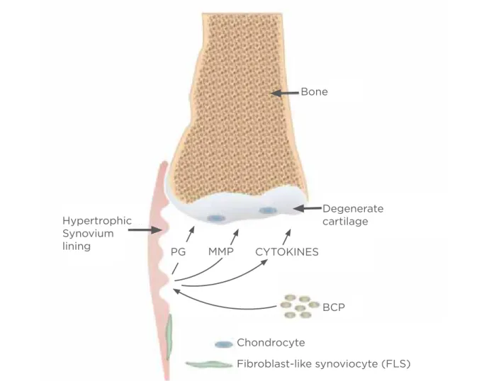
Bone
Degenerate cartilage Hypertrophic
Synovium lining
PG MMP CYTOKINES
BCP
Chondrocyte
Fibroblast-like synoviocyte (FLS) this method is diicult to interpret and also stains
other calcium containing particulates. Rosenthal et al.16 showed that BCP crystals could be identiied
using oxytetracycline staining in conjunction with ultraviolet light. There were fewer false-positive test results than with ARS staining and oxytetracyline did not bind to other particulates in joint luid. Estimates of the quantities of synthetic BCP crystals were also possible.
More advanced microscopic techniques for detecting BCP crystals include electron microscopy, atomic force microscopy, electron microprobe, Raman spectroscopy, radiograph difraction, scanning, a binding assay using 14C-labeled ethane-1-hydroxy-1,1-diphosphonate, and bisphosphonate-modiied superparamagnetic beads. These techniques are expensive and unfortunately not readily available. BCP crystals must irst be isolated from the synovial luid prior to analysis. Therefore, progress in appreciating the role of BCP crystals in OA has been hampered by diiculties in bedside identiication.17
BCP Crystals: Cause or Efect
Deposition of BCP crystals is a common inding in advanced OA. There is controversy in the literature as to whether these crystals cause OA, are a consequence of the degenerative process, or merely exacerbate the disease. Current evidence suggests that calcium crystal deposition contributes directly to joint degeneration and causes inlammation within the joint. Despite this, reviews of OA, written or presented, rarely include BCP crystals as a potential pathogenic factor in OA.18
Even if intra-articular BCP crystals are present as a consequence of joint damage, they can still play a role in perpetuating and aggravating the symptoms and signs of OA, especially by their efects on the synovium. Supportive evidence includes the fact that larger joint efusions are seen in knee joints containing BCP crystals when compared to joint luid from knees without crystals.19 BCP crystals
correlate strongly with the rapid progression of arthritis and the severity of radiographic OA.20
Furthermore, BCP crystals have been found not only
Figure 1: Proposed pathogenic efects of BCP crystals.
in advanced, but also in mild and moderate OA.21 If
BCP deposition was merely a consequence of bone exposure resulting from cartilage wear, how could the relative lack of BCP deposition in inlammatory, potentially destructive arthritis such as rheumatoid arthritis be explained?22 All evidence points to a
unique association between OA and BCP deposition.
In Vitro
Findings
An understanding of the molecular mechanisms involved in the pathological efects of BCP crystals, although incomplete, has been signiicantly advanced in recent years. Recent studies have emphasised the important role of the innate immune system in the pathogenesis of OA. The NALP3 (NACHT, LRR, and PYD domains-containing protein 3) inlammasome complex has been implicated in MSU and CPPD crystal induced inlammatory disease. Activation of Toll-like receptor (TLR) pathways may play an essential role in progression of OA, and BCP crystals appear to be inherently involved in this process. BCP crystals have been shown in numerous studies to have multiple biological efects on articular cells such as chondrocytes and synovial ibroblasts (Figure 1).
In vitro BCP crystals induce cellular proliferation and stimulate matrix MMP expression. MMPs accelerate the degradation of cartilage matrix components such as Type 2 collagen, ibronectin, laminin, and proteoglycan. BCP crystals can activate synovial ibroblasts through numerous pathways, including extracellular signal-related kinases (ERK) 1 and 2, nuclear factor
κ
B (NFκ
B), and protein kinase C (PKC). This in turn leads to upregulation of various inlammatory cytokines including tumour necrosis factor alpha (TNF-α
), interleukin-6 (IL-6), and IL-1β
.23-25 These cytokines target articularchondrocytes and synovial cells, inducing expression of cartilage degrading enzymes that ultimately lead to joint destruction.
Nitric oxide (NO) is a known central mediator in OA. NO is generated by the oxidation of arginine, catalysed by the nitric oxide synthases (NOSs). BCP crystals increase NO production. BCP crystal deposition in OA cartilage enhances chondrocyte hypertrophy and apoptosis. Cheung et al.26 demonstrated that treatment of cultured
chondrocytes with the NO donor sodium nitroprusside stimulated calciication.
BCP crystals are unusual in that they upregulate
followed by increased prostaglandin E2 in human ibroblasts.27 They induce apoptosis in synovial
ibroblasts and articular chondrocytes. These combined processes lead to an imbalance in anabolic versus catabolic mediators of cartilage turnover, which ultimately leads to ECM degradation.
In vivo studies have shown that accumulation of crystals in the joint leads to upregulated calciication or friction which activates NALP3 in synovial macrophages, leading to the production of IL-1
β
and IL-18. Jin et al.28 showed that HAcrystals lead to release of inlammatory cytokines in an NLRP3-dependent manner via reactive oxygen species (ROS) production, potassium elux, and lysosomal damage.
IL-1
β
in particular has been identiied as a key driver of destructive and inlammatory responses in OA as a result of its ability to upregulate aggrecanases and MMPs while also suppressing the biosynthesis of ECM. In keeping with this, IL-1β
has been shown to be increased in the articular cartilage and synovial luid of patients with OA. BCP crystals initiate IL-1β
–mediated inlammatory processes through NALP3 inlammasome-dependent as well as inlammasome-ininlammasome-dependent pathways. Non–lipopolysaccharide (LPS)-primed murine macrophages incubated with BCP crystals produce high levels of IL-1β
as well as IL-18, also an important cytokine in propagating joint damage. More importantly, longer incubation of LPS-primed macrophages with BCP crystals resulted in production of S100A8, a well-described damage-associated molecule that may further activate the macrophages through TLRs leading to production of IL-1β
. Therefore, BCP crystals may cause the production of IL-1β
both directly and indirectly through the autocrine efect of S100A8.29 Also,the spleen tyrosine kinase (SyK) and PI3 kinase appear necessary for the induction of IL-1
β
following macrophage activation by BCP crystals.The ability of BCP crystals to induce mitogenesis in many cell types including synoviocytes and macrophages may explain the macroscopic synovial proliferation found in OA. For example, BCP crystals activate human OA synovial ibroblasts (HOAS), leading to the induction of mitogenesis and MMP-1 production. Also, BCP crystals can act synergistically with IL1-
α
and TNF-α
to promote MMP production and likely subsequent joint degeneration.30 BCP crystals also induce theof metalloproteinases (TIMP).31-33 BCP crystals can
also induce proto-oncogenes, c-fos, and c-myc.34
Sun et al.35 showed that BCP crystals may stimulate
the endocytosis of various extracellular molecules, such as DNA fragments, nucleotides, and small peptides that might contribute to the pathogenesis of BCP crystal-associated diseases.
Animal Studies
Narayan et al.36 demonstrated that OCP crystals
induce inlammation in vivo through IL-1-dependent peritoneal inlammation without requiring the NALP3 inlammasome. BCP crystals injected into the peritoneal cavity of mice led to neutrophil recruitment and up-modulation of IL-1
α
, IL-1β
, and myeloid-related protein (MRP)-8-MRP-14 complex, to levels comparable with those induced by MSU crystals. This OCP crystal-induced inlammation was both IL-1α
and IL-1β
-dependent, as shown by inhibitory efects of anakinra and anti-IL-1β
antibody treatment. This study36 indicated that macrophages,rather than mast cells, are important for initiating and driving OCP crystal-induced inlammation.
Hang-Korng Ea et al.37 showed that intra-articular
BCP crystals have a direct pathogenic role in OA. BCP crystals injected into mouse knees induced synovial inlammation, cartilage degradation, and chondrocyte apoptosis. The efects observed were independent of the inlammasome-IL-1 pathway.
Two studies to date have looked at the efects of preventing BCP crystal deposition using pharmacological agents. Krug et al.38 evaluated
phosphocitrate (PC), a potential therapy for BCP crystal deposition. PC is the only agent to date that blocks the efects of BCP crystals and prevents calciication; the murine progressive anklyosis (MPA) model was used. This is a manifestation of an autosomal recessive mutation that produces an inlammatory joint disorder, associated with BCP crystal deposition, and results in fusion of the joints. Mice with MPA were treated with PC in vivo and there was a signiicant diference in disease progression and severity between the treated and the control group. Unfortunately, this model was somewhat inadequate as it resembled inlammatory arthritis more than OA.38,39
Cheung et al.40 examined a guinea pig OA model
with meniscal calciication, consistent with BCP
crystal deposition. After weekly treatment of this animal model for 3 months with a new, more potent formulation of PC containing salt and calcium (CaNaPC), the content of calciication in menisci and cartilage degeneration was examined. As a control they evaluated whether similar CaNaPC treatment had a therapeutic efect in a hemi-meniscectomy model with no known crystal involvement. Meniscal calciication correlated with the cartilage degeneration in this animal model. CaNaPC treatment led to signiicant reduction of calcium deposits and arrested OA disease progression. Similar CaNaPC treatment had no efect in the hemi-meniscectomy model in which articular calciication does not occur. These results support the hypothesis that calciication in the form of BCP crystals plays an important role in OA disease progression, and that CaNaPC is a potential therapeutic agent for CPPD and BCP crystal deposition disease.40 Unfortunately, no version of
PC has been studied in humans nor is any available for clinical use.
CONCLUSION
No disease modifying osteoarthritis drug (DMOAD) has been approved by a regulatory body for OA as no DMOAD has clearly shown deinite eicacy in patients with OA. With OA being the most prevalent rheumatic disease, afecting approximately 40 million patients in Europe, it is essential that we develop an efective treatment.41
Enhanced eforts should be made to pursue BCP crystals as a potential target for OA. Advances have been made in the understanding of BCP crystals in the pathogenesis of OA.
REFERENCES
1. Murphy LB et al. One in four people may develop symptomatic hip osteoarthritis in his or her lifetime. Osteoarthritis Cartilage. 2010;18(11):1372-9.
2. Murphy L et al. Lifetime risk of symptomatic knee osteoarthritis. Arthritis Rheum. 2008;59(9):1207-13.
3. Bijlsma JW, Knahr K. Strategies for the prevention and management of osteoarthritis of the hip and knee. Best Pract Res Clin Rheumatol. 2007;21(1): 59–76.
4. Piscitelli P et al. Socioeconomic burden of total joint arthroplasty for symptomatic hip and knee osteoarthritis in the Italian population: a 5-year analysis based on hospitalization records. Arthritis Care Res (Hoboken). 2012;64(9):1320-7.
5. Kotlarz H et al. Insurer and out-of-pocket costs of osteoarthritis in the US: evidence from national survey data. Arthritis Rheum. 2009;60(12):3546-53. 6. Wollheim FA, Lohmander LS, “Pathogenesis and pathology of osteoarthritis,” Hochberg MC et al. (eds.), Rheumatology (2008) 4th edition, Philadelphia: Elsevier, pp. 1711-28.
7. Lindbald S, Hedfors E. Arthroscopic and immunohistologic characterization of knee joint synovitis in osteoarthritis. Arthritis Rheum. 1987;30(10):1081-8. 8. Abou-Raya A et al. Methotrexate in the treatment of symptomatic knee osteoarthritis: randomised placebo-controlled trial. Ann Rheum Dis. 2014;doi:10.1136/ annrheumdis-2013-204856. [Epub ahead of print].
9. Jin X et al. Circulating C reactive protein in osteoarthritis: a systematic review and meta-analysis. Ann Rheum Dis. 2013;doi:10.1136/ annrheumdis-2013-204494. [Epub ahead of print].
10. Fuerst M et al. Calciication of articular cartilage in human osteaoarthritis. Arthritis Rheum. 2009;60(9):2694-703. 11. McCarthy GM, Cheung HS. Point: hydroxyapatite crystal deposition is intimately involved in the pathogenesis and progression of human osteoarthritis. Curr Rheumatol Rep. 2009;11(2):141-7. 12. Derfus B et al. Human osteoarthritic cartilage matrix vesicles generate both calcium pyrophosphate dihydrate and apatite in vitro. Calcif Tissue Int. 1998;63(3):258–62.
13. Jubeck B et al. Promotion of articular cartilage matrix vesicle mineralization by type I collagen. Arthritis Rheum. 2008;58(9):2809-17.
14. MacMullan P et al, “Basic calcium
Terkeltaub R (ed.), Gout and Other Crystal-induced Arthropathies (2012) 1st edition, Philadelphia: Elsevier, pp. 266-81. 15. McCarty DJ et al. “Milwaukee shoulder”--association of microspheroids containing hydroxyapatite crystals, active collagenase, and neutral protease with rotator cuf defects. I. Clinical aspects. Arthritis Rheum. 1981;24(3):464-73. 16. Rosenthal AK et al. Feasibility of a tetracycline-binding method for detecting synovial luid basic calcium phosphate crystals. Arthritis Rheum. 2008;58(10):3270-4.
17. Yavorskyy A et al. Detection of calcium phosphate crystals in the joint luid of patients with osteoarthritis - analytical approaches and challenges. Analyst. 2008;133(3):302-18.
18. Matthews GL, Hunter DJ. Emerging drugs for osteoarthritis. Expert Opin Emerg Drugs. 2011;16(3):479-91.
19. Carroll GJ et al. Hydroxyapatite crystals are a frequent inding in osteoarthritic synovial luid, but are not related to increased concentrations of keratan sulfate or interleukin 1 beta. J Rheumatol. 1991;18(6):861-6.
20. Molloy ES, McCarthy GM. Calcium crystal deposition disease: update on pathogenesis and manifestations. Rheum Dis Clin North Am. 2006;32(2):383-400. 21. Derfus BA et al. The high prevalence of pathologic calcium crystals in pre-operative knees. J Rheumatol. 2002;29(3):570-4.
22. Swan A et al. Submicroscopic crystals in osteoarthritic synovial luids. Ann Rheum Dis. 1994;53:467-70.
23. Daheshia M, Yao JQ. The interleukin 1beta pathway in the pathogenesis of osteoarthritis. J Rheumatol. 2008;35(12):2306-12.
24. Reuben PM et al. Molecular mechanism of the induction of metalloproteinases 1 and 3 in human ibroblasts by basic calcium phosphate crystals. Role of calcium-dependent protein kinase C alpha. J Biol Chem. 2002;277(17):15190-8. 25. McCarthy GM et al. Molecular mechanism of basic calcium phosphate crystal-induced activation of human ibroblasts. Role of nuclear factor kappab, activator protein 1, and protein kinase c. J Biol Chem. 1998;273(52):35161–9.
26. Cheung HS, Ryan LM. Phosphocitrate blocks nitric oxide-induced calciication of cartilage and chondrocyte-derived apoptotic bodies. Osteoarthritis Cartilage. 1999;7(4):409-12.
27. Molloy ES, McCarthy GM. Eicosanoids, osteoarthritis, and crystal deposition
2005;17(3):346-50.
28. Jin C et al. NLRP3 inlammasome plays a critical role in the pathogenesis
of hydroxyapatite-associated arthropathy. Proc Natl Acad Sci U S A.
2011;108(36):14867-72.
29. Cunningham CC et al. Osteoarthritis-associated basic calcium phosphate crystals induce proinlammatory cytokines and danger-associated molecules via activation of Syk and PI3 kinase. Clin Immunol. 2012;144:228–36. 30. McCarthy GM et al. Basic calcium phosphate crystals activate human osteoarthritic synovial ibroblasts and induce matrix metalloproteinase-13 (collagenase-3) in adult porcine articular chondrocytes. Ann Rheum Dis. 2001;60(4):399-406.
31. McCarthy GM et al. Basic calcium phosphate crystals cause coordinate induction and secretion of collagenase and stromelysin. J Cell Physiol. 1992;153(1):140-6.
32. Bai G et al. Basic calcium phosphate crystals up-regulate metalloproteinases and down-regulate tissue inhibitor of metalloproteinase-1 and -2 in human ibroblasts. Osteoarthritis and Cartilage. 2001;9(5):416-22.
33. Reuben PM et al. Induction of matrix metalloproteinase-8 in human ibroblasts by basic calcium phosphate and calcium pyrophosphate dihydrate crystals: efect of phosphocitrate. Connect Tissue Res. 2001;42(1):1-12.
34. Cheung HS et al. Induction of expression of c-fos and c-myc protooncogenes by basic calcium phosphate crystal: efect of b-interferon. Cancer Res. 1989;49(1):134-8.
35. Sun Y et al. Basic calcium phosphate crystals stimulate the endocytotic activity of cells - inhibition by anti-calciication agents. Biochem Biophys Res Commun. 2003;312(4):1053-9.
36. Narayan S et al. Octacalcium phosphate crystals induce inlammation in vivo through interleukin-1 but independent of the NLRP3 inlammasome in mice. Arthritis Rheum. 2011;63(2): 422-33.
37. Ea HK et al. Pathogenic role of basic calcium phosphate crystals in destructive arthropathies. PLoS One. 2013;8(2):e57352.
38. Krug HE et al. Phosphocitrate prevents disease progression in murine progressive ankylosis. Arthritis Rheum. 1993;36(11):1603-11.
mitogen-activated protein kinase cascade signal transduction pathway. J Biol Chem. 1997;272(30):18920-5.
40. Cheung HS et al. Phosphocitrate
blocks calciication-induced articular joint degeneration in a guinea pig model. Arthritis Rheum. 2006;54(8):2452-61. 41. Blanco FJ, Ruiz-Romero C. New targets