ISSN 2278- 4136
ZDB-Number: 2668735-5
IC Journal No: 8192
Volume 2 Issue 1
Online Available at www.phytojournal.com
Journal of Pharmacognosy and Phytochemistry
Vol. 2 No. 1 2013 www.phytojournal.com Page | 326
Isolation and Structural Determination of an Anti Bacterial
Constituent from the Leaves of
Cassia alata
Linn.
Barnali Paul1, Prasenjit Mitra2, Tanaya Ghosh2, Ravinernath Salhan3, Takhelmayum Amumachi Singh3, Amit Chakrabarti3, Sumanta Gupta4, Basudeb Basu4, Prasanta Kumar Mitra5*
1. Professor and Head, Department of Medical Biotechnology, Sikkim Manipal Institute of Medical Sciences, Tadong, Gangtok737102,Sikkim, India. Telefax : 0353-2468908
[E-mail: [email protected]]
2. Biochem Academy, Saktigarh, Siliguri 734005, West Bengal, India.
3. Sikkim Manipal Institute of Medical Sciences, Tadong, Gangtok737102,Sikkim, India 4. Department of Chemistry, University of North Bengal, Raja Rammohanpur 734430
Siliguri, West Bengal, India .
5. Professor and Head, Department of Medical Biotechnology, Sikkim Manipal Institute of Medical Sciences, Tadong, Gangtok737102, Sikkim, India.
[E-mail: [email protected]; Telefax: 0353-2468908]
By different solvent extractions and chromatographic techniques an antibacterial constituent was isolated from leaves of Cassia alata Linn. Infra red spectroscopy, mass spectroscopy and nuclear magnetic resonance studies showed that the isolated compound was chemically 3,4 dihydroxy cinnamic acid. In vitro antibacterial activity of 3,4 dihydroxy cinnamic acid was studied against four Gram-positive and four Gram-negative bacteria using disc diffusion method. Minimum inhibitory concentration (MIC) of 3,4 dihydroxy cinnamic acid was also recorded against those bacteria by serial dilution technique. Kanamycin was used as positive control. Results showed that 3,4 dihydroxy cinnamic acid had antibacterial activity against the tested bacteria.
Keyword: Cassia alata Linn., Gram-positive and Gram-negative bacteria, 3,4 dihydroxy cinnamic acid, kanamycin
1. Introduction
Several plants have shown anti–microbial activity. Few of them are Azadirachta indica[1], Clausena anisata[2], Amona glabra[3], Semecarpus
anacardium[4], Garcinia mangostana[5], Aegle
marmelos[6], Santolina chamaecyparissus[7],
Terminalia belerica[8], etc.
Cassia alata Linn. ( family, Caesalpiniaceae)
is an erect tropical annual herb with leather compounded leaves. It grows everywhere in the state of West Bengal, India up to 6 ft tall. It has different names like ringworm weed in English, dadmari in Hindi and cakramard in
Sanskrit. Its therapeutic values as mentioned in Ayurvedic text[9] are: Leaves are anti-parasitic, used in eczema, bronchitis, asthma, ringworm and in poisonous insect bites. Bark is used to treat skin diseases. Extract of aerial parts
is CNS depressant, diuretic and
anti-inflammatory.
Vol. 2 No. 1 2013 www.phytojournal.com Page | 327
elucidate its structure. Further, antibacterial activity of the isolated compound was also studied against few positive and Gram-negative bacteria. In present communication we report the results of those experiments.
2. Materials and Methods 2.1 Plant material
Leaves of Cassia alata Linn. were collected from the medicinal plants garden of the University of North Bengal, Siliguri, India and authenticated by Prof. A. P. Das of the department of Botany 1of the said University. A voucher specimen was kept in the department for future reference.
2.2 Extraction and Isolation
1. First step: Leaves of Cassia alata Linn. were
sundried and powdered. 50g of this powder were extracted with 500 ml of 10 : 1 (v/v) acetone – ethyl alcohol mixture for 1h on a rotary shaker. It was then centrifuged. Supernatant was collected and evaporated to dryness.
Dry brown mass was obtained.
2. Second step: Dry brown mass was refluxed
with 100 ml of 1(N) HCL for 1h on a water bath at 100 degree centigrade. It was cooled and centrifuged. Supernatant was evaporated to dryness.
3. Third step: Dry brown mass thus obtained
from the supernatant was extracted with 50 ml of a mixture of water and isobutanol (2 : 1 v/v) on a rotary shaker for 1h. Isobutanol layer was separated from water layer. It was evaporated to dryness.
4. Fourth step: Brown mass obtained was
dissolved in 10 ml methanol and subjected to column chromatography using silica gel Gas adsorbent. 5 bands were separated. Bands were collected in separate beakers . Elution was done by 50% methanol – chloroform mixture. Third band had antibacterial activity against
Staphylococcus aureus.
5. Fifth step: Eluant of third band was evaporated
to dryness. The dry mass was extracted with 15 ml ethyl acetate for 10 minutes. It was then filtered. With filtrate polyamide column chromatography was done. Elution was made by ethyl formate: formic acid mixture (100: 5 v/v). Three bands were separated. Second band
showed antibacterial activity against
Staphylococcus aureus.
6. Sixth step: Eluant of second band was
evaporated to dryness. Repeated crystallization was done from ethyl acetate–cyclohexane (50:50, v/v) mixture. Crystals obtained. Yield was 3.6 mg.
2.3 Homogeneity of the active compound This was ascertained by silica gel- G thin layer chromatography by using the following solvent systems: Acetone : methanol - 50 : 50; n-butanol : acetic acid : water - 80 : 10 : 10; Chloroform : methanol : water - 60 : 20 : 20
2.4 Structure determination
FT-IR spectrum of the sample was taken in KBr
pellets using Shimadzu FT-IR 8300
Spectrophotometer. NMR spectrum was taken using Bruker AVH 300 Spectrometer operating at 300 MHz (for 1H) and 75 MHz (for 13C) and in solvent, as indicated. 13C NMR spectrum was run in 1H-decoupled mode. The High Resolution Mass Spectral data for the compound was
obtained in Mass Spectrometer (Model:
Micromass Q-Tof Micro), run under Electron Spray Ionization (ESI) Positive Mode. Melting point was observed in an open sulfuric acid bath and is uncorrected.
2.5 Anti-bacterial Screening 2.5.1 Bacteria
Four Gram-positive strains, Staphylococcus
aureu ATCC 25923, Bacillus subtilis ATCC
19659, Straptococcus pyogenes MTCC 512 and
Bacillus megaterium MTCC 302 as well as four
Gram-negative strains, Shigella flexneri MTCC
678, Escherichia coli ATCC 25922,
Pseudomonas aeruginosa ATCC 27853 and
Salmonella typhi MTCC 733 were used in this
study. Bacterial strains were collected from the department of Microbiology, North Bengal Medical College Hospital, Siliguri, West Bengl, India and maintained on nutrient agar slants at 40C.
2.5.2 Media
Vol. 2 No. 1 2013 www.phytojournal.com Page | 328
26.8 were used for antibacterial screening and
MIC (minimum inhibitory concentration)
determination respectively
2.6 In Vitro Antibacterial activity of the isolated compound
In vitro antibacterial screening was carried out by disc diffusion method[12]. According to this method, 20 ml quantities of nutrient agar were placed in a petri dish with 0.1 ml of 10-2 dilution of bacterial culture of 20 hours old. Filter paper discs (6 mm diameter) impregnated with 30 µg
per disc of the isolated compound was placed on bacterial seeded plates.
The isolated compound was soluble in water. Blank disc impregnated with water was used as negative control. Zone of inhibition was recorded after 20 hours of incubation at 370 C. Diameters of zone of inhibition produced by the isolated compound were compared with that of standard antibiotic kanamycin under same experimental conditions.
2.7 Minimum inhibitory concentration (MIC)
determination Minimum inhibitory
concentration is defined as the lowest concentration of antibiotic completely inhibiting visible growth of bacteria after 18 – 24 hours of incubation at 370C. This was done by the method of Mosaddik and Haque13. According to this method, the isolated compound (1.0 mg) was dissolved in 2 ml nutrient broth media to obtain a stock solution of concentration 500 µ g/ml. 3 drops of Tween 80 was added in nutrient broth to facilitate dissolution. Serial dilution technique was followed to obtain 250 µg/ml concentration of the isolated compound. One drop (0.02ml) of
prepared suspensions of organism
(106organism/ml) was added to each broth dilution. These dilutions were then incubated for 20 hours at 370C. Growth of bacteria was examined by noting turbidity of the solution. The
nutrient broth media with 3 drops of Tween 80 was used as negative control while kanamycin under same experimental condition was used as positive control.
Vol. 2 No. 1 2013 www.phytojournal.com Page | 329
2.8 Statistical analysis
The values were expressed as mean ± SEM and was analyzed using one-way analysis of variance (ANOVA) using Statistical Package for Social Sciences (SPSS) 20th versions. Differences between means were tested employing Duncan’s multiple comparison tests and significance was set at p < 0.05.
3. Results and Discussion
3.1 Homogeneity of the active Compound In all cases of thin layer chromatographic experiments using three different solvent systems single spot was obtained. Thus, it was a single compound.
Fig 2: IR spectrum of the isolated compound
3.2 Structure Elucidation
The compound was a pale yellow solid, mp. 218-221oC. NMR data were as follow :The 1H-NMR (D6-DMSO): 6.16 (d, 1H, J = 15.9 Hz), 6.75
(1H, d, J = 7.4 Hz), 6.95 (dd, 1H, J = 8.1 & 2.1 Hz), 7.02 (s, 1H), 7.41 (d, 1H, J = 15.9 Hz), 9.12(br. s, 1H), 9.52 (br. s, 1H), 12.11 (br. s, 1H) ppm. Its 13C-NMR (D6-DMSO): 115.1, 115.6,
116.2, 121.6, 126.2, 145.1, 146.0, 148.6, 168.4. From 1H-NMR spectral data (Figure-1), it appeared that there were three aromatic protons, two olefinic protons and three broad singlets. The coupling patterns of the aromatic protons primarily indicated one ortho coupled doublet and one ortho-meta coupled doublet of doublet (J = 8.1 & 2.1 Hz) and the other was possibly a
meta-doublet, though appeared as a singlet ( = 7.1 ppm). On the other hand, the olefinic protons
with coupling constant, J = 15.9 Hz, indicated that the double bond was in trans configuration. Since there were three aromatic protons as seen by 1H 3NMR spectral data, the other three positions of the aromatic ring might be substituted. Only one aromatic ring was considered because of low molecular mass of the compound. Also, there were only nine chemically non-equivalent carbons according to 13C-NMR spectrum. As one substituent might be a C–C double bond, there were two other positions, might be substituted with two hydroxyl group (OH) that appeared as broad singlets. Out of three broad singlets, one broad singlets at =12.11 ppm could be assigned for the carboxylic (COOH) proton. The carboxylic acid group might be attached with the C–C double bond, leading to propose the assigned structure as dihydroxy cinnamic acid. The FT-IR (KBr) absorption maxima, shown in Figure-2, (Vmax) at 3423, 3179,
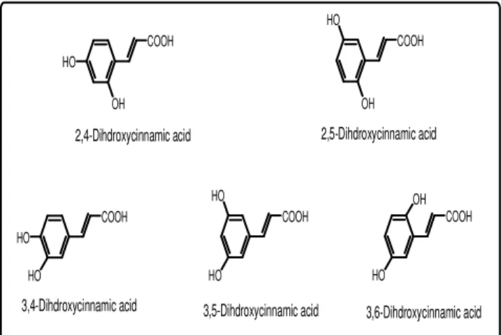
1675, 1657, 1603 cm–1 also suggested the presence of hydroxyl, conjugated carboxyl and double bonds. Considering that the compound could be a dihydroxy cinnamic acid and based on the coupling pattern of three aromatic protons (one ortho-doublets, one ortho-meta doublet of doublet and one meta-doublet), theoretically five possible structures, as shown in figure 3, might be proposed :
The 13C-NMR spectral data (Figure-4) showed that there are nine chemically non-equivalent carbons, out of which three carbons were assigned for the acrylic acid side chain carbons (– C=C–COOH). Therefore, all aromatic ring
hydrogens were non-equivalent.
Vol. 2 No. 1 2013 www.phytojournal.com Page | 330
calculated to be J=15.9 Hz). The trans -configuration of the carbon–carbon double bond was assigned based on the fact that in the case of
cis-configuration, the coupling constant (J) would have been within 6–12 Hz. The spin-spin
couplings between hydrogens were clearly shown in figure - 5 for the aromatic hydrogens (marked as Ha, Hb and Hc) as well as for the carbon– carbon double bonds, marked as Hα and Hβ.
Structure of the compound was further
corroborated by the High Resolution Mass Spectral (HRMS) data (Figure-6), run under Electron Spray Ionization (ESI) Positive Mode. In HRMS, the exact mass for compound with mf C9H8O4Na [M+Na] was calculated to be 203.1472
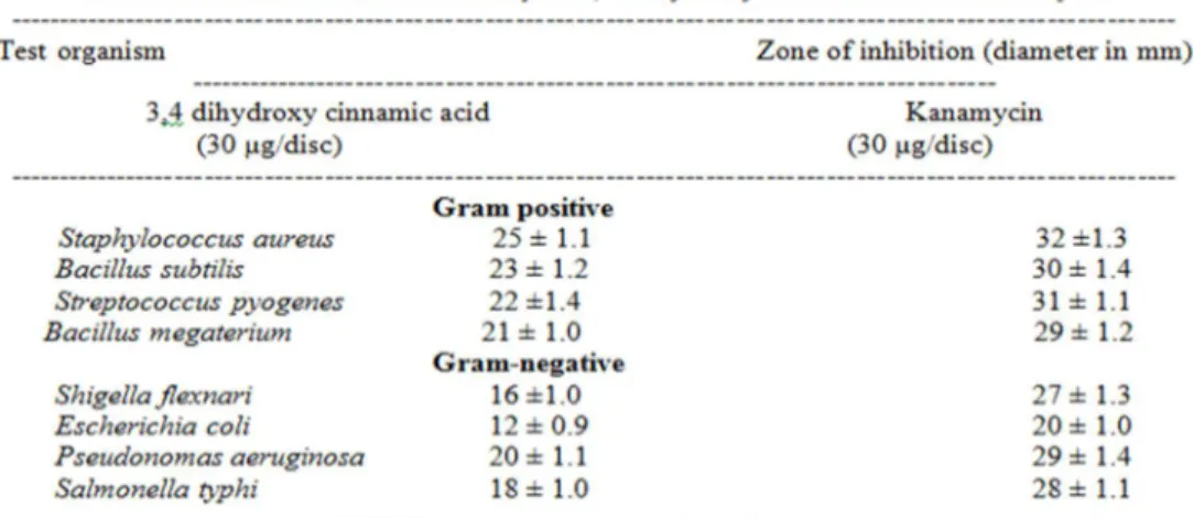
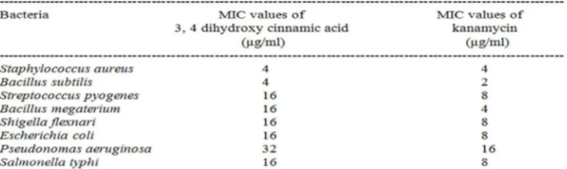
and observed as 203.1622.Therefore, the structure of the isolated compound, shown in figure-7, may be assigned as 3,4-Dihydroxycinnamic acid. Antibacterial activity of 3,4 dihydroxy cinnamic acid was studied. Results, given in Table- 1, showed that 3,4 dihydroxy cinnamic acid in disc concentration 30 µ g produced more zone of inhibitions (in between 21 ± 1.0 and 25 ± 1.1) for Gram-positive bacteria than Gram-negative bacteria (zone of inhibitions, 12 ± 0.9 and 20 ± 1.1). Kanamycin, however, produced more zone of inhibitions for both Gram-positive (29 ± 1.2 to 32 ± 1.3) and Gram-negative (20 ± 1.0 to 29 ± 1.4) bacteria under same experimental conditions. Table – 2 showed results relating to minimum inhibitory concentration (MIC) of 3,4 dihydroxy cinnamic acid and kanamycin against Gram-positive and Gram-negative bacteria. 3,4 dihydroxy cinnamic acid showed more MIC values (4-32 µ g/ml) than that of kanamycin (2-16 µg/ml). Only in case of Staphylococcus aureus
MIC value of 3,4 dihydroxy cinnamic acid was found same to that of kanamycin (4 µg/ml).
Resistance to antibiotics is becoming an increasingly difficult problem in the management bacterial infections[14]. The situation is particularly critical for Staphylococcus aureus
Vol. 2 No. 1 2013 www.phytojournal.com Page | 331
COOH
OH HO
COOH
OH HO
COOH HO
HO COOH
HO HO
COOH
HO OH
2,4-Dihdroxycinnamic acid 2,5-Dihdroxycinnamic acid
3,5-Dihdroxycinnamic acid
3,4-Dihdroxycinnamic acid 3,6-Dihdroxycinnamic acid
Fig 3: Possible structures of the isolated compound
We, in our laboratory, when screened medicinal plants for their antibacterial property noted that leaves of Cassia alata Linn. Could inhibit growth
of Staphylococcus aureus. We intended to isolate
the active compound from leaves of Cassia alata
Linn. By different solvent extractions followed by chromatography an active compound was isolated. Elucidation of structure of the active compound was undertaken by spectroscopic and other analytical data. IR spectroscopy, mass spectroscopy and nuclear magnetic resonance data suggested that the active compound was chemically 3,4 dihydroxy cinnamic acid.
Fig 4: 13C NMR spectrum of the isolated compound
In vitro antibacterial activity of 3,4 dihydroxy
cinnamic acid was carried out by disc diffusion method against four Gram-positive and four Gram-negative bacteria. Zone of inhibitions suggested that 3,4 dihydroxy cinnamic acid had antibacterial activity against the tested bacteria though it was more effective for Gram-positive bacteria than Gram-negative bacteria. Control drug kanamycin, however, produced larger zone
of inhibitions under same experimental
conditions.
COOH Hα
Hβ
HO HO
Ha
Hb
Hc
ortho -coupled
meta
-coupled
ortho-meta-dd
Ha = meta-coupled doublet
Hb = doublet of doublet
Hc = ortho-coupled doublet
trans-configuration
J (Hα - Hβ) = 15.9 Hz
Fig 5: Spin-spin couplings between hydrogen in the
structure of the isolated compound.
Minimum inhibitory concentrations (MIC) of 3,4 dihydroxy cinnamic acid against the tested bacteria were more in comparison to kanamycin. But in case of Staphylococcus aureus MIC value was recorded same (4 µg/ml) for both 3,4 dihydroxy cinnamic acid and kanamycin.
Vol. 2 No. 1 2013 www.phytojournal.com Page | 332
Fig 6: Mass spectrum of the isolated compound
4. Conclusion
3,4 dihydroxy cinnamic acid was isolated from the leaves of Cassia alata Linn. and found having antibacterial activity against four Gram-positive bacteria viz. Staphylococcus aureus, Bacillus
subtilis, Straptococcus pyogenes and Bacillus
megaterium as well as four Gram-negative
bacteria like, Shigella flexneri, Escherichia coli,
Pseudomonas aeruginosa and Salmonella typhi.
Anti-bacterial activity was comparable to that of known antibiotic kanamycin.
Figure 7. Structure of the isolated compound
5. References
1. Venugopal PV, Venugopal TV., Anti dermatophytic
activity of neen (Azadirachta indica) leaves in vitro, Indian J Phrmacol., 1994; 26: 141 – 43.
2. Chakraborty A, Chakraborty BK, Bhattacharya P.,
Clausenol and clausenine - two carbazole alkaloid from Clausena anisata, Phytochemistry 1995; 40: 295 - 99.
3. Padmaja V, Thankarmany V, Hara N., Biological
activities of Amona glabra, J. Ethnopharmacol 1995; 48: 21 – 24.
4. Nair A, Bhide SV., Antimicrobial properties of different parts of Semecarpus anacardium,, Indian Drugs 1996; 33: 323 – 28.
5. Gopalakrishnan G, Banumathi B, Suresh G.,
Evaluation of the antifungal activity of natural xanthones from Garcinia mangostana and their synthetic derivatives, J Nat Prod 1997; 60: 519 – 24.
6. Rana BK, Singh UP, Taneja V., Antifungal activity of kinetics of inhibition by essential oil isolated from leaves of Aegle marmelos, J Ethnopharmacol 1997; 57: 29 – 34.
7. Suresh B, Sriram S, Dhanaraj S, Elango K,
Chinnaswamy K., Anticandidal activity of Santolina
chamaecparissus volatile oil, J
Ethnopharmacol.1997; 55: 151 – 59.
8. Valsaraj R, Pushpangadan P, Smitt UW,
Antimicrobial screening of selected medicinal plants from India, J Ethnopharmacol. 1997; 58: 75 – 83.
9. Gurung Bejoy. The Medicinal Plants of Sikkim
Himalaya. Pub. JB. Gurung, Chakung, West Sikkim ,2002, p. 90 – 92.
10. Sakharkar PR, Patil AT., Antimicrobial activity of Cassia alata, Indian Journal of Pharmaceutical Sciences 1998; 60: 311 – 12.
11. Paul B, Mitra P, Ghosh T, Salhan RN, Chakrabarti A,
Singh TA, Das AP, Mitra PK, Effect of leaves of
Cassia alata Linnaeus (Caesalpiniaceae) on the
growth of Staphylococcus aureus Rosenbach, Pleione 2012; 6:, 341-347.
12. Rahman MM, Mosaddik MA, Wahed MI, Haque ME.,
Antimicrobial activity and cytotoxicity of Trapa bispinosa, Fitoterapia, 2000; 71:, 704-6.
13. Mosaddik MA, Haque ME., Cytotoxicity and
antimicrobial activity of goniothalamin isolated from Bryonopsis laciniosa. Phytother Res. 2003; 17: 1155-7.
14. Coates A, Hu Y, Bax R.,The future challenges facing
the development of new antimicrobial drugs, Nature Reviewing Drug Discovery 2002; 1: 895 – 910.
15. Chopra I, Antibiotic resistance in Staphylococcus
aureus : concerns, causes and cures, Expert Review of Anti – infective Therap 2003; 1: 45 – 55. 16. Centers for Disease Control and Prevention, United
Vol. 2 No. 1 2013 www.phytojournal.com Page | 333
17. Tsiodras S, Goid HS, Sakoulas G, Linezolid resistance
in a clinical isolate of Staphylococcus aureus, Lancet 2001; 358: 207 – 8.
18. Al – Bari MA, Sayeed MA, Rahman MS, Mossadik
MA, Characterization and antimicrobial activities of a phenolic acid derivative produced by Streptomyces bangladeshiensis, a novel species collected in Bangladesh, J. Respir Med. Sci. 2006; 1: 77 – 81.
19. Rahman MM, Wahed MI, Biswas MH, Sadik GM,
Haque ME, In vitro antibacterial activity of the compounds of Trapa bispinosa Roxb, Science 2001; 1: 214 – 16.
20. Projan SJ, Youngman PJ, Antimicrobials : New
solutions badly needed, Current Opinion in Microbiology 2002; 5: 463 – 65.
21. Dahanar SA, Kulkarni RA, Rege NN, Pharmacology
of medicinal plants and natural products. Indian Journal of Pharmacology 2000; 32: S 81 – S 118.
22. Chakraborty D, Mandal SM, Chakraborty J,