Article
Printed in Brazil - ©2012 Sociedade Brasileira de Química0103 - 5053 $6.00+0.00A
*e-mail: [email protected]
New Neolignans from
Krameria tomentosa
A. St.-Hil
Sara A. L. Madeiro,a Hellane F. S. de Lucena,a Caroline D. Siqueira,a
Marcelo C. Duarte,a Raimundo Braz-Filho,b José M. Barbosa Filho,a
Marcelo S. da Silvaa and Josean F. Tavares*,a
aDepartamento de Ciências Farmacêuticas, Universidade Federal da Paraíba,
CP 5009, 58051-970 João Pessoa-PB, Brazil
bLaboratório de Ciências Químicas, Universidade Estadual do Norte Fluminense,
Campos dos Goytacazes, 28013-602 Rio de Janeiro-RJ, Brazil
A investigação fitoquímica das raízes de Krameria tomentosa A. St.-Hil. levou ao isolamento de cinco neolignanas, duas delas com estruturas inéditas [1,1’-(E )-propenil-4-metóxi-3,4’-oxineolignana (ottomentosa) e ácido 2-(2’-hidróxi-4’,6’-dimetoxifenil) benzofurano-5-carboxílico (sobralina)], além de três compostos conhecidos [eupomatenoide 6, di-hidrocarinatidina e 2-(2’,4’-di-hidroxifenil)-5-(E)-propenilbenzofurano]. A caracterização estrutural dos compostos isolados foi estabelecida com base na espectroscopia no infravermelho, espectrometria de massas, ressonância magnética nuclear uni e bidimensional, além de comparação com dados espectrais descritos na literatura.
A phytochemical investigation of the roots of Krameria tomentosa A. St.-Hil. led to the isolation of five neolignans, two of them with novel structures [1,1’-(E )-propenyl-4-methoxy-3,4’-oxyneolignan (ottomentosa) and 2-(2’-hydroxy-4’,6’-dimethoxyphenyl)benzofuran-5-carboxylic acid (sobraline)] and three known compounds [eupomatenoid 6, dihydrocarinatidin and 2-(2’,4’-dihydroxyphenyl)-5-(E)-propenylbenzofuran]. The structural characterization of the compounds isolated was established based on infrared spectroscopy, mass spectrometry, one- and two-dimensional nuclear magnetic resonance, along with comparison with spectral data described in the literature.
Keywords: Krameria tomentosa,Krameriaceae, neolignans, ottomentosa, sobraline
Introduction
The genus Krameria is the only member of the
family Krameriaceae, and includes 18 herbaceous or shrub species that are predominantly represented in neotropical and ecologically restricted regions and arid or seasonally dry regions of the Americas.1-3 The presence
of neolignans and norneolignans is well documented for this genus.4-9Krameria tomentosa A. St.-Hil. (synonymy
of Krameria ovata O. Berg) is popularly known as “rhatany” and as with other species of Krameria, its roots have been long used in popular medicine in the treatment of dysentery, stomatitis, diarrhea, vaginal discharges and afflictions of the mouth.10-12 The alcoholic
extract from K. tomentosa (root) showed toxicity to
mice and fish.13 Earlier studies have demonstrated that
neolignans, norneolignans and steroids,14,15 as well as
the norlignan 2-(2’-hydroxy-4’,6’-dimethoxyphenyl)-5-[(E)-propenyl]benzofuran, inhibit acetylcholine-induced relaxation in the aorta of rats.16In the present work, it is
described the isolation and structural determination of two new neolignans, ottomentosa and sobraline (1 and 2), besides three known neolignans: eupomatenoid 6 (3), dihydrocarinatidin (4) and 2-(2’,4’-dihydroxyphenyl)-5-(E)-propenylbenzofuran (5) (Figure 1).
Results and Discussion
molecular formula C19H20O2Na (calc. 303.1356). The IR
spectrum showed absorptions in the region of 1603-1441 cm-1,
characteristic of C=C stretching of the aromatic ring, as well as absorptions of 1269-982 cm-1, characteristic
of C−O stretching. The 1H nuclear magnetic resonance
(NMR) spectrum showed in the region of hydrogens in aromatic systems two connected doublets, one at dH 7.23
(J 9.0 Hz) and the other at dH 6.85 (J 9.0 Hz), compatible
with the hydrogens of an AA’XX’ system. In the same region, there was a doublet of doublets at dH 7.04 (J 2.5 and
8.5 Hz) coupled with the doublets at dH 6.95 (J 2.5 Hz) and
at dH 6.89 (J 8.5 Hz), compatible with the hydrogens of an
AMX system. The set of signals at dH 6.33, 6.10 and 1.84,
as well as the set at dH 6.25, 6.00 and 1.80, characterized
the hydrogens of two propenyl units. The 13C APT NMR
(attached-proton-test NMR) spectrum displayed 17 signals, corresponding to 19 carbon atoms. From these, 5 were assigned to non-hydrogenated carbons, from which 3 were oxygenated, 11 to methine carbons, 2 to methyl carbons and 1 to a methoxyl carbon. According to the data described, it was possible to suggest that the phenylpropanoid units of 1 were connected forming the skeleton of an oxyneolignan,17,18 showing an oxygenated substitute, this
being a methoxyl. The correlation of the hydrogens at
dH 3.80 (OMe) with the hydrogen at dH 6.85 (H-3’/5’),
observed in the NOESY (nuclear Overhauser effect spectroscopy) spectrum, made it possible to infer that the methoxy group was connected to C-4. In the heteronuclear multiple-quantum correlation (HMQC) spectrum, direct correlations can be seen between the hydrogens at dH 7.23
(d) and 6.85 (d) with the carbons at dC 126.9 and 117.3,
assigning them to C-2’/C-6’ and C-3’/C-5’, respectively. Chemical shifts at dH 6.95 (d), 6.89 (d) and 7.04 (dd)
showed correlations with the carbons at dC 117.9, 112.8 and
122.2, respectively, in which they were thereby assigned to carbons C-2, C-5 and C-6. In the heteronuclear multiple-bond correlation (HMBC) spectrum, correlations were observed between the hydrogens at dH 6.95 (H-2) and 1.80
(H-9) with the carbon at dC 129.9 (C-7), as well as the
correlation between the hydrogen at dH 6.33 (H-7’) with
the carbon at dC 126.9 (C-2’/6’), confirming the insertion
of the propenyl group more shielded in the ring of the AMX system and less shielded in the AA’XX’ system. The shifts of the non-hydrogenated, non-oxygenated carbons were confirmed by the correlations between the signal at dH 7.04 (H-6) with the carbon at dC 150.3, assigned to
C-4, and between the hydrogens at dH 7.23 (H-2’/H-6’) and
6.85 (H-3’/H-5’) with the carbon at dC 156.8, assigned
to C-4’. Homonuclear correlation spectroscopy (COSY) showed correlations of the signal at dH 7.04 (H-6) with
6.95 (H-2) and 6.89 (H-5) and of the signal at dH 6.85
(H-3’/5’) with 7.23 (H-2’/6’). Table 1 gives a compilation of the chemical shifts and correlations observed in the spectra of one- and two-dimensional 1H and 13C NMR for
compound 1. After the analysis of all the spectral data, this compound was determined to be 1,1’-(E )-propenyl-4-methoxy-3,4’-oxyneolignan,reported here for the first time and given the trivial name ottomentosa.
Compound 2was isolated in the form of a white powder, with a melting point of 262-264 °C. The high resolution mass spectrum utilizing the ESI- ionization mode showed the peak of the deprotonated molecule at m/z 313.0708 [M − H]−
, compatible with the molecular formula C17H13O6
(calc. 313.0706). The IR spectrum revealed the presence of a wide band between 3472 and 2650 cm-1 and a band
at 1690 cm-1, characteristic of O−H and C=O stretching
of a carboxyl group, respectively. Absorption was also
observed at 1624-1458 cm-1, characteristic of C=C
stretching of an aromatic ring and between 1312-1107 cm-1
of C−O stretching. The 13C APT NMR spectrum showed
the presence of 17 signals, corresponding to 17 carbons. From these, 9 were assigned to non-hydrogenated carbons, 6 to methine carbons and 2 to methoxyl carbons.
Based on comparison with 13C NMR spectral data of
the neolignans 2-(2’-hydroxy-4’-6’-dimethoxyphenyl)-5-(E)-propenylbenzofuran and krametosan, also isolated from K. tomentosa,14 it was possible to make the following
considerations: (i) the signals at dC 153.5, 107.5, 130.1 and
157.5 were assigned to carbons C-2, C-3, C-3a and C-7a, respectively, of the benzofuran ring; (ii) the signals at
dC 100.6, 158.6, 95.1, 163.4, 91.6 and 160.8 were assigned to
carbons C-1’, C-2’, C-3’, C-4’, C-5’ and C-6’, respectively; (iii) the absence of signals at approximately dC 131.0,
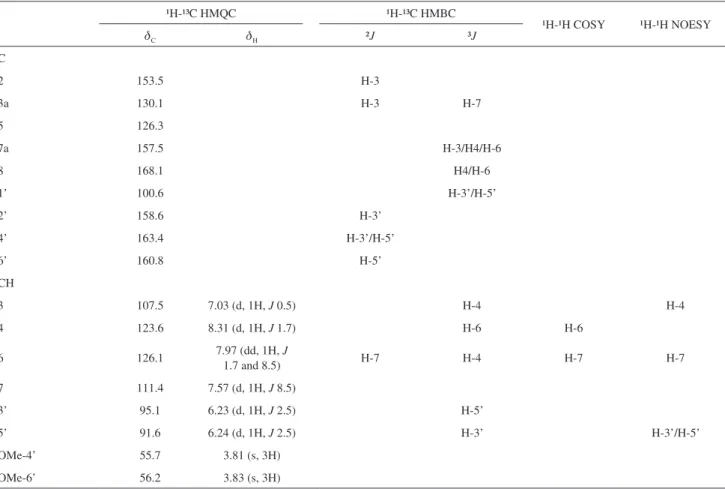
124.6 and 18.5 suggestive of a propenyl unit plus the presence of the signal at 168.0 (referring to a carbonyl) and the information obtained from the IR spectrum indicate that compound 2 is possibly the trinor-neolignan 2-(2’-hydroxy-4’,6’-dimethoxyphenyl)benzofuran-5-carboxylic acid. The 1H NMR spectrum of this compound indicated the
presence of a signal at dH 7.03 (d, J 0.5 Hz) characteristic
of H-3, as well as signals at dH 8.31 (d, J 1.7 Hz), 7.97 (dd,
J 1.7 and 8.5 Hz) and 7.57 (d, J 8.5 Hz), assigned to the
hydrogens H-4, H-6 and H-7, respectively, and signals at
dH 6.23 (d, J 2.5) and 6.24 (d, J 2.5), corresponding to the
hydrogens H-3’ and H-5’, respectively. It was also observed two singlets at dH 3.81 and 3.83, the first referring to the
methoxyl at C-4’ and the second to the methoxyl at C-6’. These assignments were confirmed by the direct correlations observed in the HMQC spectrum. The HMBC spectrum showed correlations between the hydrogens at dH 8.31
(H-4) and 7.97 (H-6) with the signal at dC 168.1, assigned
to the carbon of the carbonyl, confirming its insertion at C-5. Correlations were observed between the hydrogen at dH 8.31 (H-4) and the signal at dC 107.5, assigned to
C-3, and between the hydrogens at dH 7.03 (H-3), 8.31
(H-4) and 7.97 (H-6) and the signal at dC 157.4, assigned
to C-7a. Table 2 gives a compilation of the chemical shifts and the correlations found in the spectra of one- and two-dimensional 1H and 13C NMR for this compound,
which is reported here for the first time. This compound was given the trivial name sobraline.
C o m p o u n d 3 ( 2 ( 2 ’ , 4 ’ d i h y d r o x y p h e n y l ) -5-(E)-propenylbenzofuran) was isolated in the form of colorless crystals, showing a melting point of 181-184 °C. This substance has already been isolated from other species of the genus Krameria,4-8 but this is the first report
Table 1. Data of one- and two-dimensional 1H (500 MHz) and 13C (125 MHz) NMR of compound 1 in CDCl
3 (d in ppm, J in Hz)
¹H-¹³C HMQC ¹H-¹³C HMBC
¹H-¹H COSY ¹H-¹H NOESY
dC dH ²J ³J
C
1 131.7 H-2 H-5
3 145.2 H-2 H-5
4 150.3 H-5 H-2; H-6; OMe
1’ 132.6 H-3’/H-5’
4’ 156.8 H-3’/H-5’ H-2’/H-6’
CH
2 117.9 6.95 (d, 1H, J 2.5) H-6
5 112.8 6.89 (d, 1H, J 8.5) H-6
6 122.2 7.04 (dd, 1H, J 2.5 and 8.5) H-5 H-2
7 129.9 6.25 (dd, 1H, J 1.5 and 16.0) H-2; H-9 H-8
8 124.3 6.00 (qd, 1H, J 6.5 and 16.0) H-9 H-9
2’/6’ 126.9 7.23 (d, 1H, J 9.0) H-7’ H-8’
3’/5’ 117.3 6.85 (d, 1H, J 9.0) H-2’/H-6’
7’ 130.3 6.33 (dd, 1H, J 1.5 and 16.0) H-9’ H-8’
8’ 124.5 6.10 (qd, 1H, J 6.5 and 16.0) H-9’ H-9’
CH3
9 18.3 1.80 (dd, 3H, J 1.5 and 6.5)
9’ 18.4 1.84 (dd, 3H, J 1.5 and 6.5)
for the species Krameria tomentosa, besides being the first time that two-dimensional NMR data are described for compound 3, confirming the values provided in the literature for carbons C-3, C-3’, C-7 and C-1’. The HMQC spectrum demonstrated the direct correlation between the hydrogen at dH 7.39 and the carbon at dC 111.1,
assigning this shift to C-7, differentiating it from C-1’ at
dC 110.6, which indicates a non-hydrogenated carbon. In
the HMBC spectrum, correlations were observed between the hydrogens at dH 7.52 (H-4) and at dH 6.50 (H-5’) and
the carbons at dC 104.0 and dC 103.9 assigned to C-3 and
C-3’, respectively.
C o m p o u n d s 4 ( e u p o m a t e n o i d 6 ) a n d 5
(dihydrocarinatidin) were identified based on direct comparison with NMR data described in the literature.4,19
Conclusions
Considering the wealth of neolignans in the species of the family Krameriaceae, this study comes to confirm the predominance of this class of secondary metabolites in the family and contributes to the expansion of their chemical knowledge with the isolation of two new neolignans.
Experimental
General experimental procedures
Melting points were obtained by the digital apparatus, model MQAPF-302 from Microchemical and were not corrected. IR spectra were recorded on a BOMEM-MB 100 spectrophotometer. One-dimensional
(1H and 13C) and two-dimensional (gHMQC, gHMBC,
gCOSY and gNOESY) NMR analyses were performed on a VARIAN-System spectrometer operating at 500 MHz (1H) and 125 MHz (13C). CDCl
3 or CD3COCD3
was used as the solvent with TMS as an internal standard. HR-ESI-MS was obtained using the microTOF-II system from Bruker. Conventional chromatographic methods were used for column chromatography (CC) (silica gel 60, Merck, 0.063-0.20 and 0.04-0.063 mm). Medium pressure liquid chromatography (MPLC) was performed using the Buchi system of binary gradient flash separation, in which the chromatograph was equipped with two pump modules (C-601 and C-605), controller module (C-615), Knauer UV detector and columns packed with silica gel (Merck, 0.063-0.20 and 0.04-0.063 mm). Table 2. Data of one- and two-dimensional 1H (500 MHz) and 13C (125 MHz) NMR of compound 2 in CD
3COCD3(d in ppm, J in Hz)
¹H-¹³C HMQC ¹H-¹³C HMBC
¹H-¹H COSY ¹H-¹H NOESY
dC dH ²J ³J
C
2 153.5 H-3
3a 130.1 H-3 H-7
5 126.3
7a 157.5 H-3/H4/H-6
8 168.1 H4/H-6
1’ 100.6 H-3’/H-5’
2’ 158.6 H-3’
4’ 163.4 H-3’/H-5’
6’ 160.8 H-5’
CH
3 107.5 7.03 (d, 1H, J 0.5) H-4 H-4
4 123.6 8.31 (d, 1H, J 1.7) H-6 H-6
6 126.1 7.97 (dd, 1H, J
1.7 and 8.5) H-7 H-4 H-7 H-7
7 111.4 7.57 (d, 1H, J 8.5)
3’ 95.1 6.23 (d, 1H, J 2.5) H-5’
5’ 91.6 6.24 (d, 1H, J 2.5) H-3’ H-3’/H-5’
OMe-4’ 55.7 3.81 (s, 3H)
Silica gel TLC (thin layer chromatographic) plates PF254 7749 (Merck) stained with iodine and viewed
under UV light (254/366 nm) were used to monitor chromatographic purification procedures.
Plant material
The botanical material utilized was collected in the municipality of Santa Rita, Paraíba State, Brazil, in June 2010. Its botanical identification was carried out by Prof. Dr. Maria de Fátima Agra and a dried specimen is deposited in the Herbário Professor Lauro Pires Xavier of UFPB under No. 3271.
Extraction and isolation
The roots of K. tomentosa (3.5 kg), dried and
pulverized, were extracted with 95% EtOH at ambient temperature. The extract obtained was concentrated in a rotary evaporator under reduced pressure at 40 °C, yielding 685.0 g ethanolic extract. A portion (100.0 g)
was suspended in MeOH:H2O (7:3) and partitioned
with hexane, CH2Cl2 and EtOAc to obtain the hexane
(2.5 g), dichloromethane (5.4 g) and ethyl acetate (6.5 g) extracts. The hexane extract (2.5 g) was separated by CC, utilizing silica gel 60 (0.063-0.200 mm) and the eluents hexane and EtOAc and MeOH, pure or in binary mixtures, in increasing order of polarity, resulting in 67 fractions of 100 mL each, which were analyzed by analytical TLC. Fraction 14 yielded compound4 (16.6 mg). Fractions 1-2 (168.3 mg) were submitted to another CC utilizing similar conditions as before, providing 25 subfractions of 10 mL each. Subfractions 10-15 gave the neolignan 1 (35.4 mg). Fractions 27-35 (99.3 mg) were rechromatographed as before, from which 55 subfractions of 10 mL each were collected. Subfractions 33-37 yielded compound 5
(26.2 mg).
The dichloromethane extract (5.0 g) was submitted to MPLC, with the column packed with silica gel 60 (0.063-0.200 mm), utilizing a flow rate of 30 mL min-1 and
mobile phase of the solvents hexane and EtOAc and MeOH, pure or in binary mixtures, in increasing order of polarity. A total of 81 fractions of 100 mL each was collected, which were concentrated in rotary evaporator and combined, after analysis by analytical TLC, to form 24 groups. Fractions 42-45 provided compound 3 (22.7 mg). Fractions 67-81 (675.3 mg) were submitted to another MPLC, utilizing a column packed with silica gel 60 (0.04-0.063 mm) and flow rate of 30 mL min-1. From this column, 53 subfractions
of 100 mL each were collected, which were analyzed by analytical TLC and combined into 10 groups. Subfractions 5-6 yielded compound 2(8.5 mg).
Characterization
1 , 1 ’ - (E) - P r o p e ny l - 4 - m e t h ox y - 3 , 4 ’ - ox y n e o l i g n a n (ottomentosa) (1)
Colorless oil; IR (KBr) ν
max/cm-1 1603, 1505, 1441,
1269, 1227, 982; HR-ESI-MS at m/z 303.1372 [M + Na]+,
calcd. for C19H20O2Na, 303.1356; 1H and 13C NMR
(500 MHz and 125 MHz, CDCl3), see Table 1.
2-(2’-Hydroxy-4’,6’-dimethoxyphenyl)benzofuran-5-carboxylic acid (sobraline) (2)
Colorless crystals; mp 262-264 ºC; IR (KBr)
ν
max/cm-1 3472, 2650, 1690, 1624, 1589, 1458, 1312, 1207,
1107; HR-ESI-MS m/z 313.0708 [M − H]−
, calculated for C17H13O6, 313.0706; 1H and 13C NMR (500 MHz and
125 MHz, CD3COCD3), see Table 2.
2-(2’,4’-Dihydroxyphenyl)-5-(E)-propenylbenzofuran (3) Colorless crystals; mp 181-184 ºC; IR (KBr) ν
max/cm-1
3537, 3281, 1605, 1508, 1321, 1173; 13C NMR (125 MHz,
CD3COCD3) 110.6 (C-1’), 156.8 (C-2’), 103.9 (C-3’), 159.8
(C-4’), 108.4 (C-5’), 128.6 (C-6’), 154.8 (C-2), 104.0 (C-3), 131.3 (C-3a), 118.5 (C-4), 133.9 (C-5), 122.5 (C-6), 111.1 (C-7), 153.7 (C-7a), 132.3 (C-8), 124.4 (C-9), 18.5 (C-10);
1H NMR (500 MHz, CD
3COCD3) 6.57 (d, J 2.0, H-3’),
6.50 (dd, J 2.0 and 8.5, H-5’), 7.76 (d, J 8.5, H-6’), 7.20 (d, J 1.0, H-3), 7.52 (d, J 1.0, H-4), 7.26 (dd, J 1.0 and 8.5, H-6), 7.39 (d, J 8.5, H-7), 6.47 (d, J 1.5 and 13.0, H-8), 6.23 (dq, J 6.5 and 13.0, H-9), 1.84 (dd, J 1.5 and 6.5, H-10), 8.88 (s, OH).
Supplementary Information
Supplementary data associated with this work are available free of charge at http://jbcs.sbq.org.br as PDF file.
Acknowledgments
The authors thank CNPq (Conselho Nacional de Desenvolvimento Científico e Tecnológico), CAPES (Coordenação de Aperfeiçoamento de Pessoal de Nível Superior) and FAPESQ-PB (Fundação de Apoio à Pesquisa do Estado da Paraíba) for financial support and LMCA-Central Analitica of UFPB for providing the spectra. Dr. A. Leyva helped with the translation and editing of the manuscript.
References
2. Gimenes, M.; Lobão, C. S.; Neotrop. Entomol. 2006, 35, 440. 3. Giannini, T. C.; Takahasi, A.; Medeiros, M. C. M. P.; Saraiva,
A. M.; dos Santos, I. A.; J. Arid Environ. 2011, 75, 870. 4. Achenbach, H.; Grob, J.; Dominguez, X. A.; Cano, G.; Star,
J. V.; Brussolo, L. C.; Muñoz, G.; Salgado, F.; López, L.;
Phytochemistry1987, 26, 1159.
5. Achenbach, H.; Grob, J.; Bauereib, P.; Dominguez, X. A.; Vega, H. S.; Star, J. V.; Rombold, C.; Phytochemistry 1989, 28, 1959. 6. Achenbach, H.; Utz, W.; Dominguez, X. A.; Phytochemistry
1993, 34, 835.
7. Achenbach, H.; Utz, W.; Vega, H. S.; Touché, E. M. G.; Star, J. V.; Dominguez, X. A.; Phytochemistry 1995, 39, 413. 8. Dominguez, X. A.; Rombold, C.; Star, J. V.; Achenbach, H.;
Grob, J.; Phytochemistry 1987, 26, 1821.
9. Dominguez, X. A.; Vega, H. S.; Espinoza, G. C.; Verde, J.; Achenbach, H.; Utz, W.; Phytochemistry 1990, 29, 2651. 10. Simpson, B. B.; Krameriaceae: Flora Neotropica Monograph;
The New York Botanical Garden: New York, NY, USA, 1989. 11. Simpson, B. B.; Econ. Bot.1991, 45, 397.
12. Corrêa, M. P.; Dicionário das Plantas Úteis do Brasile das Exóticas Cultivadas; Ministério da Agricultura: Rio de Janeiro, RJ, Brasil, 1984.
13. Vieira, J. E. V.; Matos, F. J. A.; Barros, G. S. G.; Souza, M. P.; Medeiros, M. C.; Medeiros, M. J.; Rev. Bras. Farm.1968, 49, 67. 14. Silva, S. A. S.; de Castro, J. C. M.; da Silva, T. G.; da Cunha,
E. V. L.; Barbosa-Filho, J. M.; da Silva, M. S.; Nat. Prod. Lett.
2001, 15, 323.
15. Bulhões, G. C. C.; Silva, A. M.; An. Fac. Farm. Univ. Fed. Pernambuco1976, 15, 45.
16. Castro, J. C.; da Silva, M. S.; Cortes, S. F.; Lemos, V. S.; Planta Med. 2006, 72, 78.
17. Ito, K.; Iida, T.; Ichino, K.; Tsunezuka, M.; Hattori, M.; Namba, T.;
Chem. Pharm. Bull.1982, 30, 3347.
18. Moss, G. P.; Pure Appl. Chem. 2000, 72, 1493.
19. Morais, S. K. R.; Teixeira, A. F.; Torres, Z. E. S.; Nunomura, S. M.; Yamashiro-Kanashiro, E. H.; Lindoso, J. A. L.; Yoshida, M.; J. Braz. Chem. Soc. 2009, 20, 1110.