Mycoleptones A
−
C and Polyketides from the Endophyte
Mycoleptodiscus indicus
Willian J. Andrioli,
†Raphael Conti,
†Magali J. Arau
́
jo,
‡Riccardo Zanasi,
§Bruno C. Cavalcanti,
⊥Viviane Manfrim,
∥Juliano S. Toledo,
∥Daniele Tedesco,
∇Manoel O. de Moraes,
⊥Cla
udia Pessoa,
́
⊥Angela K. Cruz,
∥Carlo Bertucci,
∇Jose
́
Sabino,
○Dhammika N. P. Nanayakkara,
#Mo
̂
nica. T. Pupo,
†and Jairo K. Bastos
*
,††
Faculdade de Ciências Farmacêuticas de Ribeirão Preto, Universidade de São Paulo, 14040-903, Ribeirão Preto, SP, Brazil
‡
Departamento de Antibióticos, Universidade Federal do Pernambuco, 50670-901, Recife, PE, Brazil
§
Department of Chemistry and Biology, University of Salerno, 84084, Fisciano, Italy
⊥
Departamento de Fisiologia e Farmacologia, Universidade Federal do Ceara, 60430-270, Fortaleza, CE, Braziĺ ∥
Faculdade de Medicina de Ribeirão Preto, Universidade de São Paulo, 14040-903, Ribeirão Preto, SP, Brazil
∇
Department of Pharmacy and Biotechnology, University of Bologna, 40126, Bologna, Italy ○
Instituto de Física, Universidade Federal de Goias, 74001-970, Goiá ̂nia, GO, Brazil
#
National Center for Natural Products Research, University of Mississippi, Oxford, Mississippi 38677, United States
*
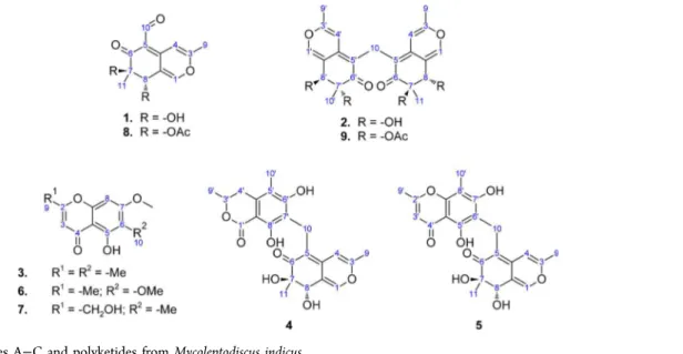
S Supporting InformationABSTRACT: Three new azaphilones with an unusual methylene bridge, named mycoleptones A, B, and C (2, 4, and5), were isolated from cultures ofMycoleptodiscus indicus, a fungus associated with the South American medicinal plant Borreria verticillata. Additionally, four known polyketides, austdiol (1), eugenitin (3), 6-methoxyeugenin (6), and 9-hydroxyeugenin (7), were also isolated. The structural characterization of compounds was carried out by nuclear
magnetic resonance spectroscopy, high-resolution mass spectrometry, electronic circular dichroism spectroscopy, time-dependent density functional theory calculations, and X-ray crystallography. Compounds1−9were weakly active when tested in antileishmanial and cytotoxicity assays.
E
ndophytic microorganisms live inside the tissues of host plants without apparently harming them and are a promising source of bioactive compounds.1−4Species of Mycoleptodiscus, such as M. indicus, M. terrestris, and M. sphericus, are commonly isolated as endophytes; they are probably latently phytopathogenic and may become phytopa-thogenic when the host plant is subjected to stress.5M. indicus, a tropical to subtropical species, occurs in leaves of different host plants, mainly monocotyledons. M. indicus is associated with large spreading lesions on leaves of Zamia spp., an American cycad, and other monocotyledonous plants.6 M. indicus has occasionally been reported to infect humans and canines, causing septic arthritis and skin infections.7
In this paper, the isolation, structural characterization, and biological activity of secondary metabolites from cultures ofM. indicus, an endophytic fungus isolated from the leaves from Borreria verticillata (Rubiaceae), are reported. Three new azaphilones, named mycoleptones A, B, and C (2, 4, and 5), were isolated and fully characterized; in addition, four known polyketides, namely, austdiol (1), eugenitin (3), 6-methox-yeugenin (6), and 9-hydroxyeugenin (7), were isolated and
identified, and the acetylated derivatives of austdiol (8) and mycoleptone A (9) were synthesized (Figure 1).
■
RESULTS AND DISCUSSIONSeven secondary metabolites ofM. indicuswere isolated, four of which were identified as austdiol, eugenitin, 6-methoxyeugenin, and 9-hydroxyeugenin by comparison of experimental spectro-scopic data with literature values.8−13
Austdiol ((7R,8S )-7,8-dihydroxy-3,7-dimethyl-6-oxo-7,8-dihydro-6H -isochromene-5-carbaldehyde) is the main toxic component of a mixture of compounds produced in moldy maize meal byAspergillus ustus and belongs to a class of compounds known generically as azaphilones.14,15 Azaphilones are yellow or orange pigments produced by fungi: their pyran oxygen atoms are easily exchanged to nitrogen atoms with ammonia, and the name is derived from the ready reaction of these metabolites with ammonia to yield vinylogous γ-pyridones.16 The skeleton of austdiol is derived from a single pentaketide chain, composed of
Received: August 22, 2013
Published: January 3, 2014
Article
pubs.acs.org/jnp
head-to-tail acetate units, and possesses two C1 units introduced from S-adenosylmethionine.17 A wide range of interesting biological activities of azaphilones, such as antimicrobial, antifungal, antiviral, antioxidant, cytotoxic, nematicidal, and anti-inflammatory, have been reported in the literature;14,17 their nonselective biological activities may be related to the production of vinylogousγ-pyridones.18
Three of the isolated metabolites are previously unreported azaphilones and were named mycoleptones A, B, and C (2,4, and5). Mycoleptone A (2) was isolated as a yellow powder, and the complete structural characterization was carried out by nuclear magnetic resonance (NMR) spectroscopy, high-resolution mass spectrometry (HRMS), electronic circular dichroism (ECD) spectroscopy, time-dependent density func-tional theory (TD-DFT) calculations, and single-crystal X-ray diffraction studies (Figure 2). The UV spectrum of2displays maximum absorptions at 254 nm (logε2.90) and 361 nm (log ε 2.83), revealing the presence of an extended conjugated system, characteristic of azaphilones. Compound 2 displays similar 1H and 13C NMR spectra to austdiol,8,9 with the
replacement of the aldehyde signals (δH9.99 andδC189.3) by methylene signals (δH 3.26 andδC18.5) at position C-5. The 1H NMR spectrum exhibits two signals atδ
H7.30 and 6.25 due to hydrogen atoms on the pyran ring, two hydroxyl hydrogens atδH 5.66 (OH, d, J= 4.6 Hz) and 4.93 (OH, s), a methine signal atδH4.22 (1H, d,J= 4.6 Hz), a methylene signal atδH 3.26, and two methyl signals atδH2.07 and 0.91. The13C NMR spectrum displays signals for 12 carbons atoms, 11 of which refer to two atoms each: two signals due to methyl carbon atoms (δC19.2 and 18.8),five signals from the skeleton of the pyran rings (δC157.8; 143.5; 141.0; 120.7 and 104.6), and four signals from the skeleton of the cyclohexenone, with one signal due to ketone carbonyl groups (δC197.8; IRνmax1667 cm−1), one due to quaternary unsaturated carbon atoms (δC 112.7), and two due to oxygenated carbon atoms (δC75.6 and 71.0). The remaining signal refers to a methylene group (δC 18.5). The heteronuclear multiple-bond correlation (HMBC) spec-trum shows key correlations of CH2-10 to 4a, 5, 6, C-4a′, C-5′, and C-6′, revealing the attachment of two azaphilone units through a methylene bridge at position C-5 (Table 1). Furthermore, the presence of four hydroxyl groups was confirmed by acetylation with pyridine−Ac2O to afford the tetraacetylated derivative (9). NMR spectroscopy identifies the structure of2as being formed by two austdiol units linked by a methylene group, generating a compound with the molecular formula C23H24O8as determined by HRMS ([M + H]+atm/z 429.1543). The chemical structure explains the simplicity of the 1H and 13C NMR spectra, in which the signal intensities are
doubled, except for the methylene group.
Compound 2 is a chiral molecule having four asymmetric carbons and a formal binary symmetry axis. An accurate analysis of the eight enantiomeric pairs of2reveals that two pairs are formed by nonoptically active meso structures and two more pairs are repeated twice; as a result, four pairs of enantiomers and two meso structures are possible. Since the reliable assessment of the absolute configuration of a compound is achieved when the theoretical chiroptical properties19of all the possible optically active diastereomers are determined,20 quantum-mechanical (QM) calculations were performed on four diastereomers of 2 (Figure 3): (7R,8S,7′R,8′S)-2 (2A), (7R,8S,7′R,8′R)-2 (2B), (7R,8S,7′S,8′S)-2 (2C), and (7S,8S,7′S,8′S)-2 (2D). QM calculations were carried out
Figure 1.Mycoleptones A−C and polyketides fromMycoleptodiscus indicus.
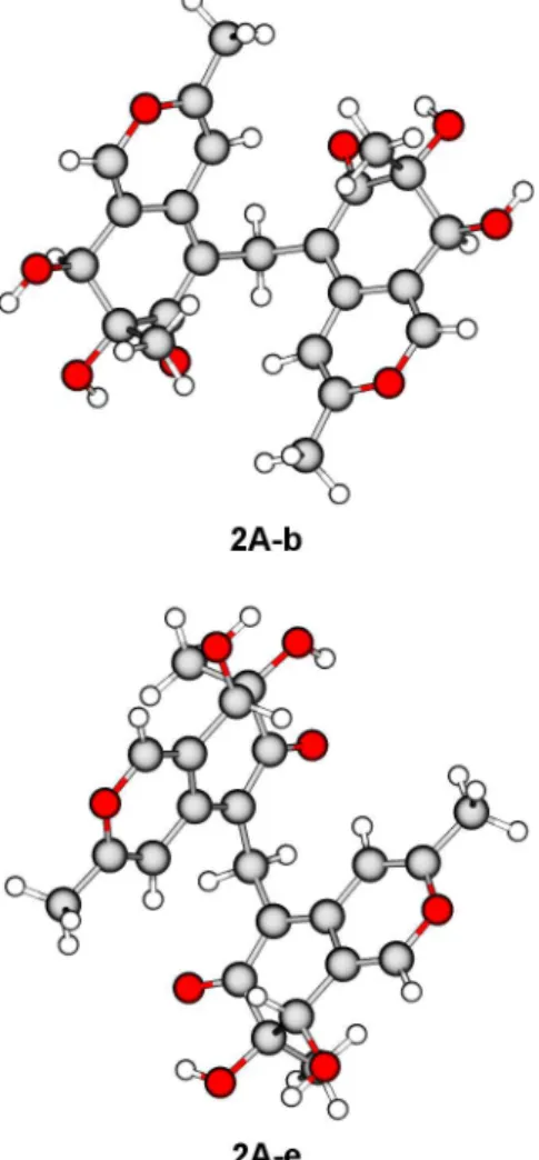
using density functional theory (DFT) and its time-dependent extension (TD-DFT); detailed results are reported in the Supporting Information. DFT geometry optimization (Table 2) yielded two equilibrium conformers having ΔEQM ≤ 2 kcal mol−1 for diastereomers 2A, 2B, and 2C, while a single equilibrium conformer within the same threshold energy was found for diastereomer2D. The most populated conformers of 2A(Figure 4) display similar geometric features: the azaphilone units are oriented in an“open folder”conformation, the pyran oxygen atoms are pointing toward opposite directions, and all the hydroxy groups are equatorial and form intramolecular hydrogen bonds with each other and with the ketone carbonyl groups. The main difference lies in the direction of the axial methyl groups on C-7 and C-7′, which point outside the molecular cavity for2A-eand inside for2A-b.
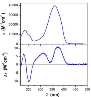
The experimental ECD spectrum of2 in methanol (Figure 5) shows four bands. The broad positive band centered at 372 nm has a strong contribution from the n→π*transitions of the ketone carbonyls, which are affected by the chiral environment around these groups. The positive band at 311 nm with a shoulder around 285 nm, the sharp negative peak at 252 nm, and the positive peak at 228 nm are due toπ→π* transitions of the conjugated system, which are affected by the chirality of the rings and by the mutual orientation of the azaphilone units. The theoretical ECD spectra of the diastereomers of 2 (Figure 6) displayed different patterns, reflecting their different stereochemistry. The comparison
between the calculated spectrum of 2Aand the experimental spectrum of2(Figure 7) shows that TD-DFT calculations are able to reproduce the experimental pattern of the transitions in the high-energy region, considering that transition energies are usually underestimated by PBE0.21The transitions in the low-energy region are not reproduced, suggesting that QM calculations are unable to describe the chiral environment of the ketone moieties, in particular hydrogen-bonding inter-actions with hydroxy groups and solvent molecules, with adequate accuracy. On the other hand, the theoretical ECD spectra of the remaining diastereomers (reported in the Supporting Information) are unable to reproduce the experimental ECD pattern in the high-energy region: on this basis, a (7R,8S,7′R,8′S) absolute configuration can be predicted for2.
Compound2crystallizes with two independent molecules in the asymmetric unit (Figure 2); the X-ray geometric parameters selected for comparison with the precursor austdiol are listed in Table 3. The C-5−C-10 distance was elongated upon change of hybridization to sp3for atom C-10, and the C-5−C-4a distance was shortened, increasing the double-bond character. The pyran rings are almost planar with an rmsd from the mean plane of 0.03 Å, while the cyclohexenone rings adopt a twisted envelope conformation with atoms C-7 and C-7′asflap atoms. The X-ray structure of2shows that the azaphilone units have the same absolute configuration as austdiol,22 retaining the stereochemistry of the chiral centers as (7R,8S); moreover, Table 1. NMR Spectroscopic Data (400 MHz) for Mycoleptones A−C
mycoleptone A (2)a mycoleptone B (
4)b mycoleptone C (
5)a
position δCmult. δH(Jin Hz) HMBCc δCmult. δH(Jin Hz) HMBCc δCmult. δH(Jin Hz) HMBCc
1 143.5, CH 7.30, s 3, 4a, 8, 8a 144.8, CH 7.43, d (1.8) 3, 4a, 8, 8a 145.7, CH 7.52, s 3, 4a, 8, 8a 2
3 157.8, C 160.1, C 160.5, C
4 104.6, CH 6.25, s 3, 4a, 5, 8a, 9 104.4, CH 6.63, s 3, 5, 8a, 9 105.5, CH 7.03, s 3, 8a
4a 141.0, C 143.7, C 144.3, C
5 112.7, C 112.8, C 112.1, C
6 197.8, C 199.3, C 200.5, C
7 75.6, C 4.93, s (OH) 76.1, C 75.4, C
8 71.0, CH 4.22, d (4.6); 5.66, d
(OH) (4.6) 1, 6, 7, 8a, 10 71.5, CH 4.42, d (1.8) 7, 8a, 11 70.7, CH 4.25, s 1, 6, 7, 8a
8a 120.7, C 121.1, C 121.3, C
9 19.2, CH3 2.07, s 3, 4 18.3, CH3 2.22, s 3, 4 19.5, CH3 2.25, s 3, 4
10 18.5, CH2 3.26, s 4a, 5, 6, 4a′,
5′, 6′ 20.7, CH2 3.53, 3.60, d(15.0) 4a, 5, 6, 6
′,
7′, 8′ 18.6, CH2 3.74, 3.46, d(15.0) 4a, 5, 6, 5
′, 6′, 7′
11 18.8, CH3 0.91, s 6, 7, 8 17.5, CH3 1.10, s 6, 7 19.0, CH3 1.00, s 6, 7, 8
1′ 143.5, CH 7.30, s 3′, 4a′, 8′, 8a′ 171.2, C
2′ 167.2, C
3′ 157.8, C 75.0, CH 4.55, m 107.9, CH 6.13, s 2′, 4a′, 9′
4′ 104.6, CH 6.25, s 3′, 4a′, 5′,
8a′, 9′ 32.2, CH2 3.17, dd (16.0,3.0) 5
′, 8a′
2.60, dd (16.0,
11.0) 3
′, 5′, 8a′, 9′ 181.8, C
4a′ 141.0, C 110.1, C 103.1, C
5′ 112.7, C 136.8, C 156.6, C 13.06, s (OH) 4a′, 5′, 6′
6′ 197.8, C 160.2, C 107.8, C
7′ 75.6, C 4.93, s (OH) 116.5, C 161.1, C
8′ 71.0, CH 4.22, d (4.6); 5.66, d (OH) (4.6) 1
′, 6′, 7′, 8a′,
10′ 160.9, C 105.0, C
8a′ 120.7, C 100.1, C 153.5, C
9′ 19.2, CH3 2.07, s 3′, 4′ 19.6, CH3 1.44, d (6.2) 3′, 4′ 20.2, CH3 2.37, s 2′, 3′
10′ 18.8, CH3 0.91, s 6′, 7′, 8′ 6.9, CH3 2.06, s 4a′, 5′, 6′ 8.5, CH3 2.00, s 7′, 8′, 8a′
aIn DMSO-d
6.bIn CD3OD.cHMBC correlations are from hydrogen(s) stated to the indicated carbon.
DFT conformer 2A-b shows a close resemblance to the crystallographic structure of2. On the basis of the foregoing evidence, the structure of mycoleptone A (2) was therefore elucidated as (7R,8S,7′R,8′S)-5-[(7′,8′-dihydroxy-3′,7′ -dimeth-yl-6′-oxo-7′,8′-dihydro-6′H-isochromen-5′ -yl)methyl]-7,8-dihy-droxy-3,7-dimethyl-7,8-dihydro-6H-isochromen-6-one.
The molecular formula of mycoleptone B (4) was determined as C23H24O8by a combination of HRMS,1H and 13C NMR, homonuclear correlation (COSY), heteronuclear
multiple-quantum correlation (HMQC), and HMBC spectros-copies. The 1H NMR spectrum contains five doublets, four singlets, two double doublets, and one multiplet; some similarities with the 1H NMR spectrum of 2 are observed, with the presence of two hydrogen atoms on the pyran ring at δH7.43 (1H, d,J= 1.8 Hz) and 6.63 (1H, s), an oxygenated sp3
methine group atδH4.42 (1H, d,J= 1.8 Hz), a CH2group at δH3.60 (1H, d,Jgem= 15.0 Hz) and 3.53 (1H, d,Jgem= 15.0 Hz) due to the methylene bridge at position C-10 connecting the azaphilone unit to a dihydroisocoumarin moiety, and two methyl signals atδH2.22 and 1.10. For the dihydroisocoumarin moiety, the 1H NMR spectrum displays an oxygen-bearing methine hydrogen atδH4.55 (1H, m), a methylene group atδH 3.17 (1H, dd,J= 16.0, 3.0 Hz) and 2.60 (1H, dd,J= 16.0, 11.0 Hz), and two methyl groups atδH2.06 (3H, s) and 1.44 (3H, d, J= 6.2 Hz). The13C NMR spectrum of4displays signals for 23 carbon atoms: one signal due to the methylene bridge (δC 20.7), 11 signals belonging to the azaphilone moiety as in2,
Figure 3.Diastereomers of mycoleptone A.
Table 2. Geometric Parameters, Energy Values, and Fractional Equilibrium Populations for the Most Populated Equilibrium Conformers of the Diastereomers of Mycoleptone A, As Obtained after DFT Geometry Optimization at the B97D/6-311+ +G(2d,2p)/IEFPCM(MeOH) Level
conformer α1(deg)a α2(deg)b β(deg)c d(Å)d EQM(Hartree) ΔEQM(kcal mol−1) χQMe
2A 2A-e −57.348 −57.348 −95.326 7.453 −1492.147 316 09 0.000 0.5934 2A-b 66.788 66.788 101.136 7.806 −1492.146 929 07 0.243 0.3939 2B 2B-f −56.583 −55.703 −92.428 7.308 −1492.149 394 96 0.000 0.6540 2B-b 68.871 69.671 105.736 8.045 −1492.148 758 18 0.400 0.3332
2C 2C-e 62.947 57.866 94.503 7.444 −1492.149 386 42 0.000 0.5886
2C-f −60.999 −66.239 −101.552 7.806 −1492.149 010 68 0.236 0.3953 2D 2D-b −68.275 −68.275 −104.848 8.007 −1492.150 185 72 0.000 0.9709
aC-10
−C-5−C-12−C-5′ dihedral angle. bC-10′
−C-5′−C-12−C-5 dihedral angle. cO-2
−C-5−C-5′−O-2′ dihedral angle. dO-2
−O-2′ distance.
e
Calculated using Boltzmann statistics at 298.15 K and 1 atm.
and 11 signals belonging to the dihydroisocoumarin moiety. The13C NMR data reveal six resonances for sp2carbon atoms being part of a benzene ring, two of them characterized by a downfield shift due to the hydroxy substituents (C-6′ at δC 160.2; C-8′atδC160.9) and the presence of a carbonyl group atδC171.2 (C-1′; IRνmax 1652 cm−1). The structural features of the lactone ring were determined from the1H−1H COSY spectrum based on the couplings between H2-4′and H-3′, as well as between H-3′ and H3-9′. The HMBC correlations of CH2-4′toδC136.8 (C-5′),δC100.1 (C-8a′),δC75.0 (C-3′), andδC19.6 (C-9′) proved the connection of the two units. In addition, the HMBC spectrum displays relevant correlations from CH2-10 (δH3.60, 3.53) toδC199.3 (C-6), 143.7 (C-4a), and 112.8 (C-5) from the azaphilone moiety and toδC160.9 (C-8′), 160.2 (C-6′), and 116.5 (C-7′) from the dihydroiso-coumarin moiety, characterizing the connection between the two units by a methylene bridge to afford the compound (7R,8S)-5-[(6′,8′-dihydroxy-3′,5′-dimethyl-1′
-oxoisochroman-7′-yl)methyl]-7,8-dihydroxy-3,7-dimethyl-7,8-dihydro-6H -iso-chromen-6-one.
The molecular formula of mycoleptone C (5) was deduced as C23H22O8by HRMS analysis in combination with 1H and 13C NMR spectroscopies. The1H NMR spectrum displays nine
singlets and two doublets; as in compounds 2 and 4, the methylene group atδH3.74 (1H, d,J= 15.0 Hz) and 3.46 (1H, d, J = 15.0 Hz) connects two moieties, the azaphilone and chromone units. The azaphilone moiety showsfive uncoupled hydrogen atoms atδH7.52 (H-1), 7.03 (H-4), 4.25 (H-8), 2.25 (H-9), and 1.00 (H-10); the chromone moiety displays four uncoupled hydrogen atoms at δH 13.06 (H-5′), 6.13 (H-3′), 2.37 (H-9′), and 2.00 (H-10′), the sharp singlet atδH 13.06 being interpreted as a hydrogen-bonded phenol hydroxy group. The 13C NMR spectrum confirms 23 carbon signals, namely, four methyl, one methylene, four methine, and 14 quaternary carbon signals. The chromone moiety displays one ketone carbonyl group (δC 181.8; IR νmax 1690 cm−1) and four quaternary aromatic (δC153.5, 107.8, 105.0, and 103.1), two oxygenated aromatic (δC161.1 and 156.6), two unsaturated (δC 167.2 and 107.9), and two methyl (δC 20.2 and 8.5) carbon atoms. The structure of the chromone portion was determined on consideration of the molecular formula, double-bond equivalents, and the downfield shifts of the carbon signals at C-2′and C-8a′(δC167.2 and 153.5). The HMBC spectrum of 5revealed relevant correlations of CH2-10 (δH3.74, 3.46) toδC 200.5 (C-6), 144.3 (C-4a), and 112.1 (C-5) from the azaphilone moiety and toδC 161.1 (C-7′), 156.6 (C-5′), and 107.8 (C-6′) from the chromone moiety, revealing the
Figure 5.Experimental UV and ECD spectra of mycoleptone A (19
μM in MeOH, 1 cm path length).
Figure 6. Theoretical ECD spectra of the diastereomers of mycoleptone A at the PBE0/6-311++G(2d,2p)/IEFPCM(MeOH)// B97D/6-311++G(2d,2p)/IEFPCM(MeOH) level (χQM-based con-formational averaging, Δσ = 0.3 eV). Solid: 2A. Dash-dotted: 2B. Dotted:2C. Dashed:2D.
Figure 7. Theoretical ECD spectrum of 2A and comparison with experimental data. Solid: experimental ECD spectrum of2(19μM in MeOH, 1 cm path length). Dotted: theoretical ECD spectrum of2Aat the PBE0/6-311++G(2d,2p)/IEFPCM(MeOH)//B97D/6-311++G-(2d,2p)/IEFPCM(MeOH) level (χQM-based conformational averag-ing,Δσ= 0.3 eV).
Table 3. Selected Bond Distances and Angles for Mycoleptone A and Austdiol, As Determined by X-ray Crystallography
parameter mycoleptone A (2) austdiol (1) C-1−C-8a 1.337(8) Å 1.338(3) Å C-3−C-4a 1.341(8) Å 1.345(3) Å C-5−C-4a 1.373(7) Å 1.399(2) Å C-5−C-6 1.433(8) Å 1.432(3) Å C-5−C-10 1.533(7) Å 1.455(2) Å C-4a−C-5−C-6 118.6(5)° 124.3(2)°
connection of both skeletons and featuring 5 as (7R,8S )-5-[(5′,7′-dihydroxy-2′,8′-dimethyl-4′-oxo-4′H-chromen-6′ -yl)-methyl]-7,8-dihydroxy-3,7-dimethyl-7,8-dihydro-6H -isochro-men-6-one.
The presence of a methylene bridge is very uncommon among natural compounds, and only a few azaphilone dimers have been reported showing such a feature.15Dicitrinones A−
C23 were isolated from Penicillium citrinum, a volcano ash-derived fungus: the presence of a single sp3 carbon bridge, either a methine or methylene group, was proposed to derive from the carboxyl group of citrinin through decarboxylation, reduction, and Friedel−Crafts alkylation mediated by a polyketide biosynthetic pathway. The methylene bridge in aspergilone B,24an azaphilone dimer that was isolated from a marine-derived fungus of theAspergillusspecies, was hypothe-sized to derive from a formaldehyde biosynthetic pathway; the same hypothesis was proposed for the biosynthesis of xyloketal F,25a dimer isolated from a mangrove fungus of the Xylaria species.
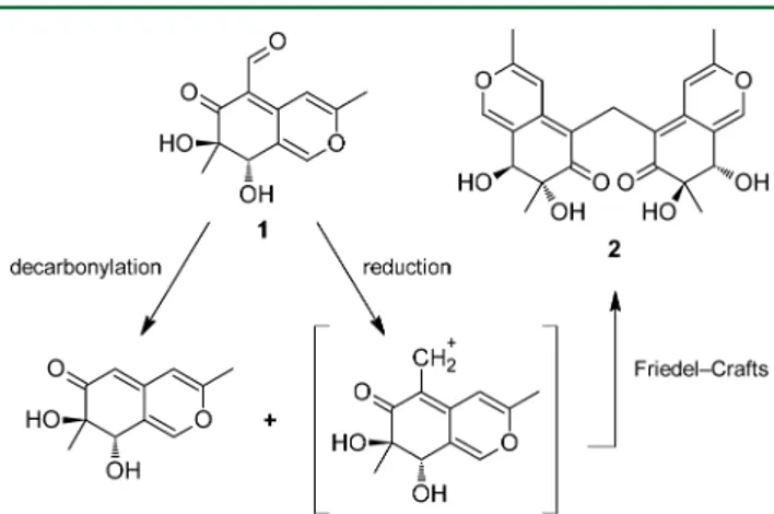
The condensation of two austdiol units to form mycoleptone A (2) is consistent with a possible biosynthetic pathway similar to the one proposed for dicitrinone C23 (Figure 8). One
austdiol unit may undergo decarbonylation, while the aldehyde group of the second austdiol unit may be reduced to a reactive carbocation species: the two units may then react by Friedel−
Crafts alkylation. Similar pathways may also explain the condensation of the dihydroisocoumarin and chromone units with an austdiol unit in mycoleptones B (4) and C (5), respectively. Due to the limited number of methylene-bridged azaphilones discovered to date, however, a general biosynthetic pathway cannot be postulated, and the biologically mediated mechanisms of such reactions are unknown.
The results of antileishmanial and cytotoxicity tests on the isolated compounds are shown in Table 4. Compounds 1−9 exhibit no significant activity againstLeishmania donovani and
Leishmania major compared to the standard antileishmanial drugs Geneticin (L. major), amphotericin B, and pentamidin (L. donovani). Mycoleptone B (4) was the most cytotoxic metabolite (PC3 cells: IC50= 7.1±3.8μM), but its activity was lower than that of doxorubicin, the reference compound for cytotoxicity assays.
■
EXPERIMENTAL SECTIONGeneral Experimental Procedures. Optical rotations were measured in CHCl3, MeOH, or DMSO using a Jasco DIP-370 digital polarimeter at room temperature; IR spectra were recorded with KBr discs using a Bruker Tensor 27 FTIR spectrometer. UV spectra were measured in CHCl3, MeOH, and DMSO on a Varian Cary 50 Bio UV−visible spectrophotometer. The HPLC system consisted of a Shimadzu SCL-10Avp multisolvent delivery system, a SPD-M10Avp photodiode array detector, an Intel Celeron computer for analytical system control, data collection, and processing, and a Shim-pack CLC-ODS(M) reversed-phase column (250×4.5 mm i.d.; 5μm particle size) protected by a Pelliguard LC-18 cartridge. The NMR spectra were acquired on a Bruker Avance DRX-400 spectrometer operating at 400 MHz for 1H NMR and 100 MHz for 13C NMR in CDCl
3, CD3OD, or DMSO-d6; multiplicity determinations (DEPT) and 2D NMR spectra (COSY, HMQC, and HMBC) were obtained using standard Bruker pulse programs. The chemical shift values (δ) are given in parts per million (ppm), and the coupling constants are in Hz. HRMS were obtained by direct injection using a Bruker Bioapex FTMS spectrometer with electrospray ionization (ESI). UV and ECD spectra of2in MeOH (sample concentration: 19μM) were recorded in the 500−215 nm spectral range on a Jasco J-810 spectropolarimeter, using a 1 cm path length cell at room temperature: measurements
Figure 8.Possible pathway for the biosynthesis of mycoleptone A.
Table 4. Biological Activities of Compounds 1−9
cytotoxicity (IC50,μM)
compound L. majorμM)(LD50, L. donovaniμM)(IC50, HCT-8 HL-60 MDA-MB435 PC3 SF-295 lymphocytehuman austdiol (1) 20.5 80.5 12.9±3.5 38.8±9.3 34.3±4.3 ND 28.0±7.2 >113 mycoleptone A (2) 28.5 >93.4 >58 >58 >58 10.0±6.5 >58 >58 eugenitin (3) 39.9 >181 >113 >113 >113 >113 >113 >113 mycoleptone B (4) 21.7 NDd >58 >58 >58 7.1±3.8 >58 >58
mycoleptone C (5) ND ND ND ND ND ND ND ND
6-methoxyeugenin (6) 34.5 >169 >105 >105 >105 20.5±11.9 >105 >105 9-hydroxyeugenin (7) ND ND >105 >105 >105 >105 >105 >105 austdiol diacetate (8) 19.8 59.3 13.9±2.6 22.1±2.6 26.9±0.3 ND 26.3±1.2 >78 mycoleptone A
tetraacetate (9) ND ND >42 >42 >42 33.3±29.0 >42 >42 Geneticin (G418)a 3.43
pentamidineb 0.0041
amphotericin Bb 0.140
doxorubicinc 0.10±0.01 0.05±0.01 1.51±0.20 0.41±0.05 0.70±0.20 0.70±0.20
aStandard antileishmanial drug against
L. major.bStandard antileishmanial drugs against
L. donovani.cReference compound for cytotoxicity assays.
d
were carried out at 0.2 nm intervals using a 1 nm spectral bandwidth, a 20 nm min−1scan rate, and a 4 s time constant.
Fungal Material.The fungus was isolated as an endophyte from the medicinal plantBorreria verticillata(L.) G. F. W. Meyer, belonging to the Rubiaceae family, and is native to South America. Five specimens ofB. verticillatawith a healthy appearance were collected from the campus of the Federal University of Pernambuco (UFPE) (34°56′57″W; 8°2′53″S) in January 2006, approximately 43 cm in height and bearing flowers. Then, the samples were taken to the
laboratory and processed in 24 h. A voucher specimen of the plant (No. 42.234/UFPE) was identified and deposited at the Herbarium
Professor Geraldo Mariz, UFP, Department of Botany, UFPE, Recife, Brazil. The isolate was identified as Mycoleptodiscus indicusbased on
sequence analysis of the ITS region of the rDNA (GenBank accession number GU220382.1).
Cultivation, Extraction, and Isolation.M. indicuswas grown on potato dextrose agar plates for 7 days at 30°C. Then, 10 plugs were transferred tofive Erlenmeyerflasks (500 mL), each containing 100
mL of potato dextrose broth prepared with distilled water. Flasks were shaken on a rotary shaker at 30°C and 120 rpm for 48 h. Next, 10 mL was transferred to each of 50flasks containing 90 g of solid medium
(rice-oat). These were grown for 30 days. On day 30, the mycelial mass was macerated with ethanol overnight andfiltered. Thefiltrate
was concentrated under vacuum to obtain a crude ethanol extract (22 g), which was partitioned with three times equal volumes of hexane and CH2Cl2, respectively, yielding hexane (6 g) and dichloromethane fractions (6.9 g). The CH2Cl2fraction was suspended in MeOH and centrifuged at 3000 rpm for 3 min, furnishing a precipitate fraction (3870 mg) and a soluble fraction (3030 mg). The precipitate fraction was subjected to chromatography over a silica gel column (30×3.5 cm i.d.) using a MeOH−CH2Cl2gradient to yield 67 fractions, which were combined on the basis of their TLC profiles into seven fractions:
A (19 mg), B (33 mg), C (2968 mg), D (100 mg), E (210 mg), F (285 mg), and G (109 mg). Fraction C (2968 mg) yielded austdiol (2830 mg), after crystallization in MeOH, and fraction E afforded
mycoleptone A (2, 200 mg) by precipitation. The soluble fraction (3030 mg) was subjected to gel filtration on a Sephadex LH-20
column (50×3 cm i.d.) using MeOH as the mobile phase, furnishing 10 fractions (S1−S10). Fraction S6 (280 mg) was washed with MeOH, yielding eugenitin (50 mg). The soluble portion was subjected to chromatography over a silica gel column (25×2 cm i.d.) using a MeOH−CH2Cl2 gradient. Forty-two fractions were collected and combined on the basis of their TLC profiles to yield nine fractions.
The fourth fraction (S6.4, 28 mg) was purified by preparative silica gel
TLC using hexane−ethyl acetate (70:30, v/v) as eluent to furnish mycoleptone B (4, 10 mg) and mycoleptone C (5, 2 mg). Fraction S5 (461 mg) was subjected to chromatography over a silica gel column (25×2 cm i.d.) eluted with a gradient of MeOH in CH2Cl2. Thirty fractions were collected and combined on the basis of their TLC profiles to yield 10 fractions. The fourth fraction (S5.4, 60 mg) was
purified by preparative silica gel TLC using hexane−ethyl acetate (70:30, v/v) as eluent to provide 6-methoxyeugenin (14 mg). Thefifth
fraction (S5.5, 4 mg) was subjected to reversed-phase HPLC using CH3CN−H2O (10:90 to 100:0, v/v) as mobile phase at aflow rate of 1.0 mL/min over 23 min, yielding 9-hydroxyeugenin (2 mg).
Acetylation of Austdiol and Mycoleptone A (2). A solution containing austdiol (80 mg), pyridine (2 mL), and Ac2O (3 mL) was stirred at room temperature for 8 h. H2O (6 mL) was added into the reaction mixture, which was subsequently extracted with CH2Cl2. The organic layer was evaporated to give a yellow, amorphous solid (140 mg), which was purified by preparative TLC eluted with a mixture of
CH2Cl2−MeOH (98:2, v/v) to afford the diacetylated derivative (8, 80.7 mg). Acetylation of mycoleptone A (2, 25 mg) was performed in a similar manner, yielding the tetraacetylated derivative (9, 20 mg).
Quantum-Mechanical Calculations.Conformational analysis on the selected diastereomers of2was performed in two steps. In thefirst
step, a preliminary conformer distribution was determined by molecular mechanics (MM) calculations at the MMFF94s26 level using the Spartan’02 software.27 In the second step, DFT28,29 geometry optimizations were performed on the MM conformers
having relative energies (ΔEMM) within a threshold value of 3 kcal mol−1. DFT calculations were carried out at the B97D/6-311+ +G(2d,2p) level30−33using the Gaussian 09 software package.34The
Boltzmann distribution of conformers at 298.15 K and 1 atm was then calculated from the relative electronic energies (ΔEQM). TD-DFT35 calculations were carried out at the PBE0/6-311++G(2d,2p) level31−33,36−38 using the Gaussian 09 software package.34 All calculations were performed using the IEFPCM continuum solvation model39,40 for MeOH. Rotational strengths (Rj) and excitation
wavelengths (λj) were calculated for the lowest 50 excited states on
the optimized geometries havingΔEQM ≤2 kcal mol−1. Theoretical ECD spectra were then obtained by approximation of Rj values to
Gaussian functions with a half-bandwidth atΔεmax/e (Δσ) of 0.3 eV, summation over all excited states, and conformational averaging, according to the Boltzmann distribution of conformers (χQM).41
Crystallographic Structure. Crystals of 2 suitable for X-ray analysis were obtained by crystallization from a methanol solution. A colorless prismatic crystal of approximate dimensions 0.3×0.2×0.2 mm3was used for the experiment. X-ray data were measured on an Enraf-Nonius CAD-4 diffractometer employing
graphite-monochro-mated Cu Kαradiation (λ= 1.5418 Å) at 298 K and operating in the
φ−ωscan mode. Crystal data: C23H24O8,M =428.42, triclinic, space groupP1,a =10.473(6) Å,b =11.223(3) Å,c =11.429(2) Å,α= 60.65(2)°,β= 89.83(3)°,γ= 67.75(4)°,V =1055.5(9) Å3,Z =2,D
c= 1.348 g/cm3,F(000) = 452, andμ(Cu Kα) = 0.86 mm−1. All tested crystal samples showed gemination, and the sample used for data collection was visually homogeneous, but it showed pseudomerohedral twinning, which caused partial superposition of structure factors leading to high residuals during the structure refinements. A total of
8384 reflections were collected (4273 unique plus 96.2% of Friedel
mates) in theθrange 4.56°to 73.92°and index rangeshfrom 15 to −15,kfrom−24 to 25, andlfrom−15 to 15. Cell refinement and data
reduction were performed with the XCAD4 software suite. The structure was solved and refined using the WingX with SHELX97
suite.42,43 Structure refinement was performed on F2 by full-matrix
least-squares calculations. Non-hydrogen atoms were refined
aniso-tropically, and all hydrogen atoms were placed in idealized coordinates and refined as riding atoms with isotropic parameters relative to their
parent atoms. Thefinal refinement residuals wereR1= 0.091 (wR2=
0.32) for 6964 observed reflections with I > 2σ(I), 575 variable
parameters, and three restraints, beingR1= 0.11 (wR2= 0.32) for all unique reflections with GoF = 1.41. The absolute structure Flack
parameter was 0.1(3), indicating that the chiral sense was correctly chosen, although this number was not reliable, given the large standard deviation, in the absence of strong anomalous scatterers.
Biological Assays.The microplate Alamar-Blue assay was used to determine growth inhibition againstL. donovani.44Antileishmanial (L.
major) and cytotoxicity assays against human colon cancer (HCT-8)
cells, human leukemia cancer (HL-60) cells, human melanoma cancer (MDA-MB435) cells, human prostate cancer (PC3) cells, human glioblastoma cancer (SF-95) cells, and human lymphocyte cells were assessed employing a colorimetric method.45
Austdiol (1):yellow crystal; mp 255−257°C; [α]25
D+223 (c 1.0, DMSO); UV (MeOH)λmax(logε) 212 (2.64), 255 (2.57), 333 (2.62) nm; IR (KBr) νmax 3470, 3373, 3100, 1679, 1604, 1471 cm−1;1H NMR and13C NMR reported in the Supporting Information; HRMS
m/z237.0759 [M + H]+(calcd for C
12H12O5+ H+, 237.0757).
Mycoleptone A (2):yellow crystal; mp 307−308°C; [α]25
D+351 (c 1.0, DMSO); UV (MeOH)λmax(logε) 254 (2.90), 361 (2.83) nm; IR (KBr)νmax3360, 1667, 1581, 1526, 1229, 1197, 1090, 878 cm−1;1H and13C NMR reported in Table 1; HRMSm/z429.1543 [M + H]+ (calcd for C23H24O8+ H+, 429.1544).
Eugenitin (3):White, solid powder; mp 153−156°C; [α]25
D0 (c 0.3, CHCl3); UV (MeOH)λmax(logε) 267 (2.53), 307 (2.55), 338 (2.54) nm; IR (KBr) νmax 1652, 1586, 1435, 1338, 1179 cm−1;1H NMR and13C NMR reported in the Supporting Information; HRMS
m/z221.0810 [M + H]+(calcd for C
12H12O4+ H+, 221.0808).
Mycoleptone B (4):colorless, amorphous solid; [α]25
D+71 (c0.2, MeOH); UV (MeOH)λmax(logε) 237 (2.83), 299 (2.83), 317 (2.90) nm; IR (KBr)νmax3391, 2925, 1652, 1615, 1289, 1127 cm−1;1H and
13C NMR reported in Table 1; HRMS m/z451.1363 [M + Na]+
(calcd for C23H24O8+ Na+, 451.1363).
Mycoleptone C (5): brown, amorphous solid; [α]25
D +95 (c 0.2, DMSO); UV (MeOH)λmax(logε) 224 (3.17), 297 (3.15), 321 (3.17) nm; IR (KBr)νmax2920, 2360, 1690, 1573, 1478 cm−1;1H and13C NMR reported in Table 1; HRMSm/z427.1387 [M + H]+(calcd for C23H22O8+ H+, 427.1387).
6-Methoxyeugenin (6): white, solid powder; mp 155−158 °C;
[α]25
D0 (c0.3, CHCl3); UV (MeOH)λmax(logε) 292 (2.85), 307 (2.89), 334 (2.88) nm; IR (KBr)νmax3404, 2923, 1657, 1617, 1570, 1492, 1169 cm−1;1H NMR and13C NMR reported in the Supporting Information; HRMSm/z259.0577 [M + Na]+(calcd for C
12H12O5+ Na+, 259.0577).
9-Hydroxyeugenin (7):white, amorphous solid; [α]25
D 0 (c 0.3, CHCl3); UV (MeOH)λmax(logε) 279 (2.87), 319 (2.91), 335 (2.92) nm; IR (KBr)νmax3217, 2360, 1662, 1573, 1497, 1330, 1138 cm−1;1H NMR and13C NMR reported in the Supporting Information; HRMS
m/z237.0711 [M + H]+(calcd for C
12H12O5+ H+, 237.0757).
Austdiol diacetate (8):yellow crystal; mp 255−257°C; [α]25
D+39 (c1.0, CDCl3); UV (CHCl3)λmax(logε) 262 (2.99), 281 (2.98), 362 (3.01) nm; IR (KBr) νmax 2946, 1746, 1628, 1500, 1241 cm−1; 1H NMR and13C NMR reported in the Supporting Information; HRMS
m/z321.0970 [M + H]+(calcd for C
16H16O7+ H+, 321.0969).
Mycoleptone A tetraacetate (9):white crystal; mp 260−263°C;
[α]25
D+159 (c1.0, CDCl3); UV (MeOH)λmax(logε) 285 (3.04), 328 (2.98), 343 (3.03) nm; IR (KBr)νmax3470, 3373, 3100, 1679, 1604 cm−1;1H NMR (CDCl
3, 400 MHz)δ6.95 (1H, d,H-1), 6.71 (1H, d, H-8), 6.27 (1H, s, H-4), 3.48 (2H, s, H-10), 2.20 (3H, s, 7-COOCH3), 2.18 (3H, s, H-9), 2.04 (3H, s, 8-COOCH3), 1.32 (3H, s, H-11);13C NMR (CDCl3, 100 MHz) δ190.5 (C, CO), 169.7 (C, 7-CO2Me), 169.5 (C, 8-CO2Me), 160.0 (C, 3), 142.6 (CH, 1), 142.3 (C, C-4a), 117.3 (C, C-8a), 114.6 (C, C-5), 106.1 (CH, C-4), 82.5 (C, C-7), 69.1 (CH, C-8), 19.5 (CH3, C-9), 20.8 (CH3, 7-CO2CH3), 21.4 (CH3, 8-CO2CH3), 17.2 (CH2, C-10); HRMS m/z 597.1966 (calcd for C31H32O12+ H+, 597.1967).
■
ASSOCIATED CONTENT*
S Supporting InformationNMR and HRMS spectra; results for the computational study on mycoleptone A. This material is available free of charge via the Internet at http://pubs.acs.org. Crystallographic data, excluding structure factors, have been deposited with the Cambridge Crystallographic Data Centre as supplementary publication number CCDC 938460. Copies of the data can be obtained, free of charge, on application to CCDC, 12 Union Road, Cambridge CB2 1EZ, UK [fax: C44 1223 336033 or e-mail: [email protected]].
■
AUTHOR INFORMATIONCorresponding Author
*Tel: +5516 3602 4162. Fax: +5516 3633 1092. E-mail: [email protected].
Notes
The authors declare no competingfinancial interest.
■
ACKNOWLEDGMENTSThe authors thank the São Paulo Research Foundation (FAPESP) grant nos. 04/07935-6 and 07/58650-0, CAPES, CNPq, the PRIN 2008 project 2008LYSEBR_005 (MIUR, Italy), and the University of Bologna forfinancial support.
■
REFERENCES(1) Petrini, O. InMicrobial Ecology of Leaves; Andrews, J. H., Hirano, S. S., Eds.; Springer-Verlag: New York, NY, 1991; pp 179−197.
(2) Gunatilaka, A. A. L.J. Nat. Prod.2006,69, 509−526.
(3) Borges, W. S.; Borges, K. B.; Bonato, P. S.; Said, S.; Pupo, M. T.
Curr. Org. Chem.2009,13, 1137−1163.
(4) Bunyapaiboonsri, T.; Yoiprommarat, S.; Srikitikulchai, P.; Srichomthong, K.; Lumyong, S.J. Nat. Prod.2010,73, 55−59.
(5) Shearer, J. F.J. Aquat. Plant Manage.2002,40, 76−78. (6) Padhye, A. A.; Davis, M. S.; Reddick, A.; Bell, M. F.; Gearhart, E. D.; Von Moll, L.J. Clin. Microbiol.1995,33, 2796−2797.
(7) Garrison, A. P.; Procop, G. W.; Vincek, V.; Moon, J.; Morris, M. I.; Doblecki-Lewis, S.; Cleary, T. J.; Brust, D.; Rosa-Cunha, I.Transpl.
Infect. Dis.2008,10, 218−220.
(8) Steyn, P. S.Tetrahedron1973,29, 107−120.
(9) Vleggaar, R.; Steyn, P. S.; Nagel, D. W. J. Chem. Soc., Perkins
Trans. 11974, 45−49.
(10) Feng, Y.; Blunt, J. W.; Cole, A. L. J.; Munro, M. H. G.J. Nat.
Prod.2002,65, 1681−1682.
(11) Fox, C. H.; Huneck, S.Phytochemistry1969,8, 1301−1304. (12) Joshi, B. S.; Ravindranath, K. R.J. Chem. Soc., Perkin Trans. 1 1977, 433−436.
(13) Li, G. Y.; Li, B. G.; Yang, T.; Liu, G. Y.; Zhang, G. L.Helv. Chim. Acta2008,91, 124−129.
(14) Osmanova, N.; Schultze, W.; Ayoub, N.Phytochem. Rev.2010,9, 315−342.
(15) Gao, J. M.; Yang, S. X; Qin, J. C.Chem. Rev.2013,113, 4755− 4811.
(16) Arai, N.; Shiomi, K.; Tomoda, H.; Tabata, N.; Yang, D. J.; Masuma, R.; Kawakubo, T.; Omura, S.J. Antibiot.1995,48, 696−702. (17) Colombo, L.; Gennari, C.; Severini Ricca, G.; Scolastico, C.; Aragozzini, F.J. Chem. Soc., Chem. Commun.1981, 575−576.
(18) Park, J. H.; Choi, G. J.; Jang, K. S.; Lim, H. K.; Kim, H. T.; Cho, K. Y.; Kim, J. C.FEMS Microbiol. Lett.2005,252, 309−313.
(19) Autschbach, J.Chirality2009,21, E116−E152. (20) Polavarapu, P. L.Chirality2012,24, 909−920.
(21) Goerigk, L.; Kruse, H.; Grimme, S. InComprehensive Chiroptical
Spectroscopy; Berova, N.; Polavarapu, P. L.; Nakanishi, K.; Woody, R.
W., Eds.; Wiley & Sons: Hoboken, NJ, 2012; Vol.1, pp 643−673. (22) Lo Presti, L.; Soave, R.; Destro, R.Acta Crystallogr.2003,C59, o199−o201.
(23) Du, L.; Li, D.; Zhang, G.; Zhu, T.; Ai, J.; Gu, Q.Tetrahedron 2010,66, 9286−9290.
(24) Shao, C. L.; Wang, C. Y.; Wei, M. Y.; Gu, Y. C.; She, Z. G.; Qian, P. Y; Lin, Y. C.Bioorg. Med. Chem. Lett.2011,21, 690−693.
(25) Wu, X. Y.; Liu, X. H.; Lin, Y. C.; Luo, J. H.; She, Z. G.; Houjin, L.; Chan, W. L.; Antus, S.; Kurtan, T.; Elsasser, B.; Krohn, K.̈ Eur. J.
Org. Chem.2005,2005, 4061−4064.
(26) Halgren, T. A.J. Comput. Chem.1999,20, 720−729.
(27)Spartan’02; Wavefunction, Inc.: Irvine, CA, 2002.
(28) Hohenberg, P.; Kohn, W.Phys. Rev.1964,136, B864−B871. (29) Kohn, W.; Sham, L. J.Phys. Rev.1965,140, A1133−A1138. (30) Grimme, S.J. Comput. Chem.2006,27, 1787−1799.
(31) Krishnan, R.; Binkley, J. S.; Seeger, R.; Pople, J. A.J. Chem. Phys. 1980,72, 650−654.
(32) Clark, T.; Chandrasekhar, J.; Spitznagel, G. W.; Schleyer, P. V.
R.J. Comput. Chem.1983,4, 294−301.
(33) Frisch, M. J.; Pople, J. A.; Binkley, J. S.J. Chem. Phys.1984,80, 3265−3269.
Foresman, J. B.; Ortiz, J. V.; Cioslowski, J.; Fox, D. J.Gaussian 09, Revision A.02; Gaussian, Inc.: Wallingford, CT, 2009.
(35) Bauernschmitt, R.; Ahlrichs, R.Chem. Phys. Lett. 1996, 256, 454−464.
(36) Perdew, J. P.; Burke, K.; Ernzerhof, M.Phys. Rev. Lett.1996,77, 3865−3868.
(37) Perdew, J. P.; Burke, K.; Ernzerhof, M.Phys. Rev. Lett.1997,78, 1396.
(38) Adamo, C.; Barone, V.J. Chem. Phys.1999,110, 6158−6169. (39) Tomasi, J.; Mennucci, B.; Cancès, E. J. Mol. Struct.
(THEOCHEM)1999,464, 211−226.
(40) Tomasi, J.; Mennucci, B.; Cammi, R.Chem. Rev. 2005,105, 2999−3093.
(41) Stephens, P. J.; Harada, N.Chirality2010,22, 229−233. (42) Farrugia, L. J.J. Appl. Crystallogr.1999,32, 837−838. (43) Sheldrick, G. M.Acta Crystallogr.2008,A64, 112−122. (44) Mikus, J.; Steverding, D.Parasitol. Int.2000,48, 265−269. (45) Mosmann, T.J. Immunol. Methods1983,65, 55−63.