34
юopyright © нлм
6 by Academic Publishing House
Researcher
Published in the Russian Federation
European Journal of Medicine
Has been issued since 2013. ISSN: 2308-6513E-ISSN: 2310-3434
Vol. 11, Is. 1, pp. 34-39, 2016
DOI: 10.13187/ejm.2016.11.34
www.ejournal5.com
UDC 61
The Study of Some Anteroposterior Cranial Indicators on Cephalometric in a Vietnamese Group Age 18-25 with Normal Occlusion
1 Anh Tran Tuan 2 Dang Tran Van 3 An Nguyen Phan Hong
4 Dung Manh Truong 5 Vo Truong Nhu Ngoc 6 Phuong Nguyen Thi Thu
* Tran Tuan Anh
1-3Binh Duong Medical College, Binhduong province, Vietnam
4-6 School of Odonto-Stomatology – Ha Noi Medical University – VietNam
* Correspondence author
529 Le Hong Phong, Thu Dau Mot City, Binh Duong, Vietnam
E-mail: [email protected]
Abstract
Objective: assessement the sagittal relationship index on Cephalometric. Subjects of study: 42 Vietnamese aged 18-25 with normal occlusion. Method: clinical description on digital Cephalometric. Results and conclusionх SNь гmale унзсп± нзмфц female умзмс±нзп2); SN-Mp cắn гmale мозпр±мзнрц female умзмс±нзпндц SNB гmale умзср± нзнрц female тфзур± нзолдц NPog – POr гmale урзуп± мзнпц female упзлф±рзрсдц ьngle Yг0д гmale смзтм± нзптц female смзнл±рзулдз ьNB гmale нзмп±лзмоц female нзлр±лзмндц Uм – SN (0) (male 10мзло±мзуоц female мллзпр±мзстдц Uм –NA (0д гmale ннзрн±мзтуц nữ ннзоо± мзуудц Uм–Nь гmale рзмл±лзсмmmц female пзфф±мзнуmmдц U1-L1 (0д гmale мнозоу±нзнтц female мнлзос±нзлрдц Lм –MeGo (0дгmale фпзфу±лзулц female фпзпу±лзундц Lм – NB (0дг male фпзфу±лзулц female фпзпу±лзунд
Keywords: cephalometric, vietnamese, normal occlusion.
I. Introduction
35
II. Subjects and research method 2.1. Research objects* Sample: the research is conducted on 42 students of Hanoi Medical University age 18-25 with normal occlusion (21 male and 21 female).
* Selection criteria: Age 18-25. Have grown permanent teeth. Patients have enough 4 first molars and no milk teeth. Permanent first molars are not destroyed because of decay or they were decayed but have been filled. They have not had orthodontic treatment and other surgeries. They do not have diseases that affect dental, mandibular and facial development.
* Elimination criteria: not satisfying the above selection criteria.
2.2. Research methods: cross-section clinical description and on plaster sample. Sample selection: random sample selection according to sample selection standard. 2.2.1 Clinical examination, getting bite marks and mould
- Devices: Examination devices: bean shaped tray, examination mirror, and dental picker. Bite mark substance: Alginate. Bite mark spoon. Stone plaster. Thin-leaf wax. Spirit lamp and alcohol 900. Rubber bowl, trowel to remove bite mark substance and plaster.
Examine, take bite mark, pour mould, take occlusion wax in centric occlusion:
Take bite marks of mandible and maxilla teeth, form mould from stone plaster and get wax print in centric occlusion for all students meeting eligibility of the samples. Record information in research case history.
Criteria of plaster sample: - Have from 28 to 32 teeth.
- Teeth in complete shape, unbroken and unchipped with no vesicle. 2.2.2 Identifying the type of occlusion on sample jaw:
Sample jaw is positioned in centric occlusion with occlusion wax. Use a soft black pencil to mark: axis of external knob near first molar of maxilla, external cavity near first molar of mandible. Depending on the relationship between external knob tip near big molar of maxilla and first big molar of mandible, we have different types of occlusion in jaw area according to Angle [1] as follows:
- Normal occlusion: external tip of first permanent big molar of maxilla fits external cavity of first permanent big molar of mandible. Teeth on jaw are arranged in an even occlusion line.
2.2.3 Cephalometric films for students with normal occlusion and film analysis
After classification of occlusion deviation according to Angle on sample jaws, students whose occlusion are identified to be normal on sample jaws will be taken Cephalometric at High-quality Odonto-Stomatology Centre (building A7) – School of Odonto-Stomatology – Ha Noi Medical University- VietNam. After that we begin the analysis of Cephalometric.
2.2.3.1 Devices
Examination device: Tray, dental mirror, dental pick, gương nha khoa, kẹp gắp, explorer brooch, gum measuring device, cotton-wool, gloves, illuminating lights and sterilizing devices. Digital cephalometric cameras: ORTHOPHOS XG. Computer. Software PLANMENCA ROMEXIS CEPALOMETRIC ANALYSIS 3.8.1.R.
36
Figure 2.ь female subject’s head positioned in the Cephalostat. (Frontal View)
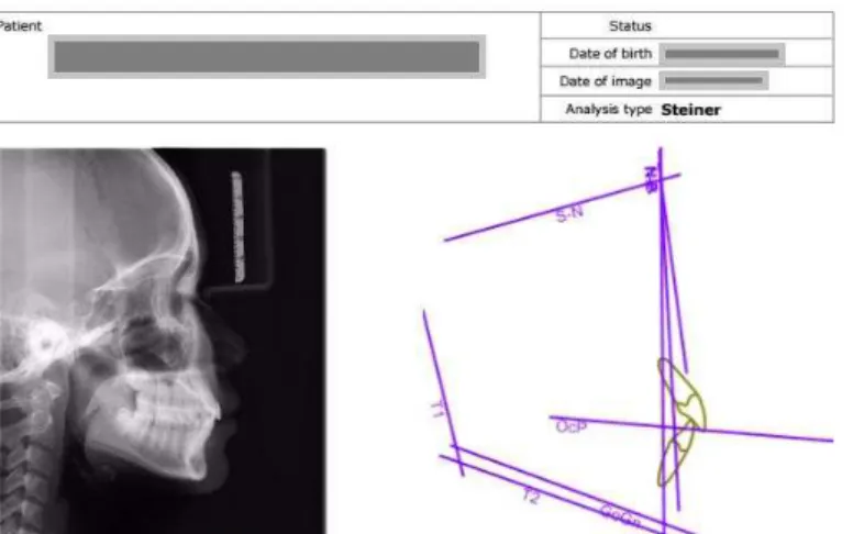
Figure 3. Results from software Planmenca Romexis Ceph. Analysis
2.2.3.2 Taking Cephalometric films for research objects
Instruct objects about the correct position when taking a film. Criteria of Cephalometric films:
- General: can see clearly hard tissues and soft tissues.
- Specificх hard and soft landmarks of subjects’ heads must be seen clearlyй Includingх external ear lob, lower part of eye socket, molars must be tight. Cranial base should reveal nasal bone, frontonatal suture. Maxillary reveals nasal cavity base, front nose spines, hollow bottom front nose, palate ceiling, butterfly slot, front teeth, the first large molars. Mandibular bone identifies chin protrusion, front, back, upper, lower part (both inside and outside) of the lower jaw, front teeth, the first large molars. Software identifies soft tissue circumference [7].
2.2.3.3 Film analysis
Identify bone standard point:
- Midpoint nest hole: S. The nose: The banks of the lower orbital Nai: Or. Previous points nose spines: ANS. Postnasal barbed point: PNS. Most low points along the curve between XHT: A. The front teeth biting edge: Is. Point the tip of the upper incisors. Point edge incisor bite below: Ii. Point the tip of the lower incisors. The first big molars of upper jaw. The first big molars of lower jaw. Most low points XHD midline: B. chin Peaks: Pog. Chin point: Me. Most point and foremost under the chin: Gn. Mandibular angle point: Go. The highest point of the outer ear canal: Po
- Determine the planes straight lines under the horizontal plane: Plane (Mp) S - N: represent front cranial base, passing through the point S and Na. Palate plane: passing points ANS and PNS. Mp FH: passing points Or and Po. Bite plane: passing the midpoint of large molar teeth bite and the midpoint of the segment exhibiting the bite level of incisor. In case front teeth is open, bite plane passing through the midpoint of first molars bite and first small molar.
37
- Identify angleз distances that need to be measured and measure such angle values v distances: SNA Angle: angle made of SN line cutting NA line at N point. SNB Angle: angle made of SN line cutting NB line at N. ANB angle = SNA – SNB. SN-OP Angle: made of SN line and bite plane. Facial angle (NPog – POr): angle made of line passing Na – Pog and FH plane. Y axis angle : acute angle made of S – Gn line and FH plane. Molar angle of maxilla and cranial base (U1-SN): angle made of SN line and line passing molar axis of upper jaw. U1 – NA Angle: angle between upper front teeth and N – A line. U1 – NA distance: distance from most prominent point of outer surface of molar to N – A line. U1-L1 Angle: angle made from line passing axis of upper front tooth and line passing axis of lower front tooth. L1-GoMe Angle: angle made of line passing axis of lower jaw front tooth and mandible plane (GoMe). L1-NB angle: angle made of line passing axis of lower front tooth and line passing Na – B. L1 – NB distance: distance from the most prominent point of front tooth of mandible to N–B line. Prominent angle of G-Sn-Pog’хcute angle identified by two lines passing G – Sn and Sn –Pog’ [н]з [с]й
Record measurements in case history.
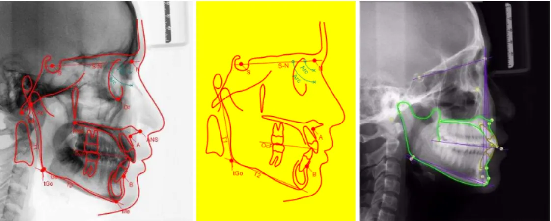
Figure 4: Landmarks used in software Planmenca Romexis Ceph.Analysis
2.2.4 Data cleaning and processing -Clean data before analysis.
-Data is entered and analyzed by SPSS 20.0 -Verify variables by t- Test and 2
2.3 DEVIATION AND SOLUTIONS 2.3.1 Deviation
Selecting research objects
Errors in examining medical history and related diseases with odonto-stomatology.
- During taking cephalometric films: Technicians adjust parameters deviation, film mode, wrong position of ear – rode. Position of objects not meeting standards.
- During data analysis: Deviation during the identification of surgery landmarks. 2.3.2 Solutions
- Master theory of occlusion classification. - Interview, clinically examine each object carefully. - Select qualified and experienced technicians.
- Give specific instructions for subjects about head position, lip position during taking films. Practice many times before taking films.
- Practice to identify landmarks on films accurately.
2.4. Research period: from February 2015 to November 2015 2.5. Research ethics.
- The research is conducted at High-quality Stomatology Centre of Odonto-Stomatology Training Institute - HMU.
- Explain to objects about research objective, responsibilities of researchers, responsibility and rights of participants.
- The study is only conducted on volunteers on the basis of co-operation, without obligation. -All collected information serves the research objectives without any other purposes.
38
treatment or other examination methods will be conducted for accurate diagnosis. -Research results will be sent to the School
III. Results
3.1. Gender distribution in research
Among the total of 42 objects, there is an equal proportion of male and female objects, each accounting for 50%.
3.2. Characteristics on cephalometric films of students with normal occlusion
3.2.1. Bone-bone corresponding indicator
Table 3.12: Bone indicators on Cephalometric (n=42)
Gender Male Female p*
Indicator
X
± SяX
± SяUpper
SNA (0) унйсп± нймф умймс±нйпн 0.5066 SN-bite plane (0) мойпр±мйнр мойсп±лйфм 0.5731
Lower
SNB (0) умйср± нйнр тфйур± нйол 0.2595 NPog – POr (0) урйуп± мйнп упйлф±рйрс 0.1653 Y axis angle (0) смйтм± нйпт смйнл±рйул 0.7129
Upper – Lower ANB (0) нймп±лймо нйлр±лймн 0.9899
* t-test Remark:
There are no statistically difference about upper jaw bone, lower jaw bone indicators on Cephalometric film between male and female (p>0.05)
3.2.2.Bone-skeletal indicator
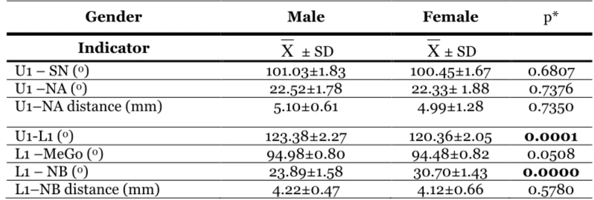
Table 3.13: Bone-skeletal indicator (n=42)
Gender Male Female p*
Indicator
X
± SяX
± SяU1 – SN (0) млмйло±мйуо мллйпр±мйст 0.6807
U1 –NA (0) ннйрн±мйту ннйоо± мйуу 0.7376
U1–NA distance (mm) рймл±лйсм пйфф±мйну 0.7350
U1-L1 (0) мнойоу±нйнт мнлйос±нйлр 0.0001
L1 –MeGo (0) фпйфу±лйул фпйпу±лйун 0.0508
L1 – NB (0) нойуф±мйру олйтл±мйпо 0.0000
L1–NB distance (mm) пйнн±лйпт пймн±лйсс 0.5780
* t-test
Remark:
Average U1-L1 angle in male is мнойоу±нйнт higher than female which is мнлйос±нйлрй This difference has statistical significance (p<0.05, t-test)
Average L1-NB angle in male is нойуф±мйру lower than female which is олйтл±мйпой This difference is statistically significant (p<0.05, t-test)
Average U1-SN value of female is higher than male and in females there is a tendency of moving forward of front teeth compared with cranial base. This suggested that front molar is a little bit outward compared with cranial complexes in male.
There are no statistical significance in remaining indicators on Cephalometric films between male and female (p>0.05).
Iv. Comments:
39
Bone-bone relation: We have not found any evidence showing statistical difference in the value of SNA angle between the two genders in the study. Also when comparing with normal SNA angle (820) there is no statistical significant difference. SNA indicator reflects the relative position of upper jaw bone compared with cranial base, the limit of this angle is 80-840, if a big angle, upper jaw bone is moving forward compared with cranial base, if small angle, this proves that front jaw bones are less developed compared with cranial base [6]. Research findings show that this angle is 81.900 (male is 82.640 and female is 81.160). Therefore, in both genders, SNA angle is still within normal limit compared with Steiner norm (820), which means that upper jaw bone in relation with cranial base of Western people is no different from Vietnamese.
SNB angle reflects the relation between the middle position of lower jaw bone and cranial base, the bigger this angle is, chin and dental alveoli bone has more protrusion compared with cranial base. Permitted limit of this angle according to Steiner is 780-820. Our study also shows that, median values of SNB angle is 80.750 and of male умйср± нйнр г0дз of female тфйур± нйол г0) and this deviation is not statistically significant. Therefore, SNB angle value of female is smaller than that of male.
ANB angle value describes the relation between upper jaw bone and lower jaw bone, with the cranial base as an intermediate. Our research finding: ANB angle value 2.090 in male is 2.140 and in female is 2.050, this is suitable with research result of Steiner about normal value of ANB angle which is 20 [3], [4].
Skeletal-bone relation: our research shows, median values of U1 – SN angle (0) male is bigger
than female, specifically: value of U1-SN angle in male млмйло±мйуог0) and in female мллйпр±мйстг0). Similarly, U1 – NA angle value (0) male is bigger than female, specifically: U1-NA angle value of male is ннйрн±мйту г0) and in female are ннйоо± мйуу г0). Our research result is similar to normal U1-SN angle value published by Steiner which is 820 and normal value of U1-NA angle is 220 [3] [4].
V. Conclusion
Bone-bone relation: the relational position of upper jaw bone compared with cranial base SNA=81.900 (male is 82.640 and female is 81.160). The relation between lower jaw bone with cranial base, this angle has median value of SNB=80.750 and in male is умйср± нйнр г0), in female is тфйур± нйол г0) and this deviation is not statistically significant. Therefore, SNB angle value in the research of female indicators is smaller than that of male. ANB angle value 2.090 in male is 2.140 and in female is 2.050.
Bone-teeth relation: our research shows that median value of U1 – SN angle (0) male is bigger than female, specifically: value of U1-SN angle in male млмйло±мйуог0д and female мллйпр±мйстг0). Similarly value of U1 – NA (0д angle in male is ннйрн±мйту г0д and in female is ннйоо± мйуу г0). The result is similar to normal value of U1-SN angle published by Steiner which is 820 and normal value of U1-NA angle is 220.
References:
1. Angle E.H. (1899). “юlassification of malocclusion”з D. Cosmos, 41, p.248 - 264.
нй Smortree Viteporn гмффрдз “The technique of cephalometric radiography”з Orthodontic
Cephalometry, Mosby – Wolfe, p.9 – 20.
3. Steiner C.C (1960). “The use of cephalometrics as an aid to planning and assessing orthodontic treatment”й ьmй Jй Orthodйз псз ppйтнм-735.
4. Steiner C.C. Cephalometrics in Clinical practice, The Angle Orthodontist Vol 29, No 1:8-29,1959.
5. Hoang Tu Hung (2005). Occlusion, Faculty of Odonto - Stomatology, University of Medicine and Health Sciences – Ho Chi Minh City: 104-111.