Acta Cryst. (1995). D51, 311-317
Structure of a Calcium-Independent Phospholipase-like Myotoxic Protein from
Bothrops asper
Venom
BY R. K. ARNI aND R. J. WaRD
Department of Physics, UNEXSP-IBILCE, Cx.P. 136, 15054--000, Sdo Jos~ do Rio Preto-SP, Brazil J. M. GUTIERRZZ
lnstituto Clodomiro Picado, Universidad de Costa Rica, San Jos~, Costa Rica AND m. TULINSKY
Department of Chemistry, Michigan State University, East Lansing, USA (Received 23 March 1994; accepted 10 October 1994)
Abstract
Myotoxin II, a myotoxic calcium-independent phospho- lipase-like protein isolated from the venom of Bothrops asper, possesses no detectable phospholipase activity. The crystal structure has been determined and refined at 2.8A to an R factor of 16.5% (F>3o') with excellent stereochemistry. Amino-acid differences between cataly- tically active phospholipases and myotoxin II in the Ca2+-binding region, specifically the substitutions T y r 2 8 ~ A s n , G l y 3 2 ~ L e u and A s p 4 9 ~ L y s , result in an altered local conformation. The key difference is that the e-amino group of Lys49 fills the site normally occupied by the calcium ion in catalytically active phospholipases. In contrast to the homologous mono- meric Lys49 variant from Agkistrodon piscivorus piscivorus, myotoxin II is present as a dimer both in solution and in the crystalline state. The two molecules in the asymmetric unit are related by a nearly perfect two- fold axis, yet the dimer is radically different from the dimer formed by the phospholipase from Crotalus atrox. Whereas in C. atrox the dimer interface occludes the active sites, in myotoxin II they are exposed to solvent.
Introduction
Phospholipases A2 (PLA2, E.C. 3.1.1.4) are small stable calcium-dependent hydrolytic enzymes that specifically hydrolyze the sn2 ester of phospholipids (Deenen & de Haas, 1964), preferentially in lamellar or micellar aggregates at membrane surfaces (reviewed by Raminez & Jain, 1991). These enzymes are widely distributed in nature and have been extensively studied (Dennis, 1983; Waite, 1987). The extracellular PLA 2,s occur abundantly in mammalian pancreatic juice and in the venoms of snakes and insects. The intracellular PLA2's, which are often membrane associated, are involved in blood platelet aggregation and inflammation (Scott et al., © 1995 International Union of Crystallography
Printed in Great Britain - all rights reserved
1990). The catalytic activity of these intracellular enzymes results in the release of arachidonic acid, a precursor of eicosanoids, and is implicated in triggering inflammatory reactions (Scott et al., 1990).
Despite the diversity of occurrence, the primary sequence of all PLA's demonstrate considerable se- quence homology and are divided into two classes on the basis of location of disulfide bridges and loop lengths. Class I PLA2's include pancreatic and elapid venom enzymes, whereas class II are found in crotalid and viper venom PLA2's (Dufton & Hider, 1983).
Recently, a sub-class of snake-venom PLA2-1ike proteins which retain cytotoxicity, but lack phospholi- pase activity have been identified and sequenced (Maraganore et al., 1984; Maraganore, Poorman & Heinrickson, 1987; Yoshizumi et al., 1990; Liu et al., 1990; Homsi-Brandenburgo, Queiroz, Santo-Neto, Rodrigues-Simon & Giglio, 1988; Francis, Gutierrez, Lomonte & Kaiser, 1991). In the catalytically active PLA2, calcium is an essential co-factor, and the non- catalytic PLA2-1ike proteins are primarily characterized by the substitution of Asp49 by Lys. In all catalytically active PLAz's, Asp49 plays a central and essential role in catalysis by contributing two carboxylate O atoms to the calcium ligation cage. The A s p 4 9 ~ L y s 4 9 substitution results in the e-amino group of lysine filling the site normally occupied by the calcium ion (Holland et al., 1990; Scott, Achari, Vidal & Sigler, 1992) resulting in the loss of calcium binding. Although initial results suggested that the Lys49 variant isolated from Agkis- trodon piscivorus piscivorus (App) demonstrated a low level of PLA2 activity (Maraganore et al., 1984), it has subsequently been shown that this is due to trace contamination by other active PLA2 species (Dhillon et al., 1987; van den Berg, Slotboom, Verheij & de Haas,
1989).
The interaction of Bothrops asper myotoxin II with liposomes of diverse composition provokes rapid and
Acta Crystallographica Section D
extensive release of the aqueous contents. Previous work (Rufini et al., 1992) has established that, (a) this process is Ca 2+ independent and occurs without detectable phospholipid hydrolysis. (b) Whereas catalytically active phospholipases are unable to induce liposome leakage in the absence of Ca 2÷, the addition of Ca 2+ results in liposome leakage with concomitant hydrolysis of phospholipids. (c) The kinetics of the Ca2+-dependent and independent liposome leakage indicate the existence of different mechanisms of interaction with the lipid bilayer. In conclusion, it is suggested that a new type of cytolytic reaction mechanism is responsible for the effects of myotoxin II in vivo (Rufini et al., 1992).
In the hope of providing an insight into this novel mechanism, we have determined the structure of myotoxin II, a myotoxic Lys49 variant from the venom of B. asper. The model we present has been determined at 2.8A resolution and refined to a crystallographic residual of 16.5% (F>3o'). The structural changes caused by amino-acid substitutions in the calcium- binding loop are examined and compared to the structure of the Lys49 variant from App. In addition, myotoxin II exists as a dimer in solution, and the structure we present here provides an insight as to the nature of the dimer interface.
Experimental methods
The Lys49 PLA2 from B. asper was isolated (Lomonte & Gutierrez, 1989) and the protein (5.0mg ml -~) was crystallized from 0.1 M citrate, 20% 2-propanol using 20% PEG 4000 as a precipitant (Arni & Gutierrez, 1993). Crystals belong to space group P2~2121 with cell constants a = 50.18, b = 67.76 and c = 88.00,~,.
X-ray diffraction data was collected from a single crystal using a Rigaku RU200 generator operated at 50kV and 150mA and a Xentronics area detector (Siemens). The data was processed using the program XENGEN (Howard et al., 1987). An Rmerge on intensities of 0.056 was obtained for 5805 unique reflections from a total of 23 021 observations. Table 1 summarizes the crystallographic data and refinement statistics.
The structure was solved by molecular-replacement techniques using the program package MERLOT (Fitz- gerald, 1988). The search model, which consisted of 887 atoms, was derived from the Lys49 variant from App (Holland et al., 1990). Structure factors were calculated in a cubic cell with P1 symmetry and edge dimensions of 120,4,. Data with I > 3o. between 8.0 and 4.0 ,~ was used coupled with a radius of integration of 23.9 ,~,. The two highest peaks in the cross-rotation function map were 6.2o" and 4.5o'. Correlation with the self-rotation results revealed the orientation of the two molecules in the asymmetric unit. The orientation angles were refined using the Lattman rotation function (Lattman & Love, 1972) with angular increments of a = 1.0, /~ -- 1.0 and y = 2.0 °. The highest peaks in the Crowther & Blow
Table 1. Summary of crystallography data and refine- ment statistics
Space group Cell constants (,~)
No. of observations No. of unique reflections Rmerge
R e f i n e m e n t statistics Resolution (.~) No. of reflections R factor (%) No. of protein atoms No. of solvent atoms
P2t2121 a = 50.18(2) b = 67.76 (3) c = 88.00 (4)
23021 5805 0.056
7.0-2.8 4915
16.5 1890 69
R.m.s. deviations from ideal values Bond lengths (/~) 0.010 Bond angles (o) 2.107
Dihedral (°) 27.948
lmpropers (°) 6.661
translation function (Crowther & Blow, 1967) with o. values between 7.9 and 5.0 produced a model with an acceptable number of close contacts. The rotational and translational parameters were optimized by utilizing the R-factor minimization procedure as implemented in MERLOT (Fitgerald, 1988) which reduced the R factor from 52.0 to 48.1% in the 6.0--3.5 ,~ resolution range.
Energy minimization and refinement using X-PLOR (Brtinger, o1988) resulted in an R factor of 23.9% in the 10.0--2.8A resolution range. The electron-density map was then examined using FRODO (Jones, 1985) and all the side-chain atoms were included. An additional round of simulated annealing resulted in a crystallographic residual of 21.2% (7.0-2.8,~). Numerous simulated- annealing omit maps were computed to permit the determination of the conformation of poorly determined loop regions (see results) and three additional rounds of
model building and refinement were carried out. The individual temperature factors were refined and the refinement converged to a final residual of 16.5% (F>3cr) for 4915 reflections between 7.0 and 2.8,~. The final model consisted of 1890 protein atoms and 69 solvent O atoms.
Results
5 0 . . .
The asymmetric unit of the myotoxin II crystal contains two monomers with near-perfect twofold rotational symmetry (see below). The ribbon diagram presented in Fig. 1 presents the fold of the myotoxin II monomer. The structure is similar to those of PLA2's isolated from other snake species (Brunie, Bolin, Gewirth & Sigler, 1985; Holland et al., 1990; White, Scott, Otwinowski, Gelb & Sigler, 1990; Freemont, Anderson, Wilson, Dennis & Xuong, 1993) and mammals (Dijkstra, Kalk, Hol & Drenth, 1981; Dijkstra, Renetseder, Kalk, Hol & Drenth, 1983; Wery et al., 1991). Using the nomencla- ture and numbering scheme of Heinrikson, Kreuger & Keim (1977), myotoxin II is classified as a class II PLA. The structure is stabilized by seven disulfide bridges (27:125, 29:45, 44:105, 50:133, 51:98, 61:91, 84:96) and
5 0
!
: i ,,
25_i..~ .... i ... = ... i ~ . . . ~ - . . ~ ...
0 ~ ~ ,,', ~.' ! , i A ' ~ • ~, / : ~.,
'0 '.~ '2o '3o ~0 ~0 ;0 '70 80 bo '.~
5O
. ~ 25 L
~ o . . . . i
~ I i ! i
,~ 100 t110 120 30 140 150 160 170 ~180 1 9 0 2(h') ...
25 ~ " ',
~ '~ ' t : t ~ . . . . ~ / ' A ,', i i ,.i 1: ' " :',i! ,' '"\ i ~ , " ' 0
L200 '21() ~220 '230 1240 i250 2 6 0 1270 ~ 8 0 '2SRI '300
Fig. 3. Superpositioning of the Ca atoms from, myotoxin II molecule 1 (thick line), myotoxin II molecule 2 (thin line), and App (dashed line).
t t
25 . . . . . . . . ' I
0 ~ . . .
~ '310 ~20 '33o ~40 350 36o 370 38o ~90 400
R e s i d u e n u m b e r
Fig. 2. Distribution of the mean temperature factors as a function of residue number. Thick and thin lines represent main-chain and side- chain atoms, respectively. The two molecules in the asymmetric unit are numbered sequentially from 1 to 121 and 201 to 321.
comprises an N-terminal a-helix (residues 2-12), linked by loops and a single helical turn to the first long a-helix (40-55). A short antiparallel ]3-sheet (75-84), referred to as the /~-wing, leads to a second long helix (90-107) which is antiparailel to the first. The loop region (25-35) corresponds to the calcium-binding loop in the enzymi- cally active PLA2's.
The deviations of bond lengths and angles from ideal values for the model are presented in Table 1. The Ramachandran plot (not shown) (Ramachandran & Sashisekharan, 1965) indicates the presence of two outliers. Fig. 2 illustrates the distribution of the temperature factors for sequentially numbered main- chain and side-chain atoms. The outliers in the Ramachandran plot lie in the same loop region in each of the two molecules (78-80 sequential numbering, 8 8 - 90 homology numbering). The loops contain a proline, possess higher than average temperature factors and are characterized by poor electron density.
Fig. 3 and Table 2 present the results of the super- positioning using the C a atoms of the homologous core (Renetseder, Brunie, Dijkstra, Drenth & Sigler, 1985) from the two monomers in the asymmetric unit of myotoxin II and the structure of the Lys49 variant from App. The structures of the two monomers are very similar, and the largest deviations between the structures of myotoxin II and the Lys49 variant from App are mainly in the calcium-binding and the C-terminal loop regions.
D i m e r interactions
314 PHOSPHOLIPASE-LIKE MYOTOXIN Table 2. Statistics derived from superpositioning the Cot
atoms o f the myotoxin II molecules 1 and 2 with App utilizing the homologous core as defined by Renetseder
et al. (1985)
First Second
molecule molecule R.m.s. deviation
Myotoxin I1 Myotoxin II 0.587
molecule ! molecule 2
Myotoxin II App 1.048
molecule I
Myotoxin II App I. 100
molecule 2
Table 3. Amino-acid side chains participating in the formation o f the dimer
Maximum contact distance considered for analysis = 3.5 ,~
From To Distance (~,)
G l u l 2 0 E I Lys280 NZ 3.2
Glu 12 OE2 Lys280 NZ 3.0
Trp77 NE1 Gin211 O 3.0
Trp77 NEI Glu212 O 3.5
Lys80 NZ Glu212 OEI 2.9
Lys80 NZ Glu212 OE2 2.9
Lys80 NZ Lys280 O 3.4
Lys80 O Lys280 NZ 3.0
monomers are related by an almost perfect twofold symmetry axis (rotation angle 179.7°). We propose this crystallographic interaction is representative of the dimeric interaction in solution.
In catalytically active PLAz's, it has been suggested that dimerization is essential and enhances enzymatic activity (Cho, Tomasselli, Heinrickson & Kezdy, 1988; Tomasselli et al., 1989). Myotoxin II presents a novel dimeric form for PLA2 molecules, as illustrated in Fig. 4(a). The fl-wing regions interact closely with each other and the putative 'catalytic' sites are exposed to the solvent. These extensive and intimate diad interactions are mainly between the amino acids located at the C- terminal end of the first helix and the loop region preceeding the first fl-strand of the /3-wing. Fig. 4(b) shows this region in more detail, and illustrates the interactions stabilizing the dimer. Table 3 provides a list of the amino-acid side chains participating in the stabilization of the dimer.
In contrast to the crystal structures of other PLA2's, where the fl-wing region is disordered (Scott et al., 1992), these regions are well defined in the electron density of myotoxin II, presumably due to the extensive interactions which stabilize the dimer (Table 3). The amino acids involved in these interactions in the structure of myotoxin II are conserved in App Lys49 and are in the same relative orientations with the exception of the indole ring of Trp77. Interestingly, App Lys49 exists as a monomer in the two crystal structures reported (Holland et al., 1990; Scott et al., 1992), and this region has not been reported to be involved in the formation of intermolecular contacts (Holland et al., 1990; Scott et al., 1992).
In the crystal structure of dimeric PLA2 from Crotalus atrox, the monomers are also related by a twofold rotational symmetry axis. However, in this case the interaction results in the formation of a very different dimer in which the catalytic residues are shielded from the solvent and face an internal cavity, and the fl-wing region is fully exposed to the solvent. Additionally, the catalytically essential residue Asp49 forms hydrogen bonds between the two molecules (Brunie et al., 1985). If this structure does indeed represent the functional dimer, and dimerization is essential for activity (Shen et al., 1975), it is unclear how the dimers dissociate or reorient
to expose the catalytic site to facilitate calcium binding (Brunie et al., 1985). The C. atrox crystallographic dimer is stabilized by five hydrogen bonds and hydrophobic interactions. Of these, out of a total of ten residues involved only three (Tyr52, Asn67 and Lys69) are conserved in myotoxin II. The myotoxin II dimer is stabilized by intermolecular hydrogen bonds involving Glnl2, Trp77 and Lys80 (see Table 3), none of these residues are conserved in C. atrox PLA2.
The calcium-binding loop
In catalytically active PLA2's, calcium is an essential cofactor. The ligated Ca 2÷ in the crystal structure of bovine pancreatic PLA2 (Dijkstra et al., 1981) is situated
. 7
~,~J 212
(a)
(b)
. . , . 77
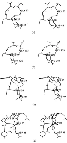
0.2,~ from the position of the e-amino group in the superimposed App Lys49 structure (Scott et al., 1992). Fig. 5 compares the same loop region from the myotoxin II [molecules 1 and 2 shown in Figs. 5(a) and 5(b), respectively] to that of App Lys49 PLA [Fig. 5(c)]. For comparison, Fig. 5(d) shows the homologous loop region from the catalytically active Asp49 Naja naja atra PLA2. The Lys49 e-amino groups in the three loop structures shown in Figs. 5(a)-(c) occupy the volume taken by the Ca 2+ ion in Fig. 5(d).
Comparison of the loop regions in the Lys49 PLA's shows that in myotoxin II Val31, Leu32 and Gly33 substitute for Trp, Gly and His, resulting in a very diferent loop conformation. Two of the three hydrogen bonds observed in the App Lys49 PLA2 (Holland et al.,
LY
33
~ Y
33
~ S 249 233
(a)
(b)
~ ~ S 249 233
~
33
49
(c)
49
31 _ ) _ _ . . _ ~ L V ~
(d)
Fig. 5. Calcium-binding loop regions (amino acids 28-33 and 49) from myotoxin II molecule 1 (a), molecule 2 (b), App (c) and the catalytically active PLA2 from Naja naja atra, which illustrates the coordination of the Ca 2÷ ion (shown as a full sphere) in which a Asp49 replaces Lys (d).
1990) are conserved in both the monomers of myotoxin II. Furthermore, in the App Lys49 structure, the coordination of the amino group is completed by a tightly bound water molecule. In myotoxin II, no solvent atoms were observed in this region.
Discussion
The structure of myotoxin II, a myotox!c Lys49 variant PLA2 from B. asper, determined at 2.8 A is very similar to the Lys49 variant from App (Holland et al., 1990; Scott et al., 1992) with which it shares 75% sequence identity (Francis et al., 1991). In agreement with previous work, we find that the substitution of Asp49 by Lys49 results in the e-amino group of Lys49 occupying the site normally filled by the calcium ion. This substitution and the consequent loss of Ca2+-binding ability, results in the lack of hydrolytic activity observed in these proteins (Maraganore et al., 1984, Francis et al, 1991). Site-directed substitution of Asp49 by Lys in porcine pancreatic PLA2 also resulted in complete loss of catalytic activity (van den Berg et al., 1989).
In all class I/II phospholipases, the amino-acid residues His48, Tyr52, Tyr73 and Asp99 are fully conserved. The Asp99/His48 pair, in conjunction with a bound water molecule, acts as a catalytic triad. Additionally, Asp99 is hydrogen bonded to the Tyr52 and Tyr73 side chains. This pattern of interconnected hydrogen bonds is referred to as the catalytic network (Kuipers, Franken, Hendricks, Verheij & de Haas, 1990). The relative positions of the amino acids in the catalytic network between the active PLA2's and the inactive myotoxin II are structurally conserved (Fig. 6). Also shown in Fig. 6 is position 68, which in N. naja atra PLA2 and myotoxin II are occupied by Pro, but in the case of App Lys49 PLA, is substituted by His. The functional significance of this substitution is unknown.
Myotoxin II forms a stable dimer in solution (Francis et al., 1991), and we propose that the arrangement of the two monomers in the crystallographic asymmelric unit is
S48
.. .IS
48
representative of the dimer in solution. The interactions between the monomers indicates a novel dimeric form in which the/5-wing regions from each monomer interact, and the proposed lipid-binding face and residues involved in catalysis are exposed to the solvent. Although the amino acids forming the contacts in the myotoxin II dimer are conserved in App Lys49, and are in approximately the same relative orientations in the two crystal structures (Holland et al., 1990; Scott et al.,
1992), they were not found to be involved in inter- molecular interactions in the crystal structure of mono- meric App. This suggests that other factors are involved in the formation and stabilization of the dimer. A number of studies have implicated the importance of the acylation of lysine residues as a mechanism to promote dimerization and a concommitant 200-fold increase in catalytic activity (Cho et al., 1988; Tomasselli et al.,
1989); however, close examination of the electron- density map does not reveal the presence of acyl groups either on the lysines at the dimer interface, or in any other regions of the molecule. Table 3 shows that the dimer is stabilized by a total of eight salt bridges and hydrogen bonds between the two monomers, which has been shown to be stable in both 0.1% sodium dodecyl sulfate at 3 5 8 K and 2 M urea (Francis et al., 1991). It is thus unlikely that such a stable structure can dissociate in solution.
Although myotoxin II is devoid of phospholipase activity, it retains potent myotoxic activity, causing myonecrosis by affecting the integrity of the plasma membrance of muscle fibers (Lomonte & Guttierez, 1989). Attempts using amino-acid sequence comparisons to identify the region of the PLA2 responsible for myotoxicity (Kini & Iwanaga, 986) have not been successful in predicting myotoxity in myotoxin II (Francis et al., 1991). The same degree of myotoxicity and edema-inducing activity exists between myotoxins I and II (Gutierrez, Ownby & Odell, 1984), suggesting that phospholipase activity is not essential for these functions. Previous comparisons of myotoxic PLA2 amino-acid sequences indicate that all have either three or four tyrosines between residues 112 and 121, a threonine at 112, and a lysine at position 38, all of which are not found in non-myotoxic PLA2 sequences (Francis et al.,
1991). Based on the structure of PLA2 from C. a t r o x , it has been suggested that Lys38 lies close to these C- terminal tyrosines, and may be implicated in myotoxic activity (Francis et al., 1991). Interestingly, we find a network of hydrogen bonds in which the fully exposed L y s l 9 and T y r l 0 9 from each monomer interact with the same amino acids from a symmetry-equivalent molecule. We are currently refining the structures of a number of other myotoxic PLA2-1ike proteins, and a comparison of the models may help to identify the region or regions conferring myotoxicity.
In conclusion, calcium ions are an essential co-factor for the hydrolysis of phospholipids by catalytically active
PLA2's. A calcium-independent mechanism of mem- brane damage, without PLA2 activity, has been demon- strated for myotoxin II (Rufini et al., 1992). This indicates a novel mechanism for membrane damage, and the availability of the model for the myotoxin II will be useful in providing further insights on this activity.
Financial assistance by FAPESP and CNPq (Brazil) to R K A and RJW, and by CONICIT (Costa Rica) to JMG is gratefully acknowledged. Atomic coordinates and struc- ture factors have been deposited with the Protein Data Bank (Bernstein et al., 1977).*
*Atomic coordinates and structure factors have been deposited with the Protein Data Bank, Brookhaven National Laboratory (Reference: I CLP, R ICLPSF). Free copies may be obtained through The Managing Editor, International Union of Crystallography, 5 Abbey Square, Chester CH1 2HU, England (Reference: GR0379). At the request of the authors, the structure factors will remain privileged until 1 May,
1997.
References
ARNI, R. K. & GUTIERREZ, J. M. (1993). Toxicon, 31, 1061-1064. BERG, C. J. VAN DEN, SLOTBOOM, A. J., VERHEIJ, H. M. & DE HAAS, G.
H. (1989). J. Cell. Biochem. 39, 379-390.
BERNSTEIN, F. C., KOETZLE, T. F., WILLIAMS, G. J. B., MEYER, E. F. JR., BRYCE, M. O., RODGERS, J. R., KENNARD, O., SIMANOUCHI, T. & TASUMI, M. (1977). J. Mol. Biol. 112, 535-542.
BRt3NGER, A. T. (1988). In Crystallographic Computing 4: Techniques and New Technologies, edited by N. W. ISAACS & M. R. TAYLOR. Oxford: Clarendon Press.
BRUNIE, S., BOLIN, J., GEWIRTH, D. & SIGLER, P. B. (1985). J. Biol. Chem. 260, 9742-9749.
CHO, W., TOMASSELLI, A., HEINRICKSON, R. L. & KEZDY, F. J. (1988). J.
Biol. Chem. 263, 11237-11241.
CROWTHER, R. A. & BLOW, D. M. (1967). Acta Cryst. 23, 544-548. VAN DEENAN, L. L. M. & DE HAAS, G. H. (1964). Advances in Lipid
Research, Vol. 2, p. 167. New York: Academic Press.
DENNIS, E. A. (1983). Phospholipase A2. The Enzymes, Vol. 16, pp. 307-353. New York: Academic Press.
DHILLON, D. S., CONDREA, E., MARAGANORE, J. M., HEINRICKSON, R. L., BENJAMIN, S. & ROSENBURG, P. (1987). Biochem. Pharmacol. 36, 1723-1730.
DIJKSTRA, B. W., KALK, K. H., HOL, W. G. J. & DRENTH, J. (1981). J.
Mol. Biol. 147, 97-123.
DIJKSTRA, B. W., RENETSEDER, R., KALK, K. H., HOL, W. G. J. & DRENTH, J. (1983). J. Mol. Biol. 168, 163-179.
DUFXON, M. J. & HIDER, R. C. (1983). Eur. J. Biochem. 137, 545- 551.
FITZGERALD, P. M. D. (1988). J. Appl. Cryst. 21,273-278.
FRANCIS, B., GUTIERREZ, J. M., LOMONTE, B. & KAISER, I. I. (1991).
Arch. Biochem. Biophys. 284, 352-359.
FREEMONT, D. H., ANDERSON, D. H., WILSON, I. A., DENNIS, E. A. & XUONG, N.-H. (1993). Proc. Natl Acad. Sci. 90, 342-346. GUTIERREZ, J. M., OWNBY, C. L. & ODELL, G. V. (1984). Toxicon, 22,
115-128.
HEINRICKSON, R. L., KREUGER, E. T. & KEIM, P. S. (1977). J. Biol. Chem. 252, 4913--4921.
HOLLAND, D. R., CLANCY, L. L., MUCHMORE, S. W., RYDEL, T. J., EINSPAHR, H. M., FINZEL, B. C., HEINRICKSON, R. L. & WATENPAUGH, K. D. (1990). J. Biol. Chem. 265, 17649-17656.
HOWARD, A. J., GILLILAND, G. L., FINZEL, B. C., POULOS, T. L., OHLENDORF, D. H. & SALEMME, F. R. (1987). J. Appl. CO'st. 20, 383- 387.
JONES, T. A. (1985). Methods Enzymol. 115, 157-171. KINI, R. M. & IWANAGA, S. (1986). Toxicon, 24, 895-905.
KUIPERS, O. P., FRANKEN, P. A., HENDRICKS, R., VERHEIJ, H. M. & DE HAAS, G. H. (1990). Protein Eng. 4, 199-204.
LATrMAN, E. E. & LOVE, W. E. (1972). Acta Cryst. B26, 1854-1857. Liu, S. Y., YOSIIIZUMI, K., ODA, N., OilNO, M., TOKUNAGA, F., IWANAGA, S. & KlltARA, H. (1990). J. Biochem. (Tokyo), 107, 400- 408.
LOMONTE, B. & GUTIERREZ, J. M. (1989). Toxicon, 27, 725-733. MARAGANORE, J. M., MERKUTA, G., CHO, W., WELCHES, W., KZEDY,
F. J. & HEINRICKSON, R. L. (1984). J. Biol. Chem. 261, 13839- 13843.
MARAGANORE, J. M., POORMAN, R. A. & HEINRICKSON, R. L. (1987). J.
Protein Chem. 6, 173-189.
RAMACItANDRAN, G. N. & SAStlISI-KIIARAN, S. (1965). Adv. Protein Chem. 23, 283-437.
RAMINFZ, F. & JAIN, M. K. (1991). Protein Struct. Funct. Genet. 9, 229-239.
RENETSEDER, R., BRUNIE, S., DIJKSTRA, B. W., DRENTH, J. & SIGLER, P. B. (1985). J. Biol. Chem. 260, 11627-11634.
RUFINI, S., CESARONI, P., DESIDERI, A., FARIAS, R., GUBENSEK, F., GUTIERREZ, J. M., LULY, R., MASSOUD, R., MORENO, R. & PEDERSEN, J. Z. (1992). Biochemistry, 31, 12424-12430.
SCOTF, D. L., ACIIARI, A., VIDAL, J. C. & SIGLER, P. B. (1992). J. Biol. Chem. 267, 22645-22657.
SCOTT, D. L., WHITE, S. P., OTWINOWSKI, Z., YUAN, W., GELB, M. H. & SIEGLER, P. B. (1990). Science, 250, 1541-1546.
SHEN, B. W., TSAO, F. H. C., LAW, J. H. & KEZDY, F. J. (1975). J. Am. Chem. Soc. 97(5), 1205-1208.
TOMASSELLI, A. G., HuI, J., FISHER, J., ZWCHER-NEELY, H., REARDON, I. M., DRIAKU, E., KEZDY, F. J. & HEINRIKSON, R. L. (1989). J. Biol. Chem. 264, 10041-10047.
WAITE, M. (1987) in The Phospholipases: Handbook of Lipid Research,
edited by M. WAITE, pp. 155-241. New York: Plenum Press. WERY, J. P., SCIlEVITZ, R. W., CLAWSON, D. K., BOBBITT, J. L., DOW, E.
R., GAMBOA, G., GOODSON, T., HERMANN, R. B., KRAMER, R. M., MCCLURE, D. B., MIHELICH, E. D., PUTNAM, J. E., SHARP, J. D., STARK, D. H., TEATER, C., WARRICK, M. W. & JONES, N. D. (1991).
Science, 352, 79-82.
WHITE, S. P., ScoTT, D. L., OTWINOWSKI, Z., GELB, M. H. & S|GLER, P. B. (1990). Science, 250, 1560-1566.