Mitochondrial Dysfunction Plus High-Sugar
Diet Provokes a Metabolic Crisis That Inhibits
Growth
Esko Kemppainen1, Jack George1, Görkem Garipler1,2¤, Tea Tuomela1, Essi Kiviranta1,
Tomoyoshi Soga3, Cory D. Dunn2, Howard T. Jacobs1,4*
1BioMediTech and Tampere University Hospital, FI-33014, University of Tampere, Tampere, Finland, 2Department of Molecular Biology and Genetics, Koç University, Sariyer, Istanbul, 34450, Turkey, 3Institute for Advanced Biosciences, Keio University, Tsuruoka, Yamagata, 997–0035, Japan,4Institute of Biotechnology, FI-00014, University of Helsinki, Helsinki, Finland
¤ Current address: Department of Biology, New York University, New York, NY, 10003, United States of America
Abstract
TheDrosophilamutanttko25texhibits a deficiency of mitochondrial protein synthesis, leading to a global insufficiency of respiration and oxidative phosphorylation. This entrains an organis-mal phenotype of developmental delay and sensitivity to seizures induced by mechanical stress. We found that the mutant phenotype is exacerbated in a dose-dependent fashion by high dietary sugar levels.tko25tlarvae were found to exhibit severe metabolic abnormalities that were further accentuated by high-sugar diet. These include elevated pyruvate and lac-tate, decreased ATP and NADPH. Dietary pyruvate or lactate supplementation phenocopied the effects of high sugar. Based on tissue-specific rescue, the crucial tissue in which this met-abolic crisis initiates is the gut. It is accompanied by down-regulation of the apparatus of cyto-solic protein synthesis and secretion at both the RNA and post-translational levels, including a novel regulation of S6 kinase at the protein level.
Introduction
Mitochondrial DNA encodes just 13 polypeptides in most metazoans, representing a small but vital subset of the subunits of the apparatus of oxidative phosphorylation (OXPHOS). In addi-tion it encodes the RNA components, i.e. 2 rRNAs and 22 tRNAs, that contribute to the sepa-rate machinery of protein synthesis inside mitochondria, that genesepa-rates these polypeptides. Defects in mitochondrial protein synthesis are a frequent primary cause of mitochondrial
dis-ease: see reviews [1,2]. Their genetic origin can be either nuclear or mitochondrial, and they
may affect mitochondrial rRNAs, tRNAs, ribosomal proteins, translation factors, aminoacyl-tRNA synthetases, aminoacyl-tRNA modifying enzymes or other accessory factors. They can show a wide range of tissue-specific symptoms that remains largely unexplained, varying dramatically also in severity and age of onset. Whilst the consequences of the primary molecular defect may be clear at the level of the translational machinery, the link to pathogenesis is poorly understood.
OPEN ACCESS
Citation:Kemppainen E, George J, Garipler G, Tuomela T, Kiviranta E, Soga T, et al. (2016) Mitochondrial Dysfunction Plus High-Sugar Diet Provokes a Metabolic Crisis That Inhibits Growth. PLoS ONE 11(1): e0145836. doi:10.1371/journal. pone.0145836
Editor:Fanis Missirlis, CINVESTAV-IPN, MEXICO
Received:August 11, 2015
Accepted:November 4, 2015
Published:January 26, 2016
Copyright:© 2016 Kemppainen et al. This is an open access article distributed under the terms of the
Creative Commons Attribution License, which permits unrestricted use, distribution, and reproduction in any medium, provided the original author and source are credited.
Data Availability Statement:All relevant data are within the paper and its Supporting Information files.
Funding:This work was supported by funding from the Academy of Finland (CoE award 272376 to HTJ); Tampere University Hospital Medical Research Fund to HTJ; the Sigrid Juselius Foundation to HTJ and EMBO (Installation Grant 2010 to CDD). The funders had no role in study design, data collection and analysis, decision to publish, or preparation of the manuscript.
Competing Interests:The authors have declared
It is assumed to have metabolic, cellular, physiological and developmental dimensions, the elu-cidation of which requires well-controlled studies in appropriate model systems.
Our analyses have focused on aDrosophilamodel of mitochondrial disease (tko25t) [3] in
which a nuclear mis-sense mutation in the gene for mitoribosomal protein S12 [4] gives rise to a quantitative defect in mitochondrial protein synthesis [5] impacting all four OXPHOS enzyme complexes dependent on mitochondrial translation products [6]. The molecular
phe-notype oftko25tresembles that seen in human disorders resulting from point mutations in
pro-teins of the mitoribosomal small subunit, including MRPS16 [7] and MRPS22 [8]. At
organismal level thetko25tmutation gives rise to a complex developmental and behavioral
phe-notype, whose main features are larval developmental delay and susceptibility to paralytic
sei-zures induced by mechanical shock, described as bang-sensitivity [6].tko25tadults show an
altered pattern of gene expression [9] that suggests a transformation of metabolism to accom-modate the OXPHOS defect engendered by the mutation. Specifically, there is up-regulation of lactate dehydrogenase and of enzymes involved in the dietary mobilization and catabolism of protein and fat that suggest a switch to glycolysis for ATP production and to other substrates for the supply of carbon skeletons for biosynthesis.
The phenotype of developmental delay and bang-sensitivity, as well as the main (bisexual)
changes in gene expression in adults, are shared with the mutant strainsesB1[10], carrying a
point mutation in the gene encoding the major isoform of the adenine nucleotide translocase
[11]. Since thesesB1mutation limits the supply of mitochondrially supplied ATP to the cell, we
infer that these common features are the signature of defective OXPHOS inDrosophila.
Further-more, thetko25tphenotype is not alleviated by expression of the non proton-motive alternative
oxidase AOX fromCiona intestinalis, nor by the non proton-motive alternative NADH
dehydro-genase Ndi1 from yeast [12], despite the fact that AOX and Ndi1 can partially compensate for the specific phenotypes produced by knockdown of subunits of OXPHOS complexes IV and I,
respec-tively [9,13,14]. This supports the idea that the developmental phenotype oftko25tresults
primar-ily from insufficient mitochondrial ATP production, rather than disturbed redox homeostasis.
Thetko25tphenotype can be partially alleviated by genetic suppressors in either nuclear or
mitochondrial DNA [15,16], which appear to increase the supply of mitochondrial translation
products above a certain threshold. These findings suggested that we might also be able to manipulate the phenotype environmentally, by culturing the flies on media containing a differ-ent balance of dietary compondiffer-ents. Specifically, we reasoned that media rich in sugars should support glycolysis and thus alleviate the mutant phenotype. If successful this could suggest a potential route to dietary management of patients with mitochondrial disease, particularly those with a global impairment of mitochondrial protein synthesis and thus of OXPHOS.
Surprisingly we found an opposite effect, namely that thetko25tphenotype was exacerbated
by sugar-rich media. Analysis of metabolite levels, gene expression, drug sensitivity and the sta-tus of proteins known to be involved in growth regulation and metabolite sensing has allowed us to construct a profile of the metabolic changes produced by the combination of mitochon-drial dysfunction and high sugar diet, and a mechanistic model for how this leads to severe growth impairment. This knowledge has broad implications for understanding disease pro-cesses and their relation to diet.
Results
High-sugar diet exacerbates the growth-retardation of flies with
mitochondrial dysfunction
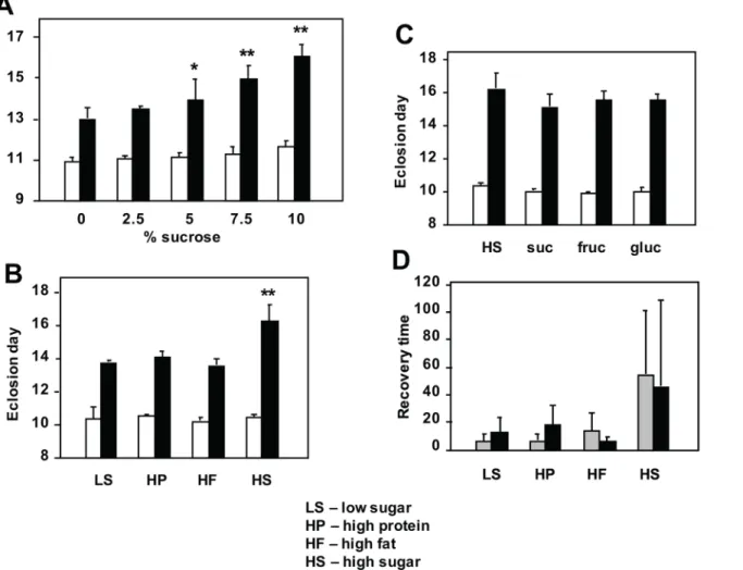
In order to test whether sugar supplementation would mitigate the developmental retardation
(3.5% dried yeast, w/v), but increasing amounts of sucrose. Surprisingly, whilst the wild-type
control strain was only minimally affected by the amount of sugar in the food,tko25tflies
devel-oped more slowly on media with increasing sucrose concentration (Fig 1Aand Panel A inS1
Fig). Males (Fig 1A) and females (Panel A inS1 Fig) were similarly affected, and the highest sucrose concentration tested (10%) doubled the developmental delay from ~2 to ~4 days at 25°C.
Because the high-sugar diets tested contain a higher caloric content than the low-sugar diets, we next tested isocaloric diets in which sugar was replaced with additional protein or fat
(or a mixture). Once again, high-sugar diet exacerbated the developmental delay oftko25tflies,
but the proportion of fat and protein as substitute calories made no difference, and the two
sexes behaved similarly (Fig 1Band Panel C inS1 Fig). Wild-type flies developed similarly on
all diets tested. The specific sugar present in the food was also immaterial: there was no
signifi-cant difference in the developmental delay oftko25tflies grown on diets containing only
sucrose, fructose or glucose, compared with the standard mixed-sugar diet (Fig 1Cand Panel
Fig 1. Modulation oftko25t
phenotype by diet.(A-C) Time to eclosion and (D) recovery time from mechanical shock (bang-sensitivity) oftko25t and wild-type flies grown on media of the indicated composition (seeSIfor details). In (A) asterisks denote data classes significantly different from flies of the same genotype grown on 0% sucrose medium (Student’sttest,*showingp<0.05,**showingp<0.01). In (B) asterisks (**) denote significant difference from flies of the same genotype grown on all other media tested (Student’sttest,p<0.01), which were not significantly different from each other. In (C) there were no significant differences from flies of the same genotype, grown on other media (Student’sttest,p>0.05). In all experiments eclosion times fortko25t
flies were also significantly different from those of wild-type flies grown on the same medium (Student’sttest,p<0.01). In (D), the wide variance inherent to the phenotype precludes a standard statistical analysis. For corresponding eclosion data of females see Panel A inS1 Fig.
D inS1 Fig). The bang-sensitivity oftko25tadults of both sexes was also alleviated by develop-ment on diets containing lower sugar content (Fig 1D). Wild-type flies were not bang-sensitive on any diet tested.
Fly larvae with mitochondrial dysfunction exhibit a global anti-sugar
response
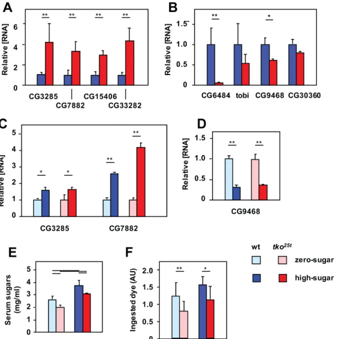
In previous transcriptomic analyses [9] we noted thattko25tadults manifested altered
expres-sion of some specific sugar transporters in the gut and Malpighian tubule (the equivalent of the mammalian kidney). Because of the unexpected observation that excess sugar is deleterious to
tko25tadults, we analyzed the expression of these genes, for clues to the mechanisms of
sugar-responsiveness oftko25t.
QRTPCR was used to profile the expression of two sets of relevant genes, in both adults and
in larvae, the life-cycle stage when the growth defect manifests intko25t[6]. We first analyzed a
set of Malpighian tubule transporters proposed to be involved in the excretion of excess sugar
[17]. Their expression was consistently elevated intko25t, both in L3 larvae (Fig 2A) and adults
(Panel A inS2 Fig), typically 3–4 fold. The most prominently expressed of these mRNAs,
CG7882 and CG3285, were also tested for responsiveness to dietary sugar at larval stage. In
both genotypes, their expression was elevated 2–3 fold on high-sugar compared with
zero-sugar food (Fig 2C). Next we analyzed a set ofα-glucosidases specific to, or highly enriched in
the gut, and putatively involved in the mobilization of dietary sugars, using a similar approach.
In both larvae (Fig 2B) and adults (Panel B inS2 Fig), these were down-regulated intko25t. A
prominently expressed representative of the set, CG9468, was further down-regulated by high-sugar diet in both genotypes (Fig 2D).
To understand the context of these changes in gene expression, we measured the total
serum sugar concentration oftko25tand control larvae grown on high-sugar versus zero-sugar
media.tko25tlarvae had significantly lower levels of total serum sugars than wild-type larvae
grown on the same medium. However, larvae cultured on high-sugar food had higher serum
sugar levels than those of the same genotype grown on zero-sugar food (Fig 2E).tko25tlarvae
also exhibited a lower rate of food consumption than control flies on the corresponding diet (Fig 2F), though on zero-sugar diet, where they grew faster, they paradoxically consumed less
food than on high-sugar diet. In all these aspects, the phenotype oftko25tlarvae is consistent
with a physiological strategy to minimize the amount of glucose, despite the initially presumed reliance on glycolysis.
Metabolic derangement of fly larvae with mitochondrial dysfunction is
exacerbated by high-sugar diet
The previous findings of up-regulation of lactate dehydrogenase (LDH) expression intko25t
adults [9] implied the use of LDH as an alternative pathway to regenerate NAD+, under condi-tions where mitochondrial respiration is limiting. We hypothesized that the resulting accumu-lation of lactate and/or the diversion of pyruvate from mitochondria may contribute to
metabolic disturbance intko25t, and underlie aspects of the mutant phenotype and its
exacerba-tion by high-sugar diet. We therefore analyzed lactate and pyruvate levels in L3 larvae. The
steady-state levels of both metabolites were found to be 2–3 fold elevated intko25tlarvae
com-pared with the wild-type control strain, when grown on either high- or zero-sugar medium (Fig 3A). When grown on high-sugar, this elevation was even more pronounced, at least for
lactate, although the differences were at the border of significance. Serum lactate was also 4–5
fold elevated intko25tlarvae (Fig 3A), and elevated lactate was maintained in adults (Panel A in
Using mass-spectrometry (S1 Table) to gain a more complete insight into the metabolic
abnormalities oftko25tlarvae, we observed a substantial ATP depletion (Fig 3B), as seen also in
adults (Panel A inS3 Fig) [9]. ATP levels were decreased in high-sugar diet in bothtko25tand
wild-type larvae, compounding the effects of genotype. NAD+and NADH levels were only
slightly altered by thetko25tmutation or by diet (Fig 3C), but we observed a striking
abnormal-ity in the level of NADPH and the NADPH/NADP+ratio (Fig 3C). In most physiological
Fig 2.tko25tflies manifest an‘anti-sugar’response.(A-D) Expression levels of various genes, based on QRTPCR, in L3 larvae of the indicated genotypes and growth conditions. (A, C) Malpighian tubule-specific sugar transporters, (B, D) gut-specificα-glucosidases; (A, B) all signals normalized to the levels in
wild-type larvae, (C, D) all signals normalized to larvae of the same genotype grown on zero-sugar medium (i.e., ignoring the differences between genotypes). All values are significantly different between genotypes or diets, as plotted (Student’sttest,p<0.01). (E) Total serum sugar concentrations in larvae of the indicated genotypes and growth conditions. Horizontal bars denote significantly different data classes (Student’sttest,p<0.01). (F) Larval feeding rate, based on dye ingestion assay, in larvae of the indicated genotypes and growth conditions. Horizontal bars denote significantly different data classes (Student’sttest,*showingp<0.05,**showingp<0.01).
contexts, there is substantially more NADPH than NADP+, which was the case in control
lar-vae grown on zero-sugar diet. The NADPH/NADP+ratio was decreased both by high-sugar
diet and by the presence of thetko25tmutation. The effects of genotype and diet were again
additive, so that intko25tlarvae grown in high-sugar the ratio was reversed. In two of the four
samples analyzed, NADPH was below the detection limit in mutant larvae grown on high
sugar (S1 Table).tko25tlarvae also showed altered levels of some amino acids (Panel C inS3
Fig), notably a deficiency of histidine, aspartic acid and asparagine (Fig 3D), and elevated levels of serine, alanine (especially on high-sugar) and threonine (only on zero-sugar). Two other
amino acid changes intko25tthat differed between diets were of citrulline and ornithine (Fig
3D), amino acids implicated in growth regulation by virtue of their role in polyamine
biosyn-thesis. Polyamines did show alterations intko25t(Panel D inS3 Fig), but the effect was the
Fig 3. Metabolites showing substantial changes intko25t
larvae.Relative levels of different metabolites in L3 larvae of the indicated genotypes and growth conditions, based on (A, B) findings from enzyme-linked assays or (C-F) mass spectrometry. Absolute values are shown for (B) ATP, (C) NAD+ and derivatives, (E) fructose 1,6-biphosphate (F1,6BP) and (F) gluconate, Values for (A) pyruvate, lactate and (D) amino acids are normalized to those for wild-type flies grown on ZS medium, enabling them to be plotted alongside for comparison. Relevant absolute values are given inS1 Table. Horizontal bars denote significantly different data classes (Student’sttest,p<0.05). SeeS7 Tablefor fuller statistical analysis of metabolite levels. (G) Triglyceride levels in L3 larvae of the indicated genotype and growth conditions, normalized to the value for wild-type larvae grown on zero-sugar medium. Horizontal bars denote significantly different data classes (Student’sttest,p<0.05). Note that we did not observe increased triglyceride levels when larvae were grown on high-sugar medium.
same, regardless of diet, whereas wild-type larvae showed clear increases in polyamine levels
when grown on high sugar. Note thatDrosophilahas only an incomplete urea cycle, though
urea was also greatly decreased intko25tlarvae (S1 Table).
In addition to elevated pyruvate and lactate, we noted substantial alterations in the level of two other glucose metabolites: the glycolytic intermediate fructose 1,6-biphospate, threefold
decreased intko25tlarvae regardless of diet (Fig 3E), and the glucose oxidation end-product
gluconate,>10-fold elevated on high-sugar diet irrespective of genotype (Fig 3F).
Finally,tko25tlarvae also showed a substantial triglyceride (TAG) accumulation not seen in
control larvae (Fig 3G).
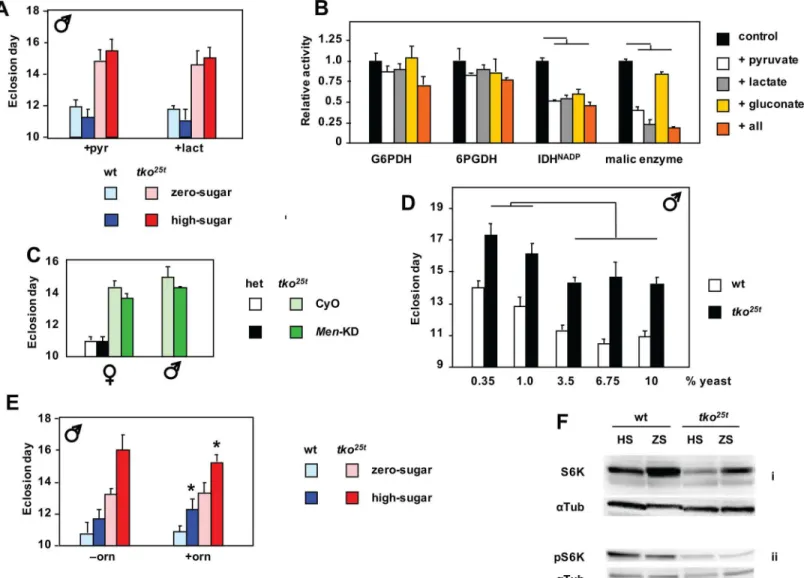
Lactate and pyruvate accumulation contribute to growth retardation and
NADPH depletion in larvae with mitochondrial dysfunction
We reasoned that the high levels of lactate and pyruvate seen intko25tlarvae may limit flux
through glycolysis, potentially accounting for a relative deficiency of ATP in animals largely dependent on glycolysis for ATP production. We therefore tested the effects of adding pyruvate or lactate to the culture medium. Lactate or pyruvate at 25 mg/ml, when added to zero-sugar
food, increased the developmental delay oftko25tflies by 1–2 days, partially phenocopying the
effect of high-sugar diet (Fig 4Aand Panel A inS4 Fig). The supplements also retarded the
devel-opment of control flies. However, when added to high-sugar diet, they had only a minimal effect. Next we considered whether the large changes in NADPH, which also correlated with the severity of the mutant phenotype on different diets, might also be influenced by excessive lac-tate and pyruvate. NADPH is required to drive biosynthetic reactions, notably fatty acid syn-thesis, but is also needed to maintain redox homeostasis. It is mainly produced by five
‘workhorse’enzymes of catabolism (Panel B ofS4 Fig), also linked to the provision of carbon
skeletons for biosynthesis. We therefore measured the activity of these enzymes in extracts
fromtko25tand control larvae, and the effects on their activities of the high levels of lactate and
pyruvate, as well as gluconate. The maximal activities of the NADPH-producing enzymes were
broadly similar in extracts fromtko25tand control larvae (Panel C inS4 Fig). However, when
we added lactate or pyruvate at the high concentrations observed intko25tlarvae grown in high
sugar, we saw inhibition of three of the enzymes that contribute substantially to NADPH pro-duction (Fig 4B). The degree of inhibition of malic enzyme by pyruvate may be an
under-esti-mate, since thein vitroassay is conducted in substrate excess, whereasin vivothe high levels of
pyruvate should decrease throughput to very low levels, via product inhibition. Gluconate also had a minor effect on NADP-linked IDH activity. Taking account of the likely inhibition of
malic enzymein vivo, and assuming that the effects of the tested metabolites on the other
enzymes are additive, this could partially explain NADPH depletion intko25tlarvae.
Further down-regulation of malic enzyme by RNAi produced no significant worsening of the developmental delay, and might even have slightly alleviated it (Fig 4C), whilst effects on
wild-type (tko25theterozygous) flies were minimal. A similar experiment to test for toxic effects
of gluconate (or the failure to detoxify glucose), by down-regulating glucose dehydrogenase (Gld), could not be meaningfully executed, since global RNAi against Gld was developmentally
lethal to both wild-type andtko25tflies, as is the null mutant [18]. However, advanced glycation
end-products did not accumulate in larvae grown on high-sugar diet (Panel B inS3 Fig),
indi-cating that they cannot account for the deleterious effects of high-sugar diet ontko25t.
How-ever, as discussed below, the accumulation of gluconate in larvae grown on high-sugar medium implies a mechanism for the additional depletion of NADPH resulting from diet.
The disturbed amino acid levels intko25tlarvae (Fig 3D) prompted us to consider whether a
this, we cultured flies on media containing a variable amount of yeast, the major source of die-tary protein, but a fixed sucrose concentration at (7.5%). Varying the amount of yeast produced
no change in the developmental delay oftko25tflies (Fig 4Dand Panel B inS1 Fig). Above a
threshold level of 3.75%, yeast supplementation had no further effect on eithertko25tor control
flies. At lower yeast concentrations, both were retarded further, but to the same extent, with
males (Fig 4D) and females (Panel B inS1 Fig) affected similarly. Similarly, high-protein diet
Fig 4. Metabolic phenotype oftko25tand its modulation.(A) Time to eclosion of male flies of the indicated genotypes and growth conditions, on medium supplemented with pyruvate (pyr) or lactate (lact). In the presence of either supplement there were no significant difference in eclosion timing betweentko25t flies grown in high-sugar versus zero-sugar medium (Student’sttest,p>0.05). See also Panel A inS4 Fig. (B) Effect on maximal activities of NADPH-producing enzymes fromDrosophila, of the presence of excess amounts the indicated metabolites (all = pyruvate, lactate and gluconate together). For comparison, all such activities are normalized to those without any additions. Horizontal bars denote values significantly different from control (Student’st test,p<0.01). (C) Time to eclosion of flies of the indicated sex, genotype and growth conditions. het–flies heterozygous fortko25t(which is recessive) and the FM7 balancer chromosome. Progeny carried either the CyO balancer chromosome marker or the RNAi construct for malic enzyme (Men-KD). (D) Time to eclosion oftko25t
and wild-type flies grown on media of the indicated composition (seeS1 Filefor details). Horizontal lines indicate significant differences between data classes on different media, for a given genotype (Student’sttest,p<0.05). On all media tested, eclosion times fortko25tflies were also significantly different from those of wild-type flies grown on the same medium (Student’sttest,p<0.01). For corresponding eclosion data of females see Panel B ofS1 Fig. (E) Time to eclosion of male flies of the indicated genotypes and dietary conditions, on medium supplemented (or not) with ornithine (orn), as shown.*denotes value significantly different than for flies of the corresponding genotype and dietary condition, with ornithineversusto without the supplement (Student’sttest,p<0.05). (F) Western blots of extracts from L3 larvae of the indicated genotypes and dietary conditions, probed for S6K or pS6K (phosphorylated at Thr-398) and theα-tubulin loading control (αTub). See also Panel F inS4 Fig.
had no detrimental effect on flies grown on low-sugar (Fig 1Cand Panels C-E inS1 Fig), and might even have had a slightly beneficial effect in the zero-sugar medium. Dietary
supplemen-tation with ornithine, found at unusually high levels intko25tlarvae grown on high-sugar diet,
had only very minor effects (Fig 4Eand Panels D and E inS4 Fig).
Growth inhibition in flies with mitochondrial dysfunction is associated
with modulation of S6K
Since the developmental outcome intko25tis still a viable fly, its developmental delay must be
the result of a coordinated signaling process with most likely both an intracellular and an
endo-crine component. In order to determine the molecular mechanism restraining growth intko25t
larvae on different diets, we interrogated the status of major signaling pathways already known to be involved in growth regulation. The best characterized such pathway responding to intra-cellular energy status is that of AMPK [19]. ATP depletion against a constant level of AMP, as
documented intko25tlarvae, should lead to activation of AMPK by phosphorylation at
Thr-172 (human numbering), which then down-regulates many downstream growth-related func-tions. Western blotting using AMPK-specific antibodies, revealed increased levels of
phosphor-ylated AMPK intko25tcompared to wild-type larvae (Panel F, sub-panels i and ii inS4 Fig)
which was further enhanced on high-sugar diet, although none of these changes was dramatic. The best-characterized response pathway to extracellular signals, including diet and insulin/ insulin-like growth factor signaling (IIS), involves the kinase Akt, whose activation by phos-phorylation at serine-505 has a growth-promoting readout that also facilitates glucose-related metabolism [20]. Accordingly, growth of wild-type larvae on high-sugar diet led to a modest
increase in the phosphorylated form of Akt (Panel F, sub-panels iii and iv inS4 Fig,) which,
taking account of the loading control, appeared to be partially abrogated intko25t. The Akt and
AMPK pathways converge on mTOR, the key regulator of cytosolic protein synthesis and growth, whose canonical readout is the phosphoryation status of S6K [21]. Consistent with this, we observed a decreased amount of the isoform of S6K phosphorylated at Thr-398 (one of
the mTOR target sites) intko25tcompared with wild-type larvae, although in both genotypes
the level of this isoform appeared marginally higher on high-sugar than zero-sugar diet (Fig 4F, panels ii). More strikingly, we observed a substantial decrease in total S6K intko25tlarvae, specifically on high-sugar diet (Fig 4F, panels i; biological replicates in Panel F, sub-panels v
and vi inS4 Fig). Analysis, by QRTPCR, of mRNA levels of the major insulin-like peptides
expressed in larvae, showed a consistent, but quantitatively minor up-regulation intko25t
(Panel G inS4 Fig; see alsoS2 Table).
Cystolic protein synthesis and secretion are key targets of the metabolic
crisis caused by mitochondrial dysfunction on high-sugar diet
Analysis of gene expression at the RNA level by global RNA sequencing revealed systematic
and diet-dependent changes intko25tlarvae that can be construed as a combined readout from
metabolic disturbance and decreased growth-signaling. We analyzed the data in two ways: firstly, according to the largest proportionate changes, and secondly, the largest absolute
changes, (S3 TableandS4 Table, respectively). We also analyzed separately the changes in
pro-tein-coding genes (Sheets A and C ofS3 Tableand Sheets A and C ofS4 Table) and non-coding
RNAs (Sheets B and D ofS3 Tableand Sheets B and D ofS4 Table), and executed all
compari-sons both by genotype (Sheets A and B ofS3 Tableand Sheets A and B ofS4 Table) and by diet
(Sheets C and D ofS3 Tableand Sheets C and D ofS4 Table: see also the full, unselected data in
As regards the non-coding RNAs, despite some striking changes in abundance, very few of them are functionally assigned at this time, and thus cannot be assessed further. The largest absolute changes in protein-coding genes were seen mostly in those which were already highly expressed, and fell into well-defined functional classes. Wild-type larvae on high-sugar diet,
compared with those grown on zero-sugar diet (Sheet C inS3 Table), showed strong induction
of genes connected with cytosolic translation, glycolysis, the structural proteins of muscle and the larval cuticle, whist the most strikingly down-regulated genes were gut-specific, and con-nected with digestive functions, including proteases, lipases, lysozymes, components of the peritrophic membrane and other chitin-binding proteins. In contrast, the vast majority of these genes were either less responsive, unaltered or even oppositely regulated by high-sugar
diet intko25tlarvae, where digestive functions were amongst the most prominently
up-regu-lated genes. Comparingtko25twith wild-type larvae (Sheet A inS3 Table), mitochondrial
tran-scripts were down-regulated on both diets, whilst the most striking differences on high-sugar diet were those already indicated above. Intriguingly, whilst many cytosolic translational
com-ponents were down-regulated intko25ton high sugar, one that was up-regulated was the
inhibi-tory factor Thor (4E-BP). Many of the genes showing the largest proportionate changes remain functionally unassigned, but genotype-specific changes were rather similar on the two diets
(Sheet A inS3 Table), whereas diet-specific changes (Sheet C inS3 Table) were largely different
between the two genotypes. One of the most highly induced genes intko25tlarvae irrespective
of diet was p24-2, whose mRNA was induced over 1000-fold compared with its level in control larvae. p24-2 is one of two fly homologues of yeast p24, a protein required for vesicle trafficking in the secretory pathway. Up-regulation of p24-2 suggests ER stress, but we saw no changes in
the level or splicing pattern of Xbp1 RNA (Panels H and I inS4 Fig), considered a marker for
the induction of the classic ER stress response. Other prominent changes intko25tlarvae
involved genes involved in antimicrobial defense. An unbiased annotation enrichment analysis of all genes showing at least two-fold changes in expression (S5 Table), executed using the
DAVID (Database for Annotation, Visualization and Integrated Discovery,http://david.abcc.
ncifcrf.gov) gene ontology database tools, reached broadly similar conclusions.
tko
25tis immune to the growth-inhibitory effects of cytosolic protein
synthesis inhibition
The strikingly opposite sugar-dependent regulation of highly expressed genes for the
machin-ery of cytosolic protein synthesis and secretion intko25tcompared with wild-type larvae could,
in principle, be construed as the cause or consequence of the metabolic disturbances and growth impairment: in other words it could be an adaptive response to limit growth under deranged metabolic conditions, or could be a maladaptive consequence of such disturbances. Thus, we reasoned that imposing an additional stress on the protein synthetic apparatus would worsen the phenotype if the above changes are maladaptive, but would be neutral or possibly even beneficial, if down-regulation of protein synthesis and secretion were part of an adaptive response.
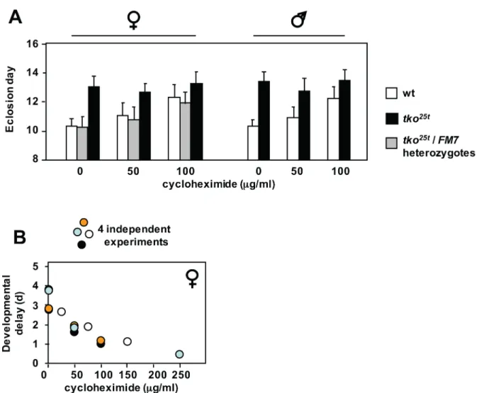
To address this, we cultured wild-type andtko25tflies on media containing low doses of
cycloheximide, an inhibitor of cytosolic translational elongation. Cycloheximide produced a
dose-dependent growth retardation in wild-type flies, but had almost no effect ontko25tmutant
flies, if anything tending to accelerate their development very slightly, especially when analyz-ing the progeny only from eggs laid on the first day of exposure to the drug, thus minimizanalyz-ing
any confounding effects on egg-laying behaviour or oogenesis (Fig 5A and 5Band Panels A-C
addition,tko25tflies were immune to the growth-retarding effects of tunicamycin seen in wild-type flies, when the drug was added to the growth medium at the highest practical dose of
12μM (Panel E inS5 Fig).
The gut is a crucial tissue for the developmental delay phenotype of
tko
25tflies
The striking down-regulation of components of the machinery of cytosolic protein synthesis
and secretion intko25tlarvae, combined with the observation that the effect of the mutation is
epistatic to the developmental delay introduced by low doses of cycloheximide, suggests that one or more tissues responsible for the synthesis of secreted proteins are key targets of the mutation. An obvious candidate is the gut, especially since many secreted gut proteins showed
oppositely altered expression intko25tcompared with wild-type larvae. We therefore tested
whether gut-specific expression of the wild-typetkogene in thetko25tbackground could
Fig 5. Effect of cycloheximide on development oftko25t
and wild-type flies.(A) Means±SD of times to eclosion of flies of the sex and genotypes indicated, on high-sugar medium, with or without cycloheximide at the indicated concentrations. Based on pairwisettests, and considering all the flies of a given sex and genotype cultured at a specific drug concentration as a single population, mean eclosion times were significantly different (p<0.05) at different cycloheximide concentrations, apart fromtko25t
flies of either sex at 100μg/ml, which were not different fromtko25t
flies cultured in the complete absence of the drug. (B) Pooled eclosion data from four independent experiments conducted with different concentration ranges of cycloheximide. Female
developmental delay showed consistent decrease with increasing cycloheximide concentration. Males showed same trend (Panel C inS5 Fig).
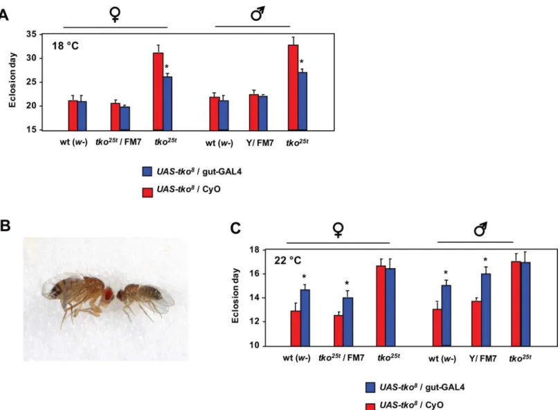
alleviate the mutant phenotype. To do this, we made use of a previously constructed transgenic
line containing a GAL4-dependent copy oftko(UAS-tko8) [22], which we combined with a
gut-specific GAL4 driver line (Kyoto 113094,S6 Fig), in thetko25tand wild-type backgrounds.
The effect was extremely temperature-dependent. At 18°C theUAS-tko8and Kyoto 113094
driver combination had no significant effect in the wild-type background, buttko25tflies
showed a clear, though partial rescue of their very long developmental delay at this temperature
(Fig 6A). At 26°C the combination ofUAS-tko8and the Kyoto 113094 driver was lethal or
semi-lethal, with just a small number of escaper flies eclosing in the wild-type background, with an ~4 day developmental delay, and exhibiting a minute phenotype (Fig 6B). At 22°C the
combination remained deleterious in the wild-type background, with flies showing a 1–2 day
developmental delay, but in thetko25tbackground there was no such effect (Fig 6C).
Fig 6. Partial rescue oftko25tby gut-specific expression oftko.(A, C) Means±SD of times to eclosion of flies of the sex and genotypes indicated, on high-sugar medium, at (A) 18°C or (C) 22°C. Asterisks denote significant differences in pairwisettests (p<0.01) between flies of a given sex and genotype carrying the gut-GAL4 driver (Kyoto line 113094) compared with the corresponding flies carrying the CyO balancer chromosome instead. (B) Minute phenotype of the few flies eclosing with the genotypeUAS-tko8
/ gut-GAL4 when cultured at 26°C.
Discussion
Our starting point in this study was the erroneous assumption that the limited capacity of
OXPHOS intko25tflies would render them highly dependent on glycolysis, and thus addicted
to sugar. However, increasing the sugar content of their diet instead led to exacerbation of the mutant phenotype. Comparisons of larvae grown on different diets revealed a set of metabolic
disturbances intko25tlarvae (high pyruvate and lactate, low ATP and NADPH) that were
gen-erally accentuated by high-sugar diet. Growth in high-sugar produced strikingly opposite changes in gene expression in wild-type and mutant larvae, notably affecting the apparatus of cytosolic protein synthesis and secretion. In addition, we observed altered cell signaling,
con-verging on the cytosolic translation machinery, whilsttko25twas immune to the growth
inhibi-tory effects of low levels of cycloheximide, an inhibitor of cytosolic translation.
Key metabolic consequences of mitochondrial dysfunction
Many of the metabolic disturbances seen intko25tlarvae are consistent with previous studies of
OXPHOS deficiency, although their exacerbation by high-sugar diet is novel. Elevated lactate, pyruvate and alanine are common findings in mitochondrial disease patients [23], considered to represent increased dependence on glycolysis under conditions where mitochondrial
NADH re-oxidation is impaired [24].tko25tlarvae showed decreased levels of the glycolytic
intermediate immediately upstream of the rate-limiting step, i.e. fructose 1,6-biphosphate (Fig 3E), consistent with increased glycolytic flux. Lactate accumulation may reflect the need for lac-tate dehydrogenase as an alternative pathway to regenerate NAD+ but, since it is accompanied by high pyruvate, may also be a consequence of decreased TCA cycle capacity due to the OXPHOS defect. Pyruvate accumulation may be partially adaptive, by feeding anaplerotic
pathways, but addition of dietary pyruvate (or lactate) totko25tpartially phenocopied the
effects of high-sugar diet, implying these end-products of glycolysis to be important agents of the metabolic crisis that limits growth, even if only via product inhibition of glycolytic flux, thus compromising the maintenance of ATP levels. The reason for the additional burden of pyruvate and lactate resulting from high-sugar diet is not immediately obvious. It may partly be a consequence of increased substrate availability for glycolysis and other catabolic pathways that converge on pyruvate. Logically, however, it also reflects maladaptive effects of sugar-dependent signaling.
Whisttko25tlarvae are able to maintain normal NAD+/NADH levels, NADP+/NADPH
homeostasis is severely disturbed (Fig 3C). As well as its role as an electron donor for biosyn-thesis, NADPH buffers oxidative stress, most importantly as a cofactor in the regeneration of
the reduced form of thioredoxin [25], which inDrosophilareplaces the functions of glutathione
reductase [26]. Mitochondria fromtko25tflies produce excess ROS [16], potentially accounting
in part for NADPH depletion. High levels of pyruvate and lactate, which we demonstrate to inhibit key NADPH-generating enzymes (Fig 4B), notably IDH and malic enzyme, may also play an influence.
The further depletion of NADPH intko25tgrown in high sugar (Fig 3C) appears to be a
compounding of these effects with an independent effect of high-sugar diet. Excess unmodified glucose is toxic because of its ability to react with the lysine side-chains of proteins, bringing
about their irreversible inactivation via the Maillard reaction [27,28]. This toxicity is believed
to play a major role in the pathology of diabetes (reviewed in [29]), and organisms use a variety
of strategies to minimize exposure of the tissues to excess glucose.Drosophilacan detoxify
glu-cose via the FAD-linked enzyme gluglu-cose dehydrogenase (Gld, CG1152, EC 1.1.99.10), which
produces gluconate as an inert end-product, avoiding the generation of peroxide [30,31].
Although the exact electron acceptor for Gld is not known [31], its regeneration should depend on the cytochrome P450 system, in which terminal oxidation is coupled to NADPH
consump-tion, in the general reaction A-H2 + 2NADPH + O2!2H2O + 2NADP+ A (where A is a
generic electron acceptor). An effect on NADPH is evident even in wild-type larvae (Fig 3C), although it does not appear to impair development in the absence of the other metabolic
dis-turbances seen intko25t. However, its compounding with the effects of OXPHOS deficiency in
tko25tleads to the highly abnormal situation of a reversal in the NADPH/NADP+ ratio that
must have serious consequences for many cellular processes. Notably, the processing of
secreted proteins in the ER is highly dependent on NADP+/NADPH homeostasis [32,33].
Dis-turbed proteostasis in the secretory pathway [34] would be predicted to affect those tissues most exposed to dietary glucose and most dependent on secretion, i.e. the gut and its associated organs, and the epidermis, which secretes the larval cuticle at each molt. This is concordant with the altered patterns of gene expression that we observed (S4 Table).
The ability of theDrosophilalarval gut to mount a classic ER stress-response is limited.
Xbp1, the key transcription factor required to effect the response, is already reported to be con-stitutively activated in the larval gut [35], consistent with our own findings (Panerls H and I of S4 Fig). Neither diet nor thetko25tmutation had any further effect on its global expression level or splicing, suggesting that there is only a limited capacity for handling further proteotoxic stress in the ER. The activation of accessory pathways for secretion, such as involving p24-2
(Sheet A inS3 Table), may be a signature of this. P24 proteins are involved in the sorting of
gly-cosylphosphatidylinositol (GPI) anchored proteins into COPII vesicles [36,37] which then
transit to the cell surface via the Golgi. The down-regulation, at the RNA level, of the apparatus
of cytosolic protein synthesis and secretion (Sheets A and C inS4 Table), combined with active
mechanisms to attenuate both processes (Fig 5andS5 Fig), such as via S6K modulation (Fig 4F
and Panel F inS4 Fig), can be rationalized as a response to proteotoxicity in the ER, due to
redox disturbance.
ATP deficiency is a frequently observed or predicted effect of OXPHOS insufficiency,
including previous reports ontko25tand otherDrosophilamutants [10,12,16,38,39].
How-ever, decreased OXPHOS capacity and limitations on glycolysis due to high pyruvate and/or lactate may not be the only reasons for ATP depletion. Protein folding and refolding are highly ATP-consuming processes, as is the proteolytic degradation of misfolded or aggregated pro-teins [40]. Disturbed ER proteostasis in the ER may therefore be a drain on already decreased levels of ATP.
The consequences of the amino acid abnormalities detected intko25tlarvae are less clear.
Deficiencies in specific amino acids are expected to trigger growth arrest and inhibition of cyto-solic protein synthesis through the TOR pathway independently of the effects of ATP
deple-tion. However, dietary supplementation with protein did not rescuetko25t. The amino acid
imbalances in mutant larvae (Fig 4D) are therefore best considered as a side-effect of pyruvate overload that, on their own, are insufficient to trigger growth arrest. Excess alanine, also seen
in patients with elevated pyruvate [23,41], probably results from the action of alanine
amino-transferase [42]. Similar arguments may apply to serine. The abnormalities in ornithine and citrulline (Fig 3D), combined with the observed changes in mRNAs for enzymes involved in
their metabolism (S3andS4Tables), are harder to interpret, as are effects on polyamine levels,
none of which offer any explanation for growth limitation intko25t(Panel C inS3 Fig).
More-over, ornithine supplementation in zero-sugar diet did not phenocopy the effects of high sugar, and may even have slightly alleviated it (Fig 4E).
Since it is difficult to manipulate the levels of pyruvate, ATP and NADPH independently, their relative contributions to the developmental phenotype are difficult to apportion.
alleviation of the phenotype (Fig 4C). However, any decrease in flux should directly decrease both NADPH and pyruvate, whilst decreased pyruvate may in turn allow an increased rate of NADPH production and facilitate glycolytic flux. Unknown allosteric effects, undetected metabolites, subcellular compartmentalization of substrates, products and enzymes, and
tis-sue-specificity of metabolic pathways may impact the diet-dependent metabolic crisis oftko25t,
summarized inFig 7.
Growth-related signaling
The metabolic crisis oftko25tdoes not lead to lethality or grossly abnormal development, but to a
decreased developmental rate, tailoring growth to resources and the ability to mobilize them, and resulting in a viable adult fly. This indicates the operation of a programmed response mechanism. We postulate that, in the wild, this would allow the fly to survive in different nutritional environ-ments, perhaps including those in which it may be exposed to toxins targeted on OXPHOS or mitochondrial translation. The response clearly has both a transcriptional and post-translational dimension, though the underlying signaling machineries may overlap, and may also operate tis-sue-specifically, accounting for the many changes in expression of genes expressed uniquely in the gut, muscle or epidermis. Furthermore, although transcriptional responses most likely under-lie the major changes in mRNA levels, we cannot exclude a contribution from non-coding RNAs
regulated in response to thetko25tmutation or the sugar content of the diet.
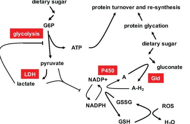
Fig 7. Summary model of the metabolic phenotype oftko25t
in high-sugar medium.High dietary sugar and mitochondrial dysfunction stimulate glycolysis, which is nevertheless limited by the build up of pyruvate and/or lactate, produced by lactate dehydrogenase (LDH, needed to regenerate NAD + when OXPHOS is impaired), restricting ATP production. Pyruvate and lactate accumulation restricts NADPH production, whilst NADPH consumption is increased by the need to maintain glutathione in the reduced state, despite increased mitochondrial ROS production, and by the need to reoxidize the electron acceptor (A) for glucose dehydrogenase (Gld) using the P450 system, preventing further peroxide generation. Glucose dehydrogenase converts excess glucose to gluconate, whilst any glycotoxic damage is compensated by the normal processes of protein refolding, turnover and re-synthesis, which consume ATP.
We found evidence for the involvement of several well-characterized signaling pathways converging on cytosolic protein synthesis, notably including the ATP-responsive AMPK and nutrient-sensitive (Akt) pathways, as well as the key target of TOR signaling, S6K (Fig 6B).
Strikingly,tko25tflies were immune to the growth-inhibitory effect of low doses of
cyclohexi-mide, consistent with the translational apparatus being a key downstream target of regulation
in the mutant. This is unlikely to be due to drug exclusion. Although induced intko25tadults
[9], the Mdr-related transporter l(2)03659 is expressed at slightly lower levels intko25tthan
wild-type larvae (S2 Table) and is not sugar-responsive. Moreover,tko25tflies are more
sensi-tive than wild-type flies to doxycycline [6], an inhibitor of mitochondrial rather than cytosolic protein synthesis.
Cycloheximide inhibits the elongation step of the ribosome cycle [43], which is normally not rate-limiting. Instead, initiation is the target of most translation-regulatory mechanisms, e.g. via the protein kinases targeting initiation factors such as eIF-2A or eIF-4E [44]. However,
it would appear that initiation is already working at close to maximal capacity in wild-type
Dro-sophilalarvae, since cycloheximide inhibits growth in a dose-dependent manner even at low
concentrations. In contrast, at doses up to 100μg/ml,tko25tflies were refractory to
cyclohexi-mide (Fig 5Band Panel C inS5 Fig), indicating a condition where initiation, rather than
elon-gation, is constrained, and is thus rate-limiting for protein synthesis and growth. The altered levels of phosphorylated isoforms of known signaling molecules is consistent with the implied
down-regulation of translational initiation intko25t, and its exacerbation by high-sugar diet.
Thus, while total levels of the AMPKα-subunit are little altered,tko25tflies show increased
amounts of the isoform phosphorylated at Thr-172, which attenuates TOR signaling [19], but a corresponding decrease in the isoform of Akt phophorylated at Ser-505, which activates TOR. The increase in AMPK phosphorylation is consistent with the observed ATP depletion [45], but the mechanism by which Akt signaling attenuated remains unknown, and could involve any of the metabolic changes associated with mitochondrial dysfunction.
Consistent with these changes,tko25tshows decreased amounts of the isoform of the key
TOR target [46] S6K, phosphorylated at Thr-398, which in turn activates translational initia-tion [47]. However, all of these changes are rather modest, raising doubts as to whether they, alone, could account for an effective halving of the growth rate during larval development.
The most important new link between S6K and growth regulation is our finding that the level of S6K protein itself (specifically, the homologue of human S6K1) is strongly decreased in
tko25tlarvae grown on high-sugar medium (Fig 4Fand Panel F inS4 Fig). A recent report of
the effects onDrosophilalifespan of different diets, combined with knockdown of an ATP
synthase subunit [48], also showed modulation of the levels of S6K protein. Although less dra-matic than the S6K changes observed here, this may indicate a parallel or accessory stress-response pathway, linking diet, mitochondrial function and regulation of cytosolic translation. Neither Sun et al. [48] nor ourselves were able to address the tissue specificity of S6K modula-tion, and the underlying molecular mechanism remains unknown, but our data raise a number of interesting, non-mutually excusive possibilities. The first is that further activation of AMPK
in susceptible tissues (Panel F inS4 Fig), resulting from the more profound depletion of ATP
(Fig 3B), triggers S6K turnover, e.g. via the ubiquitin-proteasome system. A second would invoke a regulator akin to AMPK, but activated instead by the abnormal NADPH/NADP + ratio. A third such putative regulator would be sensitive to elevated gluconate (Fig 3F), which may be considered a specific indicator of the stress of high dietary sugar in the fly. A further possibility is that S6K levels are regulated by the downstream consequences of these metabolic changes, for example in response to proteotoxic stress or translational imbalances (see
The functional differences between modified and unmodified isoforms of S6K are poorly understood, but are likely to extend beyond global regulation of translation [49]. Many of the
large quantitative differences in gene expression at the RNA level seen intko25tgrown in high
sugar were opposite to those induced by high-sugar diet in wild-type flies, notably those affecting the machineries of cytosolic protein synthesis and secretion as well as glycolysis. This suggests a regulatory event targeting the system responsible for glucose induction of gene expression which includes transcription factors such as sugarbabe [50] and Mondo/Mlx [51], but may also have a post/transcriptional component. These factors, as well as any proteins that interact with them directly or indirectly, should thus be considered as candidates for regulation by S6K.
We did not identify a specific systemic response mechanism to mitochondrial dysfunction
intko25tthat would correspond with the endocrine biomarkers GDF15 [52] or FGF21 in
mammals [53]. Neither GDF15 nor FGF21 have orthologues inDrosophila, although both are
members of well-characterized growth factor families that are well represented in the fly. No
members of these families showed significantly altered expression at the RNA level intko25t,
although this does not preclude regulation at other levels. Expression of insulin-like peptides
was also slightly increased, not depressed intko25t(Panel G inS4 Fig). Whilst the
down-regula-tion of growth intko25tmight be at least partially cell-autonomous, the fact that it appears to
emanate from a deficiency of mitochondrial protein synthesis in a specific tissue (i.e. the gut, Fig 6), and that one readout is altered feeding behaviour (Fig 2D), it is likely to involve either a known [54] or still unknown endocrine circuit centered on the gut.
Fig 8. Summary model of the growth phenotype oftko25tin high-sugar medium.NADPH depletion disturbs protein homeostasis in the ER (and may also be exacerbated by it, in a vicious cycle) consuming further ATP. ATP depletion activates AMPK, inhibiting TOR complex activation by phosphorylation (denoted as pTOR), and consequently limiting the activation of S6K and other growth-stimulatory effectors by phosphorylation. Mitochondrial dysfunction is proposed to inhibit Akt activation by an unknown process (dotted line), limiting its activation by dietary sugar. One or more of lactate/pyruvate accumulation, ATP and NADPH depletion, or their combined effects on protein homeostasis (red line), are proposed to stimulate S6K turnover, thus contributing further to the down-regulation of protein synthesis.
Tissue-specificity of the effects of mitochondrial dysfunction in
development
Five pieces of evidence implicate the gut as a crucial tissue mediating the growth retardation of
tko25tlarvae, particularly its enhancement in high-sugar diet. First, based on dye ingestion, we
observed a decreased food intake (Fig 2F). Second, specific gut alpha-glucosidases were down-regulated (Fig 2A), and further down-down-regulated in high sugar (Fig 2D), suggestive of decreased sugar absorption and accounting, at least partially, for decreased serum sugar (Fig 2E). Third, mRNAs coding for gut-specific enzymes were amongst those most responsive to diet and
geno-type, many showing clearly opposite behaviour intko25tcompared with wild-type (Sheets A
and C inS4 Table). The transcriptional readout of mitochondrial dysfunction in relation to
diet is therefore largely gut-specific. Of note also is the fact that, in high sugar,tko25tlarvae
showed down-regulation of many mRNAs coding for components of the machineries of
cyto-solic translation and secretion (Sheet A inS4 Table) and, at the protein level, of S6K (Fig 4F
and Panel F inS4 Fig), considered a key regulator of translation, whilst the gut is one of the
most important secretory organs. Fourth, the additional metabolic stress arising from NADPH depletion (Fig 3C) as a by-product of glucose oxidation must logically occur in a tissue exposed
to high sugar levels. Given the fact that serum sugar is not elevated intko25t(Fig 2E) the only
major tissues exposed to high sugar should be the epidermis and the gut. Finally, when directly
tested using a gut-specific driver at 18°C, expression of the wild-typetkogene was able partially
to rescue the developmental delay oftko25t(Fig 6A), implicating the gut as a major tissue where
insufficiency of mitochondrial protein synthesis impairs growth. The effect of GAL4 drivers is known to be temperature-dependent [55], with increased activity at 25°C compared with 18°C.
Intriguingly, gut-specific over-expression of wild-typetkoin the wild-type but not thetko25t
background at 22°C also produced a developmental delay (Fig 6C), suggesting that the expression
level of thetkogene product (mitoribosomal protein S12), which in bacteria is a key component
of the small subunit assembly pathway [56], may need to be tightly regulated in order to ensure the appropriate ratio of mitochondrial and cytosolic translational capacity. Over-expression of
tkoat even higher temperatures resulted in a phenotype of semi-lethality, with escapers having a
classic minute phenotype (Fig 6B), seen previously in mutants with cytoribosome deficiency [57]
or defective insulin signaling [58], but not intko25t. The finding further highlights the importance
of coordinated protein synthesis in cytosolic and mitochondrial compartments.
Relevance to pathophysiology of metabolic and mitochondrial disease
Despite its sensitivity to high-sugar diet,tko25tcannot be considered an exact model for
diabe-tes in humans, since serum sugar levels are lower in the mutant than in wild-type flies (Fig 2E). The expression of several of the insulin-like peptides is slightly increased at the RNA level
(Panel G inS4 Fig), but their exact functions in metabolic or growth regulation remain unclear,
and mRNA levels may not reflect those of the peptides themselves [59]. Moreover, pAkt,
con-sidered a primary mediator of insulin signaling [20,60], although down-regulated intko25t
compared with wild-type (Panel F inS4 Fig), was still modestly increased in response to
high-sugar diet (Panel F inS4 Fig).
Changes in insulin-signaling may contribute to lowered serum sugar (Fig 2E), enhanced expression of excretory sugar transporters (Fig 2A and 2C), decreased muscle biosynthesis
(Sheets A and C inS4 Table) and increased triglycerides (Fig 3G), but these may be considered
as adaptive responses to mitochondrial dysfunction rather than a pathological signature and, at
least to some degree, are seen also intko25tlarvae grown on zero-sugar medium.
Because it is also manifest on zero-sugar medium, the elevated level of triglycerides seen in
deposition represents an alternate system for storing energy needed to fuel metamorphosis, partially replacing the accumulation of muscle. Larval body wall muscle is largely turned over
during metamorphosis [61], buttko25tlarvae exhibit down-regulation of mRNAs coding for
structural proteins of muscle (Sheet A inS4 Table). The response of wild-type larvae to
high-sugar diet involves up-regulation of muscle and cuticular components, as well as glycolysis and
the cytosolic translation system (Sheet A inS4 Table). Unlike yeast or cancer cells, where
high-glucose media promote more rapid growth [62], wild-typeDrosophilalarvae do not grow more
rapidly on a high-sugar diet, possibly even slightly slower (Fig 1and Panel A inS1 Fig).
Increased biosynthesis of cuticle and muscle might be a signature of an avoidance response to a high-sugar environment. As already noted, sugar overload should be toxic to the epidermis and to the gut, due to protein glycation and/or NADPH depletion resulting from glucose oxi-dation. Increased production of cuticle proteins may protect the epidermis, whilst the accumu-lation of body wall muscle will facilitate the movement of the larva to a less stressful
environment. The metabolic consequences of mitochondrial dysfunction may limit this
response intko25tlarvae. Muscle is also one of the most energy-consuming tissues, and
devel-opmental reprogramming that limits muscle biosynthesis, storing biomass for later catabolism during metamorphosis instead as fat, may be considered an energy-sparing response to ATP depletion that is signaled via AMPK or its downstream targets.
The metabolic crisis experienced bytko25tlarvae in high sugar diet may have a more general
relevance in mitochondrial disease. Other data supports the idea that ketogenic or low
carbo-hydrate diet is beneficial for mitochondrial disease patients or animal models thereof [63,64].
The combination of ATP deletion and glycotoxic stress is likely to occur in many contexts rele-vant to human pathology. Although glucose homeostasis operates differently in mammals, some secretory tissues are naturally susceptible to glucose overload, notably the gut, and per-haps most importantly, the islet cells of the pancreas, which must respond to physiological
fluc-tuations in glucose.DrosophilaGld does not have a strict orthologue in mammals: however, it
is member of a multigene family of choline-glucose dehydrogenases that ultimately consume NADPH, whose many mammalian members remain functionally unidentified.
Our findings implicate pyruvate overload, ATP depletion and disturbed NADP/NADPH homeostasis as key elements of the metabolic crisis resulting from OXPHOS deficiency, espe-cially in the presence of high dietary sugar, with deranged protein homeostasis in the secretory pathway as an unexpected outcome. As well as suggesting possible new drug targets in mito-chondrial disease, they may also be relevant to cancer. To facilitate rapid growth, many tumors switch to aerobic glycolysis for ATP production (the Warburg effect), at the same time fre-quently down-regulating OXPHOS, commonly by accumulating mtDNA mutations. However,
if what is true ofDrosophilalarvae is also true of such tumors, high glucose levels should
pro-voke a metabolic crisis that curtails growth or may even lead to apoptosis, if NADPH/NADP + homeostasis is pushed beyond a certain limit. Since many of the commonly used drugs in cancer chemotherapy are metabolized via the NADPH-consuming cytochrome P450 system, NADPH depletion should be considered a possible mechanism by which they are really pro-voking tumor destruction. In this case, tumors that become chemo-resistant via activation of multi-drug resistance pathway might still be vulnerable to glucose overload.
In humans, pyruvate treatment has been suggested to be effective in some cases of mito-chondrial disease [65], whereas other studies found no convincing benefits from treatments such as with dichloroacetate, designed to increase mitochondrial pyruvate utilization and thus
decrease cytosolic pyruvate load (for review see [66]). Intko25tlarvae, pyruvate
mitochondria to metabolize pyruvate. Therapies targeted on pyruvate might therefore cause more harm than good, by increasing pyruvate overload in the mitochondria.
Experimental Procedures
Drosophila
stocks and maintenance
tko25t[3] in the Oregon R background was as described previously [6].w1118, standard
balanc-ers, gut-specific Kyoto GAL4 line 113094, and VDRC RNAi lines targeted onMenandGld
(#104016 and #108361, respectively) were obtained from stock centers. Except for use in spe-cific experiments, flies were maintained at room temperature in plugged plastic vials
contain-ing standard high-sugar medium (seeSIfor full details), supplemented with 0.5% propionic
acid (Sigma-Aldrich) and 0.1% (w/v) methyl 4-hydroxybenzoate (Nipagin, Sigma-Aldrich), then transferred to vials containing appropriate media for mating and larval culture in a 12 h
light-dark cycle at 25°C, except where stated. To avoid selection of suppressors,tko25tflies were
maintained using FM7 balancer and homo/hemizygotes generated as needed for experiments. In general, to avoid any confounding effects from the balancer, wild-type flies in the same
back-ground were used as controls in parallel in all experiments. UAS-tko8flies, described previously
[22] were maintained in thetko25tbackground, and their phenotype rechecked prior to
experiments.
Culture media
Fly food was created according to different recipes as detailed in SI. Most experiments used
standard high-sugar medium [67], denoted HS, and‘zero-sugar’medium, denoted ZS,
contain-ing only agar, dried yeast extract, soya and maize flours, but no added sugars. Isocaloric media with lowered sugar content but other components in varying proportions were as indicated in S1 File. Various supplements, as indicated in figures and legends, were generally added from stock solutions after medium was cooled to 50°C, including sodium pyruvate or lactate (25 mg/
ml), ornithine (5 or 20μg/ml) and cycloheximide (up to 250μg/ml). For tunicamycin (12μM),
low-melting point agarose was used instead of agar, wth cooling to 37°C before addition of drug.
Developmental time and behavioral assays
Crosses were conducted in a minimum of 3, usually 4 or 5 replicates, and mean developmental time to eclosion (at 25°C, except where indicated), as well as bang-sensitivity were measured as described previously [15]. Standard deviations were calculated based on the distributions of mean eclosion day from replicate vials (except where indicated) or, for bang-sensitivity, from the distribution of recovery times from mechanical shock of all individual flies of a given sex
and genotype tested (in batches of 30–50 flies). Larval feeding behaviour was assayed by dye
ingestion. Briefly, individual larvae grown on HS or ZS medium were placed on petri dishes of the same medium containing 0.16% erioglaucine for 20 min, washed with PBS, dried, and then
homogenized in 100μl PBS. Homogenates were centrifuged at 12,000 gmaxand dye uptake was
measured spectrophotometrically (absorbance at 630 nm) using the supernatant fractions.
Metabolite analysis
Steady-state levels of ATP, lactate and pyruvate were measured by enzyme-linked luminometry (ATP) or fluorometry in extracts from batches of 20 larvae or adult flies, using commercially
available kits according to manufacturer’s protocols (Molecular Probes, Life Technologies for
were treated overnight with trehalase (Sigma-Aldrich), following which total serum sugars were assayed using glucose (HK) reagent (Thermo Scientific). Triglyceride levels were mea-sured essentially according to Tennesen et al. [68], using triglyceride reagent (Thermo Scien-tific), with subtraction of the background signal due to free glycerol. For global metabolite analysis, batches of 15 larvae were snap-frozen in liquid nitrogen, deproteinized by chloroform extraction, centrifugation and micro-filtration, then analyzed separately for anions and cations
by CE-TOFMS in the presence of suitable standards. For full details seeS1 File.
Enzymatic analyses
Malic enzyme, glucose-6-phosphate dehydrogenase, 6-phosphogluconate dehydrogenase and
isocitrate dehydrogenase were assayed essentially as described previously [69,70,71]. Isocitrate
dehydrogenase and glutamate dehydrogenase activities were also analyzed separately using a commercially available IDH activity kit (Abcam), in order to evaluate the contribution of
NAD+/NADH- and NADP+/NADPH-dependent isoforms to the total enzyme activity. SeeS1
Filefor full details.
RNA analysis
For QRTPCR, RNA extraction fromDrosophilaadults and larvae, cDNA synthesis, PCR and
data analysis were performed as described previously [67], using primer sets shown inS6
Table. For RNA-sequencing, RNA was extracted from flash-frozen batches of 30 larvae using
miRNA Easy Mini Kit (Qiagen) and manufacturer’s instructions. Three biological replicate
samples were produced by pooling 4 independent preparations to produce each replicate. RNA sequencing was performed on Hiseq 2500 sequencers (Illumina) using paired-end library and 100 bp read-legnth and otherwise standard protocols. Expression analysis was performed using
Chipster. Sequencing reads were mapped to theDrosophilareference genome (BDGP release
5.72) using TopHat version 2.0.9, and differential expression analysis was performed using CuffDiff. The splicing pattern of Xbp1 was analysed by RT-PCR, PstI digestion and agarose gel electrophoresis [72].
Protein analysis
Total protein was extracted from batches of 20 frozen larvae in phosphate buffer containing 150 mM NaCl, 1 mM EDTA, 2 M urea, 1.3% (w/v) SDS, 10 mg/ml each Complete protease inhibitor mix (Roche) and PhosSTOP phosphatase inhibitor mix (Roche), diluted into 4 x
Laemmli loading buffer and (seeS1 Filefor details) and electrophorsesed on Criterion TGX
AnyKD precast SDS-PAGE gels (Bio-Rad). Protein was transferred to 0.45μm Hybond ECL
nitrocellulose membrane (Amersham, GE Heathcare Life Sciences). Blots were processed and
visualized by standard methods (seeS1 Filefor full details). Primary antibodies, used at 1:1,000
dilution, were against: Akt #4691 (Cell Signaling), phospho-Akt #4054 (Cell Signaling), AMPK #80039 (Abcam), phospho-AMPK #4188 (Cell Signaling), S6K #64804 (Abcam), phospho-S6K #9029 (Cell Signaling) and alpha-tubulin #52866 (Abcam, 1:10,000) with appropriate HRP-conjugated secondary antibodies (Vector Laboratories, 1:10,000): Horse Anti-Mouse IgG
#PI-2000 or Goat Anti-Rabbit IgG #PI-1000. For further details seeS1 File.
Supporting Information
S1 Fig. Supplementary data on modulation oftko25tphenotype by diet.Time to eclosion of
tko25tand wild-type flies of sex as shown, grown on media of the indicated composition (seeSI
grown on 0% sucrose medium (Student’sttest,showingp<0.05,showingp<0.01). For
corresponding eclosion data of males seeFig 1A. In (B) and (E), horizontal lines indicate
signif-icant differences between flies of a given genotype, grown on different media (Student’sttest,
showingp<0.05,showingp<0.01). For corresponding eclosion data of males grown on
high-sugar media, seeFig 4D. In (C) asterisks () denote significant difference from flies of the
same genotype grown on all other media tested (Student’sttest,p<0.01), which were not
sig-nificantly different from each other. For corresponding eclosion data of males seeFig 1B. In
(D) there were no significant differences from flies of the same genotype, grown on other
media (Student’sttest,p>0.05). In all experiments eclosion times fortko25tflies were also
sig-nificantly different from those of wild-type flies grown on the same medium (Student’sttest,
p<0.01). For corresponding eclosion data of males see Figs1and4D.
(PDF)
S2 Fig. Supplementary data on the‘anti-sugar’response oftko25tflies.Expression levels of
various genes, based on QRTPCR, in adult females of the indicated genotypes, grown on
high-sugar medium. (A) Malpighian tubule-specific high-sugar transporters, (B) gut-specificα
-glucosi-dases. All signals normalized to the levels in wild-type females. Horizontal bars denote values
significantly different between genotypes (Student’sttest,indicatingp<0.05,indicating
p<0.01).
(PDF)
S3 Fig. Supplementary data on metabolite levels intko25tand wild-type flies.Relative levels
of different metabolites in adult females or L3 larvae (as shown) of the indicated genotypes and growth conditions, based on (A) findings from enzyme-liked assays, (B) fluorescence spec-trometry or (C, D) mass specspec-trometry. Absolute values are shown for (C) amino acids. Values in (A, B) are normalized to those for wild-type larvae grown on ZS medium, enabling them to be plotted alongside for comparison. A similar plot for those amino acids exhibiting substantial
changes (here boxed in red) is shown inFig 3D. Values in (D) for polyamines are normalized
to the level of putrescine in wild-type larvae grown on ZS medium, enabling them to be plotted
alongside for comparison. Absolute values from mass spectrometry are given inS1 Table.
Hori-zontal bars denote significantly different data classes (Student’sttest,p<0.05), except in (C),
where significant differences in amino acid levels between wild-type andtko25tare shown in
Fig 3D, and presented in full inS7 Table. (PDF)
S4 Fig. Supplementary indicative data on dietary modulation oftko25tphenotype.(A) Time
to eclosion of female flies of the indicated genotypes and dietary conditions, on medium sup-plemented with pyruvate (pyr) or lactate (lact). In the presence of either supplement there were
no significant difference in eclosion timing betweentko25tflies grown on high-sugar versus
zero-sugar medium (Student’sttest,p>0.05). See alsoFig 4A. (B) Summary diagram of the
major NADPH-producing enzymes. (C) Activities of the major NADPH-producing enzymes
in extracts fromDrosophilaL3 larvae of the indicated genotypes and dietary conditions. (D, E)
Time to eclosion of female flies of the indicated genotypes and dietary conditions, on medium
supplemented (or not) with ornithine (orn), at the concentrations shown.denotes value
sig-nificantly different than for flies of the corresponding genotype and dietary condition, with
ornithineversuswithout the supplement (Student’sttest,p<0.05). (F) Western blots of
extracts from L3 larvae of the indicated genotypes and dietary conditions, probed for AMPK, pAMPK (phosphorylated at Thr-172), Akt, pAkt (phosphorylated at Ser-505) or S6K, plus the
α-tubulin loading control (αTub). See alsoFig 4F. (G) QRTPCR of mRNAs for four of the