Identification of Risk Factors for Intravenous
Infiltration among Hospitalized Children: A
Retrospective Study
Soon Mi Park1☯, Ihn Sook Jeong2☯*, Seong Sook Jun2☯*
1Department of Nursing, Pusan National University Yangsan Hospital, Yangsan, South Korea,2College of Nursing, Pusan National University, Yangsan, South Korea
☯These authors contributed equally to this work.
*jeongis@pusan.ac.kr(ISJ);jss@pusan.ac.kr(SSJ)
Abstract
This retrospective study was aimed to identify risk factors of intravenous (IV) infiltration for hospitalized children. The participants were 1,174 children admitted to a general hospital, who received peripheral intravenous injection therapy at least once, and had complete rec-ords. Data were analyzed with frequency and percentage or mean and standard deviation were calculated, and odds ratio (OR) from univariate and multiple logistic regressions. The number and % of infiltrations were 92 and 7.8%, respectively. IV infiltration risk factors were lower limb (OR = 1.72), phenytoin (OR = 11.03), 10% dextrose (OR = 6.55), steroids (OR = 6.21), vancomycin (OR = 4.10), high-concentration electrolytes (OR = 3.49), and ampicillin/ sulbactam combination (OR = 3.37). Nurses working at children’s hospitals should consider the risk of IV infiltration for children receiving IV infusion therapy and make a preventive effort to identify IV infiltration in high-risk children at an early stage.
Introduction
Peripheral intravenous (IV) catheter insertion is a common and universal medical practice to provide therapeutic IV medication [1,2]. IV infiltration and extravasation (ie., infusion leaking out of the blood vessel), are frequently observed in the clinical setting as complications related to intravenous injection [3–8]. IV infiltration can lead to problems like discomfort, the need for reinsertion of the intravenous catheters [7,9], or compartment syndrome [10], which can increase not only the period of hospitalization and medical expenses for treatment [11,12] but also permanent damage in children [13]. Considering these negative consequences of IV infiltration, it is important to prevent these outcomes. IV infiltration related factors must, therefore, be identified to determine high-risk groups and come up with appropriate manage-ment strategies.
Previous studies on the risk factors for IV infiltration simply described characteristics of children in which IV infiltration occurred, and they were based on small sized on samples, and they were uncontrolled trials or case reports [14]; therefore, they failed to evaluate risk factors a11111
OPEN ACCESS
Citation:Park SM, Jeong IS, Jun SS (2016) Identification of Risk Factors for Intravenous Infiltration among Hospitalized Children: A Retrospective Study. PLoS ONE 11(6): e0158045. doi:10.1371/journal.pone.0158045
Editor:Imti Choonara, Nottingham University, UNITED KINGDOM
Received:February 18, 2016
Accepted:June 9, 2016
Published:June 28, 2016
Copyright:© 2016 Park et al. This is an open access article distributed under the terms of the
Creative Commons Attribution License, which permits unrestricted use, distribution, and reproduction in any medium, provided the original author and source are credited.
Data Availability Statement:Data file is available at
10.6084/m9.figshare.3370210.
Funding:The authors have no support or funding to report.
based on the comparison of characteristics between the IV infiltration and non-infiltration groups [3,15–17]. In addition, these studies only took into account drug-related factors when exploring the causes of IV infiltration [3,15–17] rather than comprehensively considering physiological or device-related factors.
Therefore, the purpose of this study was to identify the physiological, device, and drug-related factors reported as to whether they have a relationship with IV infiltration occurrence using a large number of samples including children with and without IV infiltration.
Materials and Methods
Study Design
This investigation was a retrospective cohort study of children who did or did not experience IV infiltration after receiving an intravenous infusion during hospitalization at a children's hospital.
Study Subjects
The inclusion criteria were children hospitalized in three wards of a general hospital in Y city, South Korea, who received peripheral intravenous injection therapy at least once, and had complete records. Among a potential sample of 1758 children from August 1 to October 30, 2011, 1174 (66.8%) subjects met the inclusion criteria.
Study Instrument
One author developed a data collection tool which was named as 'Record on peripheral intrave-nous injection'. The tool consisted of four parts based on the existing literatures [5,14,18–22]: physiological, device, and drug-related factors along with the occurrence of IV infiltration. Physiological factors included gender, age, height, weight, and medical department of the sub-jects. Device-related factors included retention time of the intravenous injection, catheter size, and insertion site. Drug-related factors included the type of infusion and injection adminis-tered. Retention time for the intravenous injection was calculated by subtracting the starting time from the removal time. Gauge of the intravenous injection needle inserted was recorded as the catheter size, and the insertion site was noted as being in the upper or lower limb. The administered infusions consisted of 5% dextrose, 10% dextrose, 1:4 dextrose solution (SD), saline solution, total parenteral nutrition, amino acids, and lipids. All drugs administered via intravenous injection from the insertion date until the removal date were recorded, and included antibiotics, antiviral agents, 15% mannitol, blood vessel modulating agents, high-con-centration electrolytes, anticancer drugs, anticonvulsant reagents, and antitussive compounds. This tool was measured for content validity by one professor of nursing and three head nurses who had worked for over 10 years in children's hospitals, and was shown to have an over 85% item content validity index, and 90% scale content validity index by universal agreement method.
Study Procedure
This study was conducted after receiving approval from The Pusan National University Yang-san Hospital Institutional Review Board (IRB no. 05-2013-029). The obtaining informed con-sent was waived because of a retrospective study without collecting personal identifiers. To identify the risk factors, one author collected data using study instrument. If a subject received peripheral intravenous injection therapy two or more times during the hospitalization period, data from the first round of therapy were collected. Device-related factors and drug-related factors were investigated from the start of intravenous catheter insertion to occurrence of IV infiltration for subjects with IV infiltration and to catheter removal for subjects without IV infiltration.
Data Analysis
The collected data were analyzed using SPSS/WIN software (ver. 18.0). A two-tailed test was performed with a significance level (α) of 0.05. A univariate logistic regression analysis was
per-formed to examine the physiological, device, and drug-related factors associated with IV infil-tration. Prior to analysis, explanatory variables were generated or newly categorized. For instance, age was classified according to the pediatric classification system [24] as under 1 year of age (infancy), 1–5 years of age (early childhood), 6–10 years of age (school age), and 11–18 years of age (adolescence). Subjects 2 years of age or younger were classified as underweight when the body weight percentile according to gender and age was 5 percentile or lower and overweight when the percentile was 95 percentile or higher. Subjects over 2 years old were clas-sified as underweight when the body mass index percentile according to gender and age was 5 percentile or lower and overweight when the percentile was 95 percentile or higher [24].
For the medical department category, cranial nerve, allergy, respiratory system, digestive system, kidney / internal secretion, heart, and hematologic tumors were grouped into the pedi-atric medicine department, and general surgery or otorhinolaryngology grouped into the sur-gery department.
A general drug classification system was followed to evaluate drug factors. Saline solution, 5% dextrose, and 1:4 SD were classified as isotonic infusions while 10% dextrose, total paren-teral nutrition, amino acids, and lipids were classified as hypertonic infusions. Among the injections administered, vesicant [25], nafcilin, and ampicillin / sulbactam combinations regarded as moderate IV infiltration risk drugs [26] were classified as penicillin. Cefotaxime that has a risk of wide IV infiltration into blood vessel tissues and tissue damage [21] was sepa-rately classified. Dopamine and dobutamine were classified as vasopressors. Calcium gluco-nate, sodium bicarbogluco-nate, potassium chloride, and magnesium sulfate were classified as high-concentration electrolytes.
Finally, a stepwise multiple logistic regression analysis was done to identify the risk factors of IV infiltration. Explanatory variables were significant factors by the univariate analysis.
Results
Characteristics of the Subjects
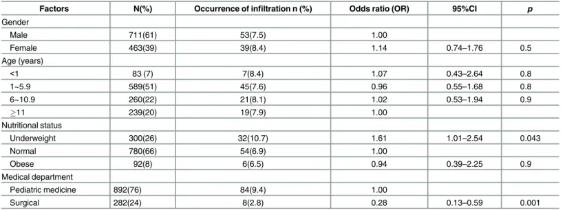
The majority (61%) of the study subjects were males with a mean age of 6.3 years and 66.6% of them had a normal body weight. Less than half (40%) of the subjects received treatment for allergies and respiratory system problems (Table 1).
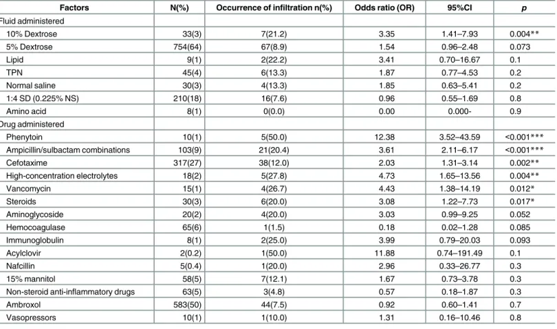
The largest portion of subjects (64%) received an infusion of 5% dextrose while 27% of the subjects were treated with cefotaxime (Table 3).
Risk factors for IV infiltration
According to the univariate analysis, physiological factors related to the occurrence of IV infil-tration were being underweight (Table 1). Patients admitted to the surgical ward were less likely to have infiltration. Significant device-related factors were retention time and insertion site (Table 2). Significant drug-related factors were 10% dextrose, use of medicines such as phe-nytoin, high-concentration electrolytes, vancomycin, ampicillin/sulbactam combinations, ste-roids, and cefotaxime (Table 3).
Multivariate analysis confirmed the findings on univariate analysis with the exception of being underweight, retention time, and use of cefotaxime. Results of the analysis identified insertion site, medical department admission, phenytoin, 10% dextrose, steroids, vancomycin, Table 1. Physiologic Factors Related to Intravenous Infiltration.
Factors N(%) Occurrence of infiltration n (%) Odds ratio (OR) 95%CI p
Gender
Male 711(61) 53(7.5) 1.00
Female 463(39) 39(8.4) 1.14 0.74–1.76 0.5
Age (years)
<1 83 (7) 7(8.4) 1.07 0.43–2.64 0.8
1~5.9 589(51) 45(7.6) 0.96 0.55–1.68 0.8
6~10.9 260(22) 21(8.1) 1.02 0.53–1.94 0.9
11 239(20) 19(7.9) 1.00
Nutritional status
Underweight 300(26) 32(10.7) 1.61 1.01–2.54 0.043
Normal 780(66) 54(6.9) 1.00
Obese 92(8) 6(6.5) 0.94 0.39–2.25 0.9
Medical department
Pediatric medicine 892(76) 84(9.4) 1.00
Surgical 282(24) 8(2.8) 0.28 0.13–0.59 0.001
doi:10.1371/journal.pone.0158045.t001
Table 2. Device-related Factors Related to Intravenous Infiltration.
Factors N(%) Occurrence of infiltration n (%) Odds ratio (OR) 95%CI p
Retention time (hours)
24.0 203(17) 13(6.4) 1.00
24.1–48.0 319(27) 28(8.8) 1.41 0.71–2.78 0.3
48.1–72.0 378(32) 26(6.9) 1.08 0.54–2.15 0.8
72.1–96.0 230(20) 18(7.8) 1.24 0.59–2.60 0.6
96.1–120 44 (3) 4(11.1) 1.83 0.56–5.96 0.3
120.1 10 (1) 3(37.5) 8.77 1.88–40.81 0.006
Catheter size(gauge)
22 70 (6) 1(1.4) 0.16 0.02–1.18 0.072
24 1104(94) 91(8.2) 1.00
Insertion site
Upper limb 992(85) 65(6.6) 1.00
Lower limb 181(15) 27(14.9) 2.50 1.55–4.04 <0.001
high-concentration electrolytes, and ampicillin/ sulbactam combinations as significant risk fac-tors (Table 4).
The ORs for use of phenytoin and 10% dextrose were 11.03 (95% CI = 2.92–41.68,p<0.001) and 6.55 (95% CI = 2.36–18.2,p<0.001), respectively. The OR for pediatric medicine admission
Table 3. Drug-related Factors Related to Intravenous Infiltration.
Factors N(%) Occurrence of infiltration n(%) Odds ratio (OR) 95%CI p
Fluid administered
10% Dextrose 33(3) 7(21.2) 3.35 1.41–7.93 0.004**
5% Dextrose 754(64) 67(8.9) 1.54 0.96–2.48 0.073
Lipid 9(1) 2(22.2) 3.41 0.70–16.67 0.1
TPN 45(4) 6(13.3) 1.87 0.77–4.53 0.2
Normal saline 30(3) 4(13.3) 1.85 0.63–5.41 0.2
1:4 SD (0.225% NS) 210(18) 16(7.6) 0.96 0.55–1.69 0.8
Amino acid 8(1) 0(0.0) 0.00 0.000- 0.9
Drug administered
Phenytoin 10(1) 5(50.0) 12.38 3.52–43.59 <0.001***
Ampicillin/sulbactam combinations 103(9) 21(20.4) 3.61 2.11–6.17 <0.001***
Cefotaxime 317(27) 38(12.0) 2.03 1.31–3.14 0.002**
High-concentration electrolytes 18(2) 5(27.8) 4.73 1.65–13.56 0.004**
Vancomycin 15(1) 4(26.7) 4.43 1.38–14.19 0.012*
Steroids 30(3) 6(20.0) 3.08 1.22–7.73 0.017*
Aminoglycoside 20(2) 4(20.0) 3.03 0.99–9.25 0.052
Hemocoagulase 65(6) 1(1.5) 0.18 0.02–1.28 0.085
Immunoglobulin 8(1) 2(25.0) 3.99 0.79–20.03 0.093
Acylclovir 2(0.2) 1(50.0) 11.88 0.74–191.49 0.1
Nafcillin 5(0.4) 1(20.0) 2.96 0.33–26.77 0.3
15% mannitol 58(5) 7(12.1) 1.67 0.73–3.78 0.3
Non-steroid anti-inflammatory drugs 63(5) 3(4.8) 0.57 0.18–1.87 0.3
Ambroxol 583(50) 44(7.5) 0.92 0.60–1.41 0.7
Vasopressors 10(1) 1(10.0) 1.31 0.16–10.46 0.8
SD = dextrose solution
*p<0.05,
**p<0.01,
***p<0.001
doi:10.1371/journal.pone.0158045.t003
Table 4. Intravenous Infiltration Risk Factors by Multiple Logistic Regression.
Factors B SE OR 95%CI p
Phenytoin 2.400 0.678 11.03 2.92–41.68 <0.001
10% Dextrose 1.879 0.522 6.55 2.36–18.20 <0.001
Steroids 1.826 0.568 6.21 2.04–18.91 0.001
Vancomycin 1.410 0.638 4.10 1.17–14.30 0.027
High-concentration electrolytes 1.250 0.578 3.49 1.13–10.82 0.031
Ampicillin/sulbactam 1.215 0.289 3.37 1.91–5.94 <0.001
Insertion site(lower limb) 0.544 0.270 1.72 1.02–2.92 0.044
SE: standard error, OR: odds ratio, CI: confidence interval
was 3.97 (95% CI = 1.68–9.36,p= 0.002), and the OR for the lower limb as insertion site was 1.72 (95% CI = 1.02–2.92,p= 0.044).
Discussion
Risk factors of IV infiltration were lower limb insertion site, pediatric medicine admission, and administration of phenytoin, 10% dextrose, steroids, vancomycin, high concentration electro-lyte, ampicillin/sulbactam combinations in this study.
Certain fluids and drugs can easily cause venous rupture when the venous endothelium and blood vessel walls are irritated, leading to IV infiltration or extravasation. Since strong acids or strong alkalinities can damage cellular proteins, reduce durability of the venous endothelium, and inflict venous rupture, pH ranging from 5 to 9 is recommended for intravenous injection [14]. Phenytoin is as an anticonvulsant that is also a strong alkaline [27] with a pH of 12, and has been found to reduce vein durability by damaging cell proteins [21,26]. Vancomycin is a strong acid with a pH of 2.8–4.5 [27], and a study has shown several cases who developed infil-trations after the administration of vancomycin [28]. Ampicillin / sulbactam combinations have strong alkalinity with a pH of 8–10, and cause irritation to the vessel walls and facilitate the occurrence of infiltration [26]. Previous studies have reported total skin loss after adminis-tration of penicillin antibiotics [15].
Likewise, osmotic pressure within the range of 250–350 mOsm/L is permitted to be given peripherally [14,27]. High osmotic pressure exceeding 350 mOsm/L can disrupt cell functions and rupture cells according to movement of moisture from cell to interstitial space [14]. Osmotic pressure above 600 mOsm/L causes peripheral phlebitis within 24 hours in a study [4]. On the other hand, low osmotic pressure below 250 mOsm/L results in rupture of cells due to edema [14]. High-concentration electrolytes have high osmotic pressure and have strong alkalinity that can damage the vein [14,21,26], resulting in an increased risk of IV infiltration. Previous studies have reported significant local tissue necrotic cases caused by peripheral extravasation of intravenous solutions containing high-concentration electrolytes like calcium salts [16,29]. A 10% dextrose solution identified as a risk factor of IV infiltration in this study is associated with high osmotic pressure of 505 mOsm/L [18,26]. Hyperosmolar agents such as 10% dextrose or total parenteral nutrition solutions can cause skin necrosis and serious issue damage [14,18] and facilitate the occurrence of infiltration. Yosowitz et al [29] have reported 8 patients, including 6 infants who had significant local tissue necrosis by peripheral extravasa-tion of intravenous soluextravasa-tions containing calcium salts and/or 10% dextrose.
Steroids are known to induce the formation of emboli when used over a long period of time [30]. Emboli frequently arise due to repeated damage of blood vessels and coagulation activated by platelets and fibrin. When embolus formation is continued, blood flow is affected and blood vessel walls are weakened becoming vulnerable to IV infiltration [31].
According to the results, nurses in clinical settings are recommended to select an IV site in upper arm rather than in lower limb, and need to make repeated observations to identify IV infiltration occurrence when they care for children in the pediatric medicine department, and administer high risk drug/fluid such as phenytoin, 10% dextrose, steroids, vancomycin, high-concentration electrolytes, and ampicillin/sulbactam.
characteristics used by the nurses to determine IV infiltration risk factors. This includes factors such as catheter insertion skill and appropriateness of catheter fixation after insertion.
Conclusions
Risk factors related to the occurrence of IV infiltration in children were assessed in the present study. Based on the findings, nurses working at the children’hospitals should consider the risk of IV infiltration for children receiving intravenous infusion therapy and make efforts to iden-tify IV infiltration in high-risk children at an early stage to prevent damage. Furthermore, a replication study on children with different characteristics should be conducted to increase the possibility for generalization of the risk factors. A prospective study should also be performed to investigate the nurse-associated factors such as catheter insertion skill.
Author Contributions
Conceived and designed the experiments: SMP ISJ SSJ. Performed the experiments: SMP. Ana-lyzed the data: SMP ISJ. Contributed reagents/materials/analysis tools: SMP ISJ SSJ. Wrote the paper: SMP ISJ SSJ.
References
1. Wilson J. Preventing infection associated with intravascular therapy. In: Infection control in clinical prac-tice. ( 3rd ed). London: Baillière Tindall; 2006.
2. Dougherty L. Back to basics in IV therapy: an unfortunate necessity. Br J Nurs. 2008; 17(19): S3. PMID:
18974683
3. McCullen KL, Pieper B. A retrospective chart review of risk factors for extravasation among neonates receiving peripheral intravascular fluids. J Wound Ostomy Continence Nurs. 2006; 33(2): 133–139. PMID:16572012
4. Infusion Nurses Society. Infusion nursing standards of practice. J Infus Nurs. 2006; 29(suppl 1):S1– S92.
5. de Lima Jacinto AK, Avelar AF, Pedreira ML. Predisposing factors for infiltration in children submitted to peripheral venous catheterization. J Infus Nurs. 2011; 34(6):391–398. doi:10.1097/NAN.
0b013e3182306491PMID:22101633
6. Hetzler R, Wilson M, Hill EK, Hollenback C. Securing pediatric peripheral IV catheters-application of an evidence-based practice model. J Pediatr Nurs. 2011; 26(2):143–148. doi:10.1016/j.pedn.2010.12. 008PMID:21419974
7. Kim JS, Lee YR, Kim NS. Effects of the structured nursing intervention for caregivers on maintenance of intravenous infusions in infants. J Korean Acad Child Health Nurs. 2012; 18(3):135–142.
8. Sung SH, Kim HS. Risk factors of intravenous infiltration in children. Clin Nurs Res. 2007; 13(2):61–72. 9. Fang L, Fang SH, Chung YH. Factors affecting the unplanned peripheral reinsertion in pediatric
patients from a teaching hospital inTaiwan. J Infus Nurs. 2011; 34(6):366–372.http://dx.doi.org/10. 1097/NAN.0b013e318230661c1doi:10.1097/NAN.0b013e31823061c1PMID:22101630
10. Talbot SG, Rogers GF. Pediatric compartment syndrome caused by intravenous infiltration. Ann Plast Surg. 2011; 67(5):531–533.http://dx.doi.org/10.1097/SAP.0b013e3182085915doi:10.1097/SAP. 0b013e3182085915PMID:21301289
11. Amjad I, Murphy T, Nylander-Housholder L, Ranft A. A new approach to management of intravenous infiltration in pediatric patients. J Infus Nurs. 2011; 34(4): 242–249.http://dx.doi.org/10.1097/NAN. 0b013e31821da1b3. doi:10.1097/NAN.0b013e31821da1b3PMID:21734520
12. Woody G, Davis BA. Increasing nurse competence in peripheral intravenous therapy. J Infus Nurs. 2013; 36(6):413–419.http://dx.doi.org/10.1097/NAN.0000000000000013doi:10.1097/NAN. 0000000000000013PMID:24202121
13. Clifton-Koeppel R. Wound care after peripheral extravenous extravasation: what is the evidence?. Newborn Infant Nurs Rev. 2006; 6(4):202–211.
14. Doellman D, Hadaway L, Bowe-GeAddes LA, Franklin M, LeDonne J, Papke-O'Donnel L, et al. Infiltra-tion and extravasaInfiltra-tion. J Infus Nurs. 2009; 32(4):203–211. doi:10.1097/NAN.0b013e3181aac042
15. Kumar RJ, Pegg SP, Kimble RM. Management of extravasation injuries. ANZ J Surg. 2001; 71: 285– 289. PMID:11374477
16. Saravath A, Kruavit A. Extravasation injury: what is the appropriate management of extravasated skin ulcer?. Thai J Surg. 2006; 27: 19–25.
17. McDonnell PJ. Purple glove syndrome. Pennsylvania Patient Safety Authority. 2004; 3(4): 1–3. 18. Sauerland C, Engelking C, Wickham R, Corbi D. Vesicant extravasation part 1: mechanisms,
patho-genesis, and nursing care to reduce risk. Oncol Nurs Forum. 2006; 33(6):1134–1141. PMID:17149396
19. Hadaway L. Infiltration and extravasation: preventing a complication of IV catheterization. Am J Nurs 2007; 107(8):64–72 PMID:17667395
20. Goolsby TV, Lombardo FA. Extravasation of chemotherapeutic agents: prevention and treatment. Semin Oncol 2006; 33(1):139–143. PMID:16473651
21. Payne AS, Savarese DM. Extravasation injury from chemotherapy and other non-neoplastic vesicants. Internet: http://www.uptodate.com/contents/extravasation-injury-from-chemotherapy-and-other-non-antineoplastic-vesicants. 2013.
22. Montgomery LA, Hanrahan K, Kottman K, Otto A, Barret T, Hermiston B. Guideline for IV infiltrations in pediatric patients. Pediatr Nurs. 1999; 25(2):167–180. PMID:10532013
23. Flemmer L, Chan JS. A pediatric protocol for management of extravasation injuries. Pediatr Nurs. 1993; 19(4):355–358. PMID:8414723
24. Ahn HS. Pediatrics. Seoul:Mirae-n;2012.
25. Patient Safety Advisory. Extravasation of radiologic contrast. Pennsylvania Patient Safety Authority. 2004; 1(3):1–6.
26. Clark E, Giambra BK, Hingl J, Doellman D, Tofani B, Johnson N. Reducing risk of harm from extravasa-tion. J Infus Nurs. 2013; 36(1):37–45.http://dx.doi.org/10.1097/NAN.0b013e3182798844doi:10.1097/ NAN.0b013e3182798844PMID:23271150
27. Earhart A, McMahon P. Vascular access and contrast media. J Infus Nurs. 2011; 34(2): 97–105.http:// dx.doi.org/10.1097/NAN.0b013e31820b4a11doi:10.1097/NAN.0b013e31820b4a11PMID:21399455
28. Le A, Patel S. Extravasation of noncytotoxic drugs: a review of the literature. Ann Pharmacother. 2014; 48:870–886.http://dx.doi.org/10.1177/1060028014527820PMID:24714850
29. Yosowitz P, Ekland DA, Shaw RC, Rarsons RW. Peripheral intravenous infiltration necrosis. Ann Surg. 1975; 182:553–556. PMID:811181
30. Glueck CJ, Richardson-Royer C, Schultz R, Burger T, Bowe D, Padda J, et al. Testosterone therapy, thrombophilia-hypofibrinolysis, and hospitalization for deep venous thrombosis-pulmonary embolus: an exploratory, hypothesis-generating study. Clin Appl Thromb Hemost. 2014; 20(3):244–249 doi:10. 1177/1076029613499819PMID:23925401