73
1. MD, Ph.D. Hospital São José de Doenças Infecciosas and ClinicalResearch Unit, Hospital Universitário Walter Cantídio, Universidade Federal do Ceará, Fortaleza, CE, Brazil.
2. MD, Ph.D. Clinical Research Unit, Hospital Universitário Walter Cantídio, Universidade Federal do Ceará, Fortaleza, CE, Brazil.
3. MD, M.Sc. Clinical Research Unit, Hospital Universitário Walter Cantídio, Universidade Federal do Ceará, Fortaleza, CE, Brazil.
4. MD, Ph.D. Hospital Infantil Albert Sabin, Secretaria da Saúde do Ceará, Fortaleza, CE, Brazil.
Manuscript received Mar 17 2004, accepted for publication Sep 20 2004.
Suggested citation: Rey LC, Martins CV, Ribeiro HB, Lima AA. American visceral leishmaniasis (kala-azar) in hospitalized children from an ende-mic area. J Pediatr (Rio J). 2005;81:73-8.
Abstract
Objective: To study epidemiological and clinical aspects of American visceral leishmaniasis in hospitalized children in Ceará, Brazil.
Methods: A retrospective and observational study was carried out with children suffering from American visceral leishmaniasis admitted to Hospital Infantil Albert Sabin in Fortaleza. Medical records were reviewed consistently. Inclusion criteria were children with amastigote-positive smears in bone marrow or in splenic aspirates, or a positive Leishmania sp immunofluorescence assay.
Results: From January 1995 to December 2002, 450 children with American visceral leishmaniasis were identified, accounting for 9 to 27% of all reported cases in Ceará in that period, with peak hospitalization rates in 1995 and 2000. The mean age was 4.4 years (12% < 1 year and 65% < 5 years of age). The overall male: female ratio was 1.1 and 1.48 in children > 5 year (p = 0.04). Urban patients infected by American visceral leishmaniasis increased steadily over an 8-year period (χ2 p = 0.01). The main clinical complaints on
admission were fever (96%), pallor (90%) and abdominal swelling (75%). Clinical cure was defined as the absence of fever, regression of splenic and hepatic enlargement and of pancytopenia. Overall mortality was 9.2% and 21.2% in patients younger than one year. Malnutrition, edema, bleeding, jaundice, and concomitant infections were related to higher mortality.
Conclusions: Cases of American visceral leishmaniasis spiked with a 5-year interval, and affected most under-five urban children. Mortality was related to low age, signs of severe disease and concomitant infection.
J Pediatr (Rio J). 2005;81(1):73-8: American visceral leishmaniasis, kala-azar, epidemiology, treatment, children.
Resumo
Objetivos: Estudar os aspectos epidemiológicos e clínicos da leishmaniose visceral americana em crianças hospitalizadas do Ce-ará.
Métodos: Estudo retrospectivo e observacional de crianças com leishmaniose visceral americana admitidas no Hospital Infantil Albert Sabin, em Fortaleza. Os prontuários foram revistos sistematicamen-te. Os critérios de inclusão foram crianças com esfregaços positivos para Leishmania em aspirado de medula óssea ou de baço, ou teste de imunoensaio positivo para Leishmania sp.
Resultados: Entre janeiro de 1995 e dezembro de 2002, foram identificados 450 pacientes, perfazendo 9 a 27% dos casos de leishmaniose visceral americana notificados no Ceará no período, com picos de admissão em 1995 e 2000. A idade média foi de 4,4 anos (12% < 1 ano e 65% < 5 anos de idade). A relação masculino:feminino foi de 1,1 em geral e de 1,48 em < 5 anos (p = 0,04). Os pacientes urbanos aumentaram regularmente no período de 8 anos (χ2, p = 0,01). As principais queixas foram febre
(90%), palidez (90%) e aumento abdominal (75%). A cura clínica foi constatada por ausência de febre, regressão da hepato-esplenome-galia e da pancitopenia. A letalidade geral foi de 9,2%, e 21,2% em lactentes < 1 ano. Desnutrição, edema, sangramento, icterícia e infecção intercorrentes foram fatores relacionados com maior letali-dade.
Conclusões: Casos hospitalizados de leishmaniose visceral americana apresentaram picos a cada 5 anos e afetaram crianças urbanas < 5 anos. A mortalidade esteve associada à baixa idade, sinais de gravidade e infecção concomitante.
J Pediatr (Rio J). 2005;81(1):73-8: Leishmaniose visceral ameri-cana, calazar, epidemiologia, tratamento, criança.
Copyright © 2005 by Sociedade Brasileira de Pediatria
O
RIGINALA
RTICLEAmerican visceral leishmaniasis (kala-azar)
in hospitalized children from an endemic area
Leishmaniose visceral americana (calazar) em crianças hospitalizadas de área endêmica
Luís C. Rey,1 Ceci V. Martins,2 Hildenia B. Ribeiro,3 Aldo A. M. Lima4
Nota: a versão completa deste artigo em português está disponível em www.jped.com.br
Introduction
American visceral leishmaniasis (AVL) is an expanding anthroponosis in Brazil. Over 3,000 new cases are reported
every year, in 19 out of 23 states.1 The Northeast region
(nine states) accounts for 70-90% of all patients, mostly in the states of Bahia, Ceará, Piauí and Maranhão, with an
incidence rate of 25 cases per 100,000 inhabitants.1 Urban
transmission to humans, assessed through clinical reports or serological surveys, has continually increased in the last
four decades;2,3 in most regions where AVL transmission is
The shift of Leishmania (L.) chagasi transmission from rural to urban and periurban areas has been explained by migration of impoverished families of peasants with infected
dogs.2-5 Cyclic drought periods related to El Niño are also
implicated in the ecological impact of deforestation and new
human riverbank settlements.5-7 Lutzomyia (L) longipalpis
(Diptera: Psychodidae) became adapted to urban houses and to the overcrowding conditions of slums, thus increasing
its efficacy as the vector of L. chagasi.8 Cyclic urban AVL
outbreaks occurred in the city of Teresina in the years 1980
to 1986 and 1992 to19964,9 and in other state capitals such
as São Luís,10 Natal11 and Belo Horizonte.12 Other Brazilian
towns have also experienced periurban burdens of AVL.13,14
Clinical reports of AVL in Ceará are scarce and most were
published more than 30 years ago.2-3 AVL has been described
as a severe disease characterized by malnutrition, extreme pallor, abdominal enlargement with edema, bleeding, jaundice, chronic or persistent diarrhea and respiratory infections. Milder disease often was unrecognized at the local level and still is, which delays proper diagnosis and treatment. This updated hospital-based clinical study in an endemic area is therefore necessary for clinicians at the primary and local level of health care. The aim of the study was to analyze epidemiological and clinical aspects of AVL in children admitted to a reference (tertiary-care) public hospital in Fortaleza.
Patients and methods
The study was conducted in Fortaleza, the state capital of Ceará, Northeastern Brazil. Hospital Infantil Albert Sabin (HIAS) is a tertiary-care pediatric facility with nearly 200 beds and qualified specialists.
All infants or children admitted to HIAS with an amastigote-positive bone marrow or splenic aspirate were included in the study. Those with positive Leishmania immunofluorescence assay findings were also included. Any patients who were admitted to HIAS and were infected with AVL but did not reside in the state of Ceará were excluded.
Handwritten medical records were used to screen AVL patients treated between 1995 and 1998, and a computerized database system was used to screen those
treated between 1999 and 2002. All selected medical records were consistently reviewed, and a structured protocol form was employed to collect epidemiological data (age, sex, place of residence, housing conditions) and clinical data (duration of signs and symptoms, physical examination, diagnostic tests, laboratory data, treatment and outcome). Visceral enlargement (in cm) was measured from the lower rib to the distal edge of the affected organ. Prothrombin activity (in %) was recorded as indicated by the manufacturer (Option, bioMérieux Brasil, Rio de Janeiro, Brazil). The study protocol was approved by the Ethics and Research Committee of the referred hospital.
The data were validated by reviewing 20% of the selected hospital files and by double-typing the data entries
into an IBM-compatible computer. Epi-Info 6.0415 and SPSS
software were used for the statistical analysis. Continuous variables such as age, weight, liver and spleen measurements, and laboratory data, were described by means and 95% confidence intervals (95%CI), when appropriate, and compared using Students t-test for independent or dependent variables. Paired data were measured before and after treatment. Contingency tables containing categorical variables, such as sex and outcome, and dichotomized continuous variables were analyzed through the chi-square test or Fishers exact test, when necessary, and the odds ratio was calculated using Cornfields 95% confidence intervals. Stratified (bivariate) analyses of contingency tables were used to identify possible interactions among clinical findings (low age, jaundice, bleeding, edema, infection) and their association with hospital outcome (childs death or discharge). In these particular situations, the Mantel-Haenszel-adjusted odds ratio was considered. In all cases, a p < 0.05 was considered as significant.
Results
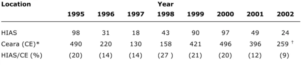
From January 1995 to December 2002, 450 patients with confirmed AVL were admitted to HIAS in Fortaleza. The rate of confirmed cases in this facility varied from 9
to 27% of all cases reported in that state1 during the study
period (Table 1). Peak hospitalization rates occurred in 1995 and between 1999 and 2000, decreasing in the subsequent years.
Table 1 - Cases of American visceral leishmaniasis reported in the State and confirmed at Hospital Infantil Albert Sabin, 1995-2002
* Source: COVEV/CGDT/DEVEP/SVS-MS.1 † Preliminary data.
Location Year
1995 1996 1997 1998 1999 2000 2001 2002
HIAS 98 31 18 43 90 97 49 24
Ceara (CE)* 490 220 130 158 421 496 396 259
Ninety-seven children lived in Fortaleza (22%) and 120 (27%) in six towns in the metropolitan area of Fortaleza (MAF): Caucaia, Eusébio, Maranguape, Pacajus, Maracanau and Pacatuba. The number of towns with hospital admissions due to AVL was highest in 1995 and 2000 (98 and 97 out of 184 towns, respectively,) and lowest in 1997 (only 18 towns).
No relative increase was observed throughout the study period in AVL patients from Fortaleza and from the MAF
(
χ
2 = 9.49, df = 7, p = 0.22) (Figure 1). However, there wasa significant increase in the hospital admission of AVL children living in all urban areas in the study period
(
χ
2 = 18.08, df = 7, p = 0.01) (Figure 2).Of the 438 families for which information was available, 255 (58%) lived in brick houses and 183 (42%) in adobe or mud-and-stick houses (t-test p < 0.001). By comparing the ratio of adobe vs brick houses in 1995 and 2000 (outbreak years), it is observed that the proportion shifted from 1.07 (48/45) to 0.53 (33/62), showing a significant increase in
the number of brick houses (
χ
2 = 5.46, p = 0.01), whichindicates an increase in the urban origin of our patients.
The mean age of the 450 AVL cases was 54 months (95%CI: 50-57 months), (57 months for boys and 50 for girls) (p < 0.05). Patients were younger than one year (12% or 52/450) and younger than five (65% or 294/450).
Fifty-three percent (240/450) of the patients were male and 47% were female (male:female ratio= 1.1). The male:female ratio was as high as 1.48 (t-test p = 0.04) in children older than five years.
Tables 2 and 3 show the nutritional status and clinical findings regarding AVL patients admitted to HIAS. The average length of the disease prior to hospital admission was 7.9 weeks (95%CI: 7.1 to 8.7 weeks). Clinical manifestations of severe disease (bleeding, edema, jaundice) were not associated with longer duration of the disease. Infants less than one year of age showed more
hemorrhagic disorders than older patients (
χ
2 = 10.0,p = 0.0007).
Figure 1 - Counties of residence of American visceral leishmaniasis patients admitted at Hospital Infantil Albert Sabin, by year, 1995-2002
Figure 2 - Place of residence of admitted American visceral leishmaniasis patients at Hospital Infantil Albert Sabin, by year, 1995-2002
70
60
50
40
30
20
10
1995 1996 1997 1998 1999
Year
N
o
.
o
f
c
a
s
e
s
2000 2001 2002
0
Fortaleza metropolitan area Other counties
1995 1996 1997 1998 1999 2000 2001 2002
70
60
50
40
30
20
10
0
Rural Urban
Year
N
o
.
o
f
c
a
s
e
s
Table 2 - Clinical aspects of American visceral leishmaniasis at HIAS, 1995-2002
* WAZ: weight-for-age z score; well nourished: WAZ > -1; mild malnutrition: -1 > WAZ > -2; moderate: -2 > WAZ > -3; severe: WAZ < -3.
† Cumulative data.
Nutritional status (n = 432) n % 95% CI
(WAZ score) *
Well nourished 168 39 34-44
Mild malnutrition 153 35 31-40 Moderate malnutrition 81 19 15-23 Severe malnutrition 30 7 5-10
Symptoms and signs (n = 450)
Fever 432 96 94-98
Pallor 387 86 82-89
Abdominal swelling 342 76 72-80
Anorexia 221 49 44-54
Weakness 210 47 42-51
Weight loss 197 44 39-49
Edema 128 28 24-33
Hemorrhage 42 9 7-13
Jaundice 33 7 5-10
Non-specific complaints were cough (51 children), diarrhea (41), shortness of breath (13), limb and joint pain (six), febrile seizures (three), and alopecia (three cases). Community and nosocomial infections were detected in 49% of all patients. Patients younger than one year presented more infectious episodes than older
patients (
χ
2 = 5.0, p = 0.03).Before treatment, splenic enlargement was detected in 99% (436) of the 450 patients and liver enlargement in 95% (406) of the children; 25% of the patients had serum hemoglobin < 5.0 g/dl, 25% had a total leukocyte count
Of 445 bone marrow aspirates performed, 73% were amastigote-positive (329 smears). A second bone marrow aspirate was performed in 53 out of 116 amastigote-negative smears, and 53% (28) were also positive for Leishmania. In 31 amastigote-negative bone marrows, a splenic aspirate was performed, and 81% (24 specimens) were amastigote-positive. The remaining 71 patients had a confirmed diagnosis of AVL by Leishmania-specific immunofluorescence assay (IFA). The IFA was performed in 179 patients during their hospital stay, and 95 out of 111 amastigote-positive blood marrow or splenic smears were seropositive (sensitivity of 86%). Three cases of amastigote-negative bone marrow and splenic aspirate were diagnosed only through Leishmania-specific IFA. The mean hospital stay before therapy began was 6 days (median: 5 days), but a treatment delay of 45 days was detected.
Once AVL was confirmed, 98% (439/450) of the children were treated with meglumine antimoniate; 29% also received allopurinol as an adjuvant to antimoniate. After a mean 25-day course of pentavalent antimony, only 4% (18 patients) required amphotericin B as second-line treatment. Of these, two died soon after amphotericin B therapy was started. A total of 388 patients (91%) responded successfully to treatment (cured or clinically improved). Children admitted but not treated constituted 6.2% (28) of the patients. Five of these patients died before any specific treatment was started, and 23 were transferred to another hospital. The mean hospital stay at the HIAS was 28 days.
Before or during hospitalization, 220 children (49%, 95%CI: 44-54%) presented at least one infectious episode.
The most frequent infections were pneumonia (108 cases), sepsis (32), otitis media (27), infectious dermatitis (25) and diarrhea (four cases).
Infection
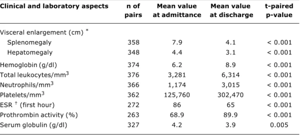
All patients improved their clinical and laboratory conditions with the treatment (Table 3). The mean decre-ase in splenic and hepatic size amounted to 48% and 32%, respectively. Hemoglobin level rose 44%, total leukocyte count 48%, neutrophils 61%, platelets 58% and prothrombin activity increased 22% at hospital dis-charge; a 24 and 7% reduction was observed for ESR and serum globulin levels, respectively.
AVL mortality was 8.7% (39/450) (95%CI: 6.3-11.8) in the study period. Mortality was 21.2% in children younger than one year and 7.6% in children older than one year [MantelHaenszel (MH)adjusted OR = 3.49; 95%CI: 1.5 -8.0, p < 0.001]. Moderate to severe malnutrition [measured by weight-for-age (WAZ) z-score < -2] was related to a 13.2% mortality rate, compared to 7.0% in children with WAZ > -2 z-scores, (MH-adjusted OR = 2.04; 95%CI: 0.93-4.36, p = 0.04). Other clinical findings on admission related to hospital mortality were: jaundice (MH-adjusted OR = 3.76, 95%CI: 1.41-9.76, p = 0.002), hemorrhage (MH-adjusted OR = 7.22, 95%CI: 3.14-16.57, p < 0.001), edema (MH-adjusted OR = 3.07, 95%CI: 1.49-6.35, p < 0.001), and community or nosocomial infection (MH-adjusted OR = 3.34, 95%CI: 1.5 - 7.6, p < 0.001). The mean hospital stay of patients who died was 14 days, thus shorter than the 28 days of discharged children (t-test p < 0.001).
Clinical and laboratory aspects n of Mean value Mean value t-paired
pairs at admittance at discharge p-value
Visceral enlargement (cm) *
Splenomegaly 358 7.9 4.1 < 0.001
Hepatomegaly 348 4.4 3.1 < 0.001
Hemoglobin (g/dl) 374 6.2 8.9 < 0.001
Total leukocytes/mm3 376 3,281 6,314 < 0.001 Neutrophils/mm3 366 1,174 3,015 < 0.001 Platelets/mm3 362 125,760 302,470 < 0.001
ESR (first hour) 272 86 65 < 0.001
Prothrombin activity (%) 263 68.9 89.9 < 0.001
Serum globulin (g/dl) 327 4.2 3.9 0.005
Table 3 - Clinical aspects and laboratory mean values of children at admittance and discharge
Causes of death in 39 patients were: infection in 29 cases (74%), hemorrhage in six (15%), hepatic failure in three (8%) and cardiac arrhythmia in one case (3%). Fatal infections were septicemia and pneumonia.
Discussion
Retrospective studies using medical records of children with AVL can provide useful epidemiological and clinical data, particularly in endemic areas, and should be performed on a regular basis. The cyclic increase in AVL cases at HIAS from 1995 to 2002 suggests that L. chagasi transmission and the burden of disease is out of control in that state. Also, the high rate of patients in a single facility should be a matter of concern and may indicate a high rate of unreported cases throughout the state, or concentration of AVL management in a few reference health facilities, mainly in Fortaleza. In states where AVL
is uncommon as São Paulo16 and Federal District of
Brasília,17 the presence of AVL patients from endemic
areas of Brazil is frequent because of the local difficulties to receive appropriate care; the mean time of disease previous to hospital admission is 5.6 months in São Paulo, and 3 months in Brasília and in São Luís (state of
Maranhão).18
Outbreaks of AVL have occurred in state capitals as Natal, where 60% out of 1,500 cases were reported in the
state in 1991;11 differently, in our study, we were not able
to identify a specific burden of the disease in Fortaleza, where 20% of our patients live. We have detected, however, a consistent increase in urban AVL in the state (59% of all hospitalized patients were urban settlers), as described in
prospective studies.19 On the other hand, 72% of AVL
patients treated in Brasília dwell in rural areas, and most of
them came from the state of Bahia.17
At HIAS, in Ceará, AVL affected mainly children younger
than five years, a finding reported since the 1980s;2
patients younger than five years corresponded to 49% in
Natal, 64% in Maranhão,11,18 but to only 28% in Belo
Horizonte,12 a new expanding area of AVL in Brazil. The high
proportion of infants younger than one year old in our hospital (12%) could be explained by the indoor transmission of L. chagasi in urban and periurban areas, as in Belo
Horizonte (5%)12 and in São Luís.18
The severity of clinical findings is related to both a delay in providing medical assistance and to the young age of the patients. It appears that the shorter the duration of the disease, the better the clinical aspect of AVL. Children from
a hyperendemic area of AVL in Jacobina20 showed less
severe disease than patients admitted to the Teaching Hospital of Salvador. In São Paulo, the higher proportion of edema and jaundice in hospitalized patients is probably due to the long period of disease, as many patients live in other
states, especially Bahia and Piauí.16.
As occurs in hospital-based series of children with AVL, the disease was clinically apparent. In community-based studies of endemic areas, serological screening for Leishmania sp revealed eight to 16 asymptomatic seropositive
patients for each clinical case, which reflects the need of
local surveillance and treatment as early as possible.21
According to our data, a poorer outcome was strongly related to severe clinical signs prior to admission.
Previous nutritional status is crucial for the clinical
outcome of AVL.20 Moderate to severe malnutrition
(weight-for-age z score below -2) in Ceará accounted for 9.2% in children < 5 years, and mild malnutrition (-1.9 to
-1.0 z score), to 24% of these children.22 Our data show
a higher rate of malnutrition compared to community-based studies, as reported in AVL cases admitted to
hospitals in Brasília and São Paulo.16,17 In Jacobina,
Bahia, prospective studies showed that malnutrition was an important condition for the burden of clinical AVL as well as a consequence of long-term disease, and was less
severe in early-diagnosed patients.20,23
The diagnosis of AVL could be established earlier if more rapid and accurate tests for AVL were available. Leishmania
culture16 is a time-consuming alternative (2 to 4 weeks),
and time is crucial in advanced (severe) disease and for infants with AVL. Leishmania cultures are not routinely
performed in Fortaleza, Natal, São Luís or Salvador.11,18
The length of antimoniate therapy varied from 20 to 40 days in Fortaleza. Assessment of cure in AVL is controversial. Amastigote-negative smears have low negative predictive values, and immunoassays will become negative months after treatment. Physical examination and improved laboratory findings are therefore important criteria for cure. Absence of fever, physical activity, recovery of appetite, and weight gain are indirect signs. A 50% decrease in splenic and (to a lesser degree) hepatic size, and a substantial increase in blood cell counts are crucial. In our series, the treatment with meglumine antimoniate showed a high cure rate and low
toxicity, as also reported in Natal.11 In our study, the
decrease in visceral swelling was less than 50%, although we were unable to obtain information on visceral measurements from many discharged patients, probably because they had little or no visceral enlargement.
Therapeutic failure with antimoniate was seen in 15% of cases of an age-mixed group of patients in São Paulo,
with different disease duration and treatment regimens.16
The concomitant use of allopurinol and antimony salt was frequent at HIAS in patients showing poor clinical improvement. However, no study on the efficacy of allopurinol in AVL has been published in Brazil, and protocols are necessary to validate the use of this drug in this condition.
Therapy with pentavalent antimony at the Teaching Hospital of Salvador showed to be effective after 30 days of treatment. In the endemic area of Jacobina, where clinical AVL is detected early, recovery is achieved with a
15-day treatment course.24
The mean specific mortality rate in our study is similar to reports from other urban health centers as Belo
Horizonte7 (8-17% mortality), São Paulo 7%,16 São
Luís10 (6.7%), Natal11 (9%), and Brasília17 (9.2%). In
Jacobina, but 14% in patients at the Teaching Hospital of
Salvador.24 Visceral leishmaniasis in southern Europe is
milder, shows rare hemorrhages, no jaundice or edema, and 100% treatment efficacy with meglumine antimoniate, although it is considered by many to be caused by the
same Leishmania species.25
To reduce AVL morbidity and mortality, it would be necessary to screen and treat AVL earlier, at the local or regional level. In this regard, rapid and simple diagnostic
tests for clinical AVL, as recombinant K39 strip tests26,27
should be available. Clinical trials with oral miltefosine (hexadecylphosphocholine), a new and highly effective drug used to treat Indian kala-azar, should also be
performed with AVL.28,29
Corresponding author: Luís C. Rey
Unidade de Pesquisa Clínica, Instituto de Biomedicina Faculdade de Medicina, Universidade Federal do Ceará Av. José Bastos, 3390 sala 90
CEP 60436-160 Fortaleza, CE Brazil
Phone: +55 (85) 9982.3925/261.1013 E-mail: [email protected] References
1. Brasil. Ministério da Saúde. Secretaria de Vigilância em Saúde. Departamento de Vigilância Epidemiológica. Manual de vigilância e controle da leishmaniose visceral. Ministério da Saúde, Secretaria de Vigilância em Saúde, Departamento de Vigilância Epidemiológica. Brasília:Ministério da Saúde, 2003. 120 p. Disponível em: htttp://dtr2001.saude.gov.br/editora/produtos/ livros/pdf/03_1193_M.pdf.
2. Deane LM, Deane MP. Leishmaniose visceral urbana (no cão e no homem) em Sobral, Ceará. Hospital 1955;47:75-87. 3. Alencar JE. Leishmaniose visceral no Brasil. Revista Médica da
Universidade Federal do Ceará. 1978;129:17-8.
4. Arias JR, Monteiro PS, Zicker F. The reemergence of visceral leishmaniasis in Brazil. Emerging Infectious Diseases 1996;2:145-6.
5. Mendes WS, Silva AAM, Trovão JR, Silva AR, Costa LML. Expansão espacial da leishmaniose visceral americana em São Luis, Maranhão, Brasil. Rev Soc Bras Med Trop 2002;35:227-31. 6. Franke CR, Ziller M, Staubach C, Latif M. Impact of the El Niño/ Southern oscillation on visceral leishmaniasis, Brazil. Emerging Infectious Diseases 2002;8:914-7.
7. Thompson RA, Lima JWO, Maguire JH, Braud DH, Scholl DT. Climatic and demographic determinants of American visceral leishmaniasis in northeastern Brazil using remote sensing technology for environmental categorization of rain and region influences on leishmaniasis. Am J Trop Med Hyg 2002;67:648-55. 8. Campbell-Lendrum D, Dujardin JP, Martinez E, Feliciangeli MD, Perez JE, Passerat de Silas LN, et al. Domestic and peri-domestic transmission of American cutaneous leishmaniasis: changing epidemiological patterns presents new control opportunities. Mem Inst Oswaldo Cruz. 2001;96:169-2.
9. Costa CHN, Pereira HF, Araújo MV. Epidemia de leishmaniose visceral no estado do Piauí, Brasil, 1980-1986. Rev Saúde Públ. 1990;24:361-72.
10. Mendes WS, Silva AA, Trovão JR, Silva AR, Costa JM. Expansão espacial da leishmaniose visceral americana em São Luis, Maranhão, Brasil. Rev Soc Bras Med Trop. 2002;35:227-31. 11. Jerônimo SM, Oliveira RM, Mackay S, Costa RM, Sweet J,
Nascimento ET, et al. An urban outbreak of visceral leishmaniasis in Natal, Brazil. Trans R Soc Trop Med Hyg. 1994;88:386-8. 12. Silva ES, Gontijo CM, Pacheco RS, Fiuza VO, Brazil RP. Visceral
leishmaniasis in the metropolitan region of Belo Horizonte, state of Minas Gerais, Brazil. Mem Inst Oswaldo Cruz. 2001;96:285-91. 13. Marzochi MC, Marzochi KB, Carvalho RW. Visceral leishmaniasis
in Rio de Janeiro. Parasitol Today. 1994;10:37-40.
14. Cunha S, Freire M, Eulalio C, Cristóvão J, Netto E, Johnson Jr WD, et al. Visceral leishmaniasis in a new ecological niche near a major metropolitan area of Brazil. Trans Royal Soc Trop Med Hyg. 1995;89:155-8.
15. Dean AG, Dean JA, Coulombier D, Brendel KA, Smith DC, Burton AH, et al. Epi-Info, version 6: a word-processing, database and statistics program for public health on IBM-compatible microcomputers. 1995; Atlanta, Georgia, USA: Centers for Disease Control and Prevention.
16. Pastorino AC, Jacob CM, Oselka GW, Carneiro-Sampaio M. Leishmaniose visceral: aspectos clínicos e laboratoriais. J Pediatr (Rio J). 2002;78:120-7.
17. Campos Jr D. Características clínico-epidemiológicas do calazar na criança. Estudo de 75 casos. Jornal de Pediatria (Rio J). 1995;71:261-5.
18. Silva AR, Viana GM, Varoniol C, Pires B, Nascimento MD, Costa JM. Leishmaniose visceral (calazar) na ilha de São Luiz, Maranhão, Brasil: evolução e perspectivas. Rev Soc Bras Med Trop. 1997;30:359-68.
19. Jerônimo SM, Teixeira MJ, Sousa AQ, Thielking P, Pearson RD, Evans TG. Natural history of Leishmania (Leishmania) chagasi infection in Northeastern Brazil: long-term follow-up. Clin Infect Dis. 2000;30:608-9.
20. Badaró R, Jones TC, Carvalho EM, Sampaio D, Reed SG, Barral A, et al. New perspectives on a subclinical form of visceral leishmaniasis. J Infect Dis. 1986;154:1003-11.
21. Costa CH, Steward JM, Gomes RB, Garcez LM, Ramos PK, Bozza M, et al. Asymptomatic human carriers of Leishmania chagasi. Am J Trop Med Hyg. 2002;66:334-7.
22. Victora CG, Barros FC, Tomasi E, Ferreira FS, MacAulliffe J, Silva AC, et al. A saúde das crianças dos estados do Ceará, Rio Grande do Norte e Sergipe, Brasil: descrição de uma metodologia para diagnósticos comunitários. Rev Saúde Pública 1991;25:218-25. 23. Harrison LH, Naidu TG, Drew JS, Alencar JE, Pearson RD. Reciprocal relationships between undernutrition and the parasitic disease visceral leishmaniasis. Rev Infect Dis. 1986;8:447-53. 24. Badaro R, Jones TC, Lorenço R, Cerf BJ, Sampaio D, Carvalho EM, et al. A prospective study of visceral leishmaniasis in an endemic area of Brazil. J Infect Dis. 1986;4:639-49.
25. Maltezou HC, Siafas C, Mavrikou M, Spyridis P, Stavrinadis C, Karpathios T, et al. Visceral leishmaniasis during childhood in Southern Greece. Clin Infect Dis. 2000;31:1139-43.
26. Burns JM Jr, Shreffler WG, Benson DR, Ghalib HW, Badaro R, Reed SG. Molecular characterization of a kinesin-related antigen of Leishmania chagasi that detects specific antibody in African and American visceral leishmaniasis. Proc Ntl Acad Sci USA. 1993;90:775-9.
27. Badaró R, Benson D, Eulálio MC, Freire M, Cunha S, Neto EM, et al. RK39: a cloned antigen of Leishmania chagasi that predicts active visceral leishmaniasis. J Infect Dis. 1996;173:758-61. 28. Bhattacharya SK, Jha TK, Sundar S, Thakur CP, Engel J,
Sindermann H, et al. Efficacy and tolerability of miltefosine for childhood visceral leishmaniasis in India. Clin Infect Dis. 2004;38:217-21.