Arq Bras Oftalmol. 2007;70(1):19-21
Tomografia de coerência óptica em pacientes com retinopatia da prematuridade
Vision Institute, Department of Ophthalmology. 1Médica oftalmologista, estagiária do 2º ano do Setor de
Retina e Vítreo do Departamento de Oftalmologia da Universidade Federal de São Paulo - UNIFESP - São Paulo (SP) - Brasil.
2Médica oftalmologista, estagiária do 3º ano do Setor de Retina e Vítreo do Departamento de Oftalmologia da UNIFESP - São Paulo (SP) - Brasil.
3Tecnóloga do Setor de Retina e Vítreo do Departamento de Oftalmologia da UNIFESP - São Paulo (SP) - Brasil. 4Tecnóloga do Setor de Retina e Vítreo do Departamento de Oftalmologia da UNIFESP - São Paulo (SP) - Brasil. 5Professor Livre docente do Departamento de
Oftalmo-logia da UNIFESP - São Paulo (SP) - Brasil. 6Professor Titular do Departamento de Oftalmologia da
UNIFESP - São Paulo (SP) - Brasil.
7Chefe do Setor de Retina e Vítreo do Departamento de Oftalmologia da UNIFESP - São Paulo (SP) - Brasil.
Address to correspondence: Aline do Lago. Rua Sena Madureira, 1265, Apto. 51 - São Paulo (SP) Zip code 04021-051
E-mail: [email protected] Recebido para publicação em 02.11.2005 Última versão recebida em 26.05.2006 Aprovação em 10.08.2006
None of the authors has a proprietary interest in any of the products mentioned in this article.
Aline do Lago1 Lícia Matieli2 Michele Gomes3 Natalia Tamie Baba4 Michel Eid Farah5 Rubens Belfort Junior6
Nilva Simeren Bueno de Moraes7
INTRODUCTION
Retinopathy of prematurity (ROP) is a proliferative disease(1) and is still
a major cause of blindness in children in the developed world despite preventive strategies of screening examinations and current treatment of threshold disease(2). Studying the premature infant shortly after birth with
Stratus optical coherence tomography (OCT)(3), give us an opportunity to
evaluate the macula area as it exists in utero and help us to better under-stand the last stages of fetal development.
Foveal maturation appears to be a slow process, although it has been considered to be the first region to undergo maturation(3). Human macula
does not complete its development until a number of weeks after birth(4-5).
Macular alterations are present only in advanced ROP, and usually consist of temporal displacement or tractional retinal detachment(6). In ROP, the
presence of a line or a ridge in the peripheral retina may act as a barrier retarding peripheral migration of cells even in the posterior pole and thus delay macula development.
In this study, our aim was to investigate the morphologic characteristics of the macula through OCT in premature patients with retinopathy of prematurity.
METHODS
Twelve patients with retinopathy of prematurity grades I, II and III were prospectively evaluated from May to October 2004 by an ophthalmic exami-nation with indirect ophthalmoscopy.
Stratus optical coherence tomography findings in
patients with retinopathy of prematurity
Keywords: Retinopathy of prematurity; Tomography, optical coherence; Macula lutea; Fovea centralis
Purpose: To describe morphological features of the macula in patients
with retinopathy of prematurity. Methods: Twelve premature babies with
retinopathy of prematurity grades I, II and III underwent dilated fundus examination and optical coherence tomography evaluation. Results: In
all thirteen eyes of the twelve premature patients optical coherence tomography revealed a condensed retinal pigmented epithelial layer in the macular-foveal area shown by increased reflectivity. In these eyes the retinal layers were not well differentiated. Foveal depression was clearly evident in 23%. Conclusions: In premature patients with retinopathy of
prematurity, optical coherence tomography revealed poorly differentiated layers in the macular region with increased reflectivity in retinal pigmented epithelial-choriocapillaris zone.
Arq Bras Oftalmol. 2007;70(1):19-21
20 Stratus optical coherence tomography findings in patients with retinopathy of prematurity
Screening check-ups were scheduled according to UK gui-delines that recommend ophthalmologic examination in all in-fants of less than 1500 grams birth weight and less than 32 weeks of gestational age(7). Informed consent was obtained
from each patient’s mother. Each baby was documented ac-cording to the International Classification of ROP(8). Babies’
pupils were dilated with 0.5% cyclopentolate and 2.5% pheny-lephrine eye drops instilled at least 30 minutes before examina-tion. Topical anesthetic eye drops (0.1% proxymetacaine hy-drochloride) were instilled immediately before examination. A lid speculum and a scleral indenter were used to assist the positioning of the eye. After the examination, the babies were submitted to OCT evaluation. The fovea was measured four times for the 6 radial scans. Fundoscopy, classification and OCT were peformed on the same day.
RESULTS
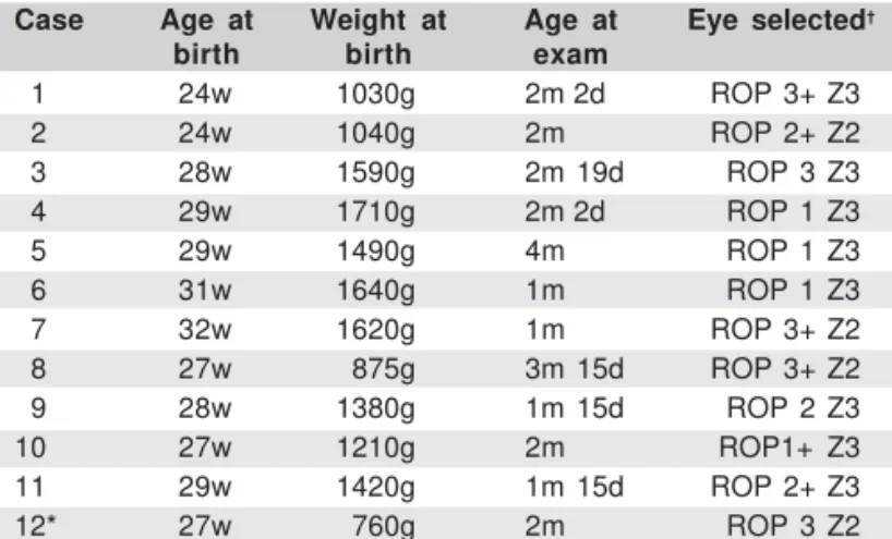
OCT images were obtained from fourteen eyes of thirteen premature babies (Table 1). Nominal categorization of ROP was applied according to the International Classification of ROP(7). Mean age and weight were 28 weeks and 1271.1 grams
respectively (range examination at from 24 to 32 weeks and 760 to 1710 grams). Mean age was 2 months and two days. ROP I was identified in four eyes (30.7%), ROP II in 23.1% (3/13 eyes) and six eyes had ROP III (46.2%). Although OCT images of one patient (31 weeks, 1060 grams), were analyzed, they were not included in the final results in table 1 because the baby had no ROP.
The fovea, with its characteristic depression, was easily recognizable in the retinal profile in patient #5 (Figure 1). OCT findings in patient #8 revealed the photoreceptor layer as a poorly reflective band immediately above the retinal pigment epithelium and that the band was thicker in the area of the foveal depression (Figure 2). The characteristic external portion of the
retina with its highly reflective band that corresponded to the retinal pigment epithelium and the less reflective structure which matched up with Bruch's membrane and choriocapillaris layer were also observed underneath (Figure 3). This preterm infant (31 weeks, 1060 grams), who had no ROP signals, was examined at 3 months of age and became our control.
DISCUSSION
The macula of the fetal retina is one of the last parts of the eye to complete its development(3). OCT has an important
his-Table 1. Patient data regarding age and weight at birth, classifi-cation and age at examination
Case Age at Weight at Age at Eye selected†
birth birth exam
01 24w 1030g 2m 2d ROP 3+ Z3
02 24w 1040g 2m ROP 2+ Z2
03 28w 1590g 2m 19d ROP 3 Z3
04 29w 1710g 2m 2d ROP 1 Z3
05 29w 1490g 4m ROP 1 Z3
06 31w 1640g 1m ROP 1 Z3
07 32w 1620g 1m ROP 3+ Z2
08 27w 875g 3m 15d ROP 3+ Z2
09 28w 1380g 1m 15d ROP 2 Z3
10 27w 1210g 2m ROP1+ Z3
11 29w 1420g 1m 15d ROP 2+ Z3
12* 27w 760g 2m ROP 3 Z2
w: weeks; g: grams; m/d: month/day; †According to the international Classification
of ROP; *both eyes
Figure 1 - Optical coherence tomography shows the foveal depression of patient #5
Figure 2 - Optical coherence tomography shows the poorly reflective band immediately above the retinal pigment epithelium layer (#8)
Arq Bras Oftalmol. 2007;70(1):19-21
Stratus optical coherence tomography findings in patients with retinopathy of prematurity 21
tological/clinical correlation with the inner parts of the retina in many diseases of the posterior segment. By studying the premature infant shortly after birth with Stratus OCT, it is possible to observe in vivo the structures of the last stages of the fetal retina(9).
OCT allows the retina to be studied in cross-sections from the vitreous/internal limiting membrane interface to the cho-riocapillaris and the inner choroidal layers(9).
Some authors observed that at 22 weeks of gestation, the fovea is a circular or elliptical zone approximately 1.5 mm in diameter comprising five to seven layers of cells in the gan-glion cell layer, a thin nerve fiber layer, a well-defined inner plexiform, inner nuclear and outer plexiform layers and an outer nuclear layer as well containing exclusively cone nuclei(4). The first recognizable depression in the central
region of the macula is first evident at 24-26 weeks of gesta-tion, apparently caused by thinning of the ganglion cell and inner nuclear layers. By the seventh month of gestation the inner nuclear layer becomes markedly thinned and the foveal pit appears more prominent. At 8 months two layers of gan-glion cells remain; this is reduced to a single layer in the neonate. Between birth and 45 months of age the diameter of the cones continues to decrease. Fovea reaches maturity from 11 to 15 months to five years(7).
Yuodelis and Hendrickson affirmed that the foveal pit is more pronounced at the seventh month of gestation by histo-logical evaluation and that the macula continues its develop-ment as late as five years from birth(5). The patients presented
in table 1 had ROP disease between grades I to III according to the International Classification(8). None of them showed
ma-cula detachment at fundoscopy or by OCT. In all cases the OCT features of the macular region had a strong correlation with the known histological aspects of the normal macula of a preterm baby, according to the literature(3-5). Easily
recogni-zable foveal depression and the external portion of the neural retina were found in three babies. One of them had no ROP (Figure 3). The highly reflective band that corresponded to the retinal pigment epithelium and the photoreceptor layer as well was observed in all babies.
CONCLUSION
These findings demonstrated that Stratus OCT may be a useful exam to acess the macula morphology of normal prema-ture babies and with ROP (class I, II, III) without any interven-tional procedure. OCT enables the diagnosis and quantifies
retinal features. It could be useful to follow the development of the macula anatomy in those patients.
We are unaware of previous reports of Stratus OCT images of premature infants with and without retinopathy of prematu-rity and we could find no reference to them in a computerized search using MEDLINE.
RESUMO
Objetivo: Descrever os aspectos morfológicos da mácula em
pacientes com retinopatia da prematuridade (ROP). Métodos:
Doze pacientes com retinopatia da prematuridade graus I, II and III foram submetidos a mapeamento de retina e avaliação por tomografia de coerência óptica. Resultados: Em todos os
treze olhos de 12 pacientes a tomografia de coerência óptica mostrou a camada do epitélio pigmentar hiperrefletiva, sendo a área macular com maior intensidade. Nesses olhos as cama-das da retina não estavam totalmente diferenciacama-das. A depres-são foveal ficou claramente evidente pela tomografia de coe-rência óptica em 23%. Conclusão: Nos pacientes prematuros
com retinopatia da prematuridade, a tomografia de coerência óptica mostrou as camadas da retina pouco diferenciadas com aumento da refletividade na área macular do complexo epitélio retiniano pigmentar-coriocapilar.
Descritores: Retinopatia da prematuridade; Tomografia de
coerência óptica; Macula lutea; Fóvea central
REFERENCES
1. Liarth JCS, Gonçalves JOR, Gonçalves EA, Meneses ES, Soares FM. Laser de diodo no tratamento da retinopatia da prematuridade. Arq Bras Oftalmol. 2001;64(5):411-3.
2. Smith LE. Pathogenesis of retinopathy of prematurity. Growth Horm IGF Res. 2004;14 Suppl A:S140-4.
3. Hendrickson A. A morphological comparison of foveal development in man and monkey. Eye. 1992;6(Pt 2):136-44.
4. Hendrickson AE, Yuodelis C. The morphological development of the human fovea. Ophthalmology. 1984;91(6):603-12.
5. Yuodelis C, Hendrickson A. A qualitative and quantitative analysis of the human fovea during development. Vision Res. 1986;26(6):847-55. 6. Isenberg SJ. Macular development in the premature infant. Am J Ophthalmol.
1986 Jan 15;101(1):74-80.
7. Retinopathy of prematurity: guidelines for screening and treatment. The report of a Joint Working Party of The Royal College of Ophthalmologists and the British Association of Perinatal Medicine. Early Hum Dev. 1996;46(3):239-58. 8. An international classification of retinopathy of prematurity. The Committee for the Classification of Retinopathy of Prematurity. Arch Ophthalmol. 1984; 102(8):1130-4.
9. Brancato R, Lumbroso B. Guide to optical coeherence tomography Interpre-tation. Rome: Innovation-News-Communication; 2004.