Evaluation of toxic retinopathy caused by antimalarial
medications with spectral domain optical
coherence tomography
Avaliação da frequência de retinopatia por antimaláricos com o
exame tomografia de coerência óptica de domínio espectral
Renata Tavares de Souza Cabral1, Evandro Mendes Klumb2, Maria Isabel Noronha Neta Couto3, Sueli Carneiro4
1. Sector of Ophtalmology, Retina and Vitreo Departament, Hospital Universitário Pedro Ernesto, Universidade do Estado do Rio de Janeiro, Rio de Janeiro, RJ, Brazil.
2. Department of Medical Clinic, Faculdade de Ciências Médicas, Universidade do Estado do Rio de Janeiro, RJ, Brazil. 3. Centro Universitário Augusto Motta, Rio de Janeiro, RJ, Brazil.
4. Department of Medical Specialties, Faculdade de Ciências Médicas, Universidade do Estado do Rio de Janeiro, Rio de Janeiro, RJ, Brazil.
Submitted for publication: January 10, 2018 Accepted for publication: May 27, 2018
Funding: No specific financial support was available for this study.
Disclosure of potential conflicts of interest: None of the authors have any potential conflicts of interest to disclose.
Corresponding author: Renata Tavares de Souza Cabral. Rua Santos Dumont, 904 - A - Macapá, AP - 68901-270 - Brazil E-mail: [email protected]
Approved by the following research ethics committee: Hospital Universitário Pedro Ernesto (CAAE: 06114912.2.0000.5259).
ABSTRACT | Purpose: To investigate the frequency of toxic retinopathy in patients with lupus erythematosus and rheuma-toid arthritis with long-term use of chloroquine diphosphate or hydroxychloroquine through spectral domain optical coherence tomography and the outcomes of ophthalmological exams (visual acuity - Snellen’s table, color vision test - Ishihara’s table, fundos-copy, and retinography - red-free). Methods: A cross-sectional study was carried out involving the ophthalmologic evaluation of patients using regular chloroquine diphosphate or hydroxychlo-roquine for a period of 1 year or longer. The patients completed a questionnaire on their opinions and treatment regularity. The same patients underwent ophthalmologic examination and spectral domain optical coherence tomography. Results: The prevalence of toxic retinopathy caused by antimalarials was 4.15% (9 of 217 patients), 7.4% (4 of 54 patients) following chloroquine diphosphate usage, and 0.82% (1 of 121 patients) following hydroxychloroquine usage. Only patients with advanced stage maculopathy presented abnormalities during the ophthalmologic exam: the color vision test was altered in 11.1%, and visual acuity and fundoscopy were altered in 33.3%. Identification of early toxic retinopathy, detected in six patients, was possible using spectral domain optical coherence tomography. The mean
duration of antimalarial drug usage among patients with toxic retinopathy was 10.4 years. Only 31% of the patients reported some symptoms during treatment, and although 24% were afraid to use the medication, they did so as prescribed. Conclusion: Use ofspectral domain optical coherence tomography was essential for the diagnosis of early-stage antimalarial toxic retinopathy in patients with the following characteristics: asymptomatic, antimalarial use 7 days a week for a period of more than 5 years, and normal clinical ophthalmologic examination.
Keywords: Retinopathy/etiology; Antimalarials/adverse effects; Hydroxychloroquine/adverse effects; Chloroquine/adverse effects; Tomography, optical coherence/methods
possível por meio da tomografia de coerência óptica de domínio espectral. A duração média do tempo de uso de drogas antimalá-ricas entre os pacientes com retinopatia tóxica foi de 10,4 anos. Apenas 31% dos pacientes relataram algum sintoma durante o tratamento e apesar de 24% terem medo de usar a medicação, eles o fizeram conforme prescrito. Conclusão: O uso da tomografia de coerência óptica de domínio espectral foi essencial para o diagnóstico de retinopatia tóxica antimalárica em estágio inicial em pacientes com as seguintes características: uso assintomático, antimalárico 7 dias por semana por um período maior que cinco anos e exame oftalmológico clínico normal.
Descritores: Retinopatia/etiologia; Antimaláricos/efeitos adversos; Hidroxicloroquina/efeitos adversos; Cloroquina/efeitos adversos; Tomografia de coerência óptica
INTRODUCTION
Toxic retinopathy (TR) caused by antimalarials, first described in 1951, is a rare adverse effect that causes irreversible vision loss(1-4). Currently, additional
ophthal-mologic tests enable the early diagnosis of maculopathy, even in asymptomatic patients(4-10). The prevalence of TR
differs among antimalarials. For chloroquine diphospha-te (CQ), a frequency ranging from 2.5% to 10% has been reported(11-13).
The risk factors for antimalarial TR described by the American Academy of Ophthalmology in 2016 were ca-tegorized as major and minor. The minor risk factors are age over 60 years, presence of liver disease, and generic factors (anomaly in ABCA423-2016 or P45024-2016 gene 4). On the other hand, the major risk factors are period of exposure greater than 5 years, nephropathy, con comitant use of tamoxifen, previous presence of ma-culopathy, and dose/kg of antimalarials (for hydroxychlo-roquine - HCQ, greater than 5.0 mg/kg of actual weight and for CQ, greater than 2.3 mg/kg of actual weight)(4).
Some articles consider cumulative dose as the most important risk factor. A daily dose of CQ greater than 250 mg/day or greater than 4 mg/kg/day and of HCQ greater than 400 mg/day or greater than 6.5 mg/kg/day translates to an accumulated risk greater than 460 g for CQ and greater than 1000 g for HCQ(2,3,5,8,14-19).
In the pre-macula stage, spectral domain optical cohe-rence tomography (SD-OCT) can detect alterations in the photoreceptor layer, at the junction of the internal and external segments of the retina (ISOS), and in the retinal pigment epithelium (RPE). It is possible to identify dis-crete irregularities in the bilateral perifoveal region. In the initial phase of TR, small faults can be detected in the photoreceptor layers, ISOS, and RPE in the perifoveal region in addition to the above irregularities.
At the moderate maculopathy stage, loss of the phy-siological depression of the fovea occurs with interrup-tions of the RPE and ISOS in the parafoveal and perifo-veal macular regions, characterizing the aspect in comet and macula in target. In the moderate and advanced stages, a typical fundoscopic TR lesion in the form of a bull’s eye can be identified, resulting from antimalarials. The advanced stage of maculopathy occurs with increased atrophy, foveal disruption, and visual acuity loss(15).
There is still no gold standard test for the diagnosis of toxic maculopathy, which makes it difficult to assess the sensitivity and specificity of diagnostic methods. The additional ophthalmologic tests described for the diagnosis of toxic maculopathy are multifocal electrore-tinography (mfERG), which has a sensitivity of 92.9% and specificity of 86.9%; computerized visual field, which has a sensitivity of 92.5% and specificity of 85.7%; and SD-OCT, which has a sensitivity of 78.6% and specificity of 98.1%(5,14).
This article describes the frequency of TR assessed using SD-OCT in patients with lupus erythematosus (LE) and rheumatoid arthritis (RA) with long-term use of chloroquine diphosphate (CQ) or hydroxychloroquine (HCQ) and the outcome of clinical ophthalmological exams (visual acuity tests - Snellen’s table, color vision test - Ishihara’s table, fundoscopy, and retinography - red-free).
METHODS
A cross-sectional study of outpatients at the Ophthal-mology Service from November 2012 to October 2014, who were regular users of CQ and/or HCQ for a period longer than 1 year, was conducted. The study protocol was approved by the Research Ethics Committee, and the patients signed a free and informed consent form.
Three hundred patients answered a questionnaire about the frequency, regularity, and duration of medi-cation use, the presence of adverse events, their level of knowledge about the drugs used, and their degree of sa-tisfaction with the treatment. Patients with ocular disease such as optic neuropathy, glaucoma, vascular occlusion, or degenerative maculopathy were excluded. After exclu-sion, 217 patients were eligible for the study.
RESULTS
Of the 217 patients, 208 (95.85%) had normal ophthal-mological and SD-OCT results. The prevalence of TR caused by antimalarials was 4.15% (95% CI: 0.015 to 0.068), occurring in 9 of 217 patients. These nine pa-tients were all women between 41 and 60 years of age, who regularly used one antimalarial tablet per day (5-7 days a week according to the medical prescription). The prevalence of toxic maculopathy was 7.4% from CQ usa-ge (4 of 54 patients; 95% CI: 0.0042 to 0.143) and 0.82% from HCQ usage (1 in 121 patients; 95% CI: 0.00786 to 0.02438). The prevalence when CQ was later replaced with HCQ was 9.52% (95% CI: 0.0064 to 0.1836), oc-curring in 4 of 42 patients.
The prevalence of TR was higher in the 41-to- 50-year-old age group (55.6%). TR developed in 6 of 153 patients (66.7%) with a diagnosis of LE and in 3 of 64 patients (33.3%) with RA. Among the nine patients with TR, one patient used HCQ, and eight used CQ (four of them only at the beginning of the treatment, after which they switched to HCQ). The prevalence of TR was similar in both diseases. Most of the patients had used CQ at some point.
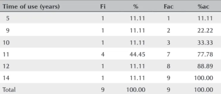
The duration of antimalarial medication use by pa-tients with TR varied from 5 to 14 years, with a mean duration of 10.4 years and a standard deviation of 2.45 (Table 1).
Of the 19 patients who did not take antimalarial me-dication at the time of the research but had previously taken it regularly, 36.8% (7) claimed that the medication was stopped by their physicians because of complications, 31.6% (6) reported that they switched to a different me-dication without stating the reason, and 31.6% (6) sus-pended their own medication out of fear of taking the drug. However, most patients with TR (66.7%, 6) stated that they were not afraid to use the medication and that
they were satisfied with it. Only one patient with HCQ use of more than 10 years developed TR, diagnosed at a moderate stage.
Only 31% of antimalarial users reported some symp-toms during treatment, such as altered skin color (40), blurred vision (31), headache (24), and tinnitus (22). Three (33.3%) TR patients had symptoms (altered skin color in two patients, dizziness, red eyes, headache, tinnitus, and blurred vision), and the other six (66.7%) remained asymptomatic throughout treatment.
The results were normal for 66.7% (6) of the patients with TR in the visual acuity test, for 88.9% (8) in the color vision test, and for 66.7% (6) in fundoscopy (Table 2). Most patients with TR (55.6%) had an ophthalmologic exam annually but did not undergo additional exams. One patient who had been using the drug for 5 years had the test performed just before starting treatment, and one third of patients had periodic clinical exams but not SD-OCT. Therefore, they were not diagnosed with maculopathy and continued to use antimalarials.
SD-OCT was useful for the classification of TR. Of the nine patients with TR caused by antimalarials, 44% (4) presented premacular maculopathy (Figure 1), 22% (2) at the initial stage (Figure 2), 22% (2) at the moderate stage (Figure 3), 11% (1) at the advanced stage (Figure 4), and none at the terminal stage (Table 3).
This exam enabled a diagnosis of early-stage TR in six patients who were still asymptomatic and had normal routine eye examinations.
DISCUSSION
When CQ and HCQ users were analyzed separately, the prevalence of TR caused by CQ (7.4%) was lower than that reported by Bernstein (10%) and higher than that demonstrated by the meta-analysis conducted by Ruiz Irastorza (ophthalmology examination and OCT).
Table 1. Time of use of the antimalarial medication by patients with toxic maculopathy
Time of use (years) Fi % Fac %ac
05 1 011.11 1 011.11
09 1 011.11 2 022.22
10 1 011.11 3 033.33
11 4 044.45 7 077.78
12 1 011.11 8 088.89
14 1 011.11 9 100.00
Total 9 100.00 9 100.00
Of the patients with maculopathy, 44.4% (4) used antimalarial medication for 11 years.
Table 2. Ophthalmological examination of patients with maculopathy associated with antimalarials: visual acuity, color vision, Ishihara’s table, fun-doscopic examination, and spectral domain optical coherence tomography
Normal Altered Normal Altered
Ophthalmological exam Fi Fi Total % % Total
Visual acuity 6 3 9 66.70 33.30 100
Color vision table 8 1 9 88.90 11.10 100
Fundoscopy 6 3 9 66.70 33.30 100
SD-OCT* 0 9 9 0 100 100
However, it was higher than that reported by Ruiz Iras-torza et al. (0.1%). Both drugs are recommended for the treatment of RA and LE. However, HCQ is described as having less toxicity, and therefore, it is more often prescribed(9,11,13,16,17).
The main reason for discontinuation of treatment, reported by a little more than a third of the patients, was ophthalmological complications. However, the majority had been misdiagnosed with TR using normal SD-OCT and could return to treatment. On the other hand, a third of patients with TR expressed fear of using the medication, but continued to use it anyway according to the prescription(9-12,20,21).
More than half of patients with TR used the medica-tion 7 days a week, taking 1 tablet per day according to the medical prescription without reaching the maximum dose. Durcan et al., in 2015, reported that the blood level of the prescribed medication may not correspond to the quantity administered to the patient because of non-adherence to treatment. Adherence to treatment in LE and RA ranges from 31.7%to 45.9%(6,21,22-24).
The TR prevention protocol recommends a prior ophthalmic examination before the start of treatment or in the first year of exposure. This is the baseline ophthalmic examination. The second examination should Figure 1. Spectral domain optical coherence tomography examination
with premacular maculopathy. Presence of discrete irregularities at the junction of the internal and external segments of the retina in the perifoveal region.
Figure 2. Spectral domain optical coherence tomography examination with previous maculopathy. Presence of irregularities at the junction of the in-ternal and exin-ternal segments of the retina, and retinal pigment epithelium.
Figure 3. Spectral domain optical coherence tomography examination with moderate maculopathy. We note interruption of the representative lines on the parafoveal macular region: at the junction of the internal and external segments of the retina; retinal pigment epithelium.
Figure 4. Infraredimage obtained by spectral domain optical coherence tomography: maculopathy in the form of a bull’s eye, alterations found in moderate maculopathy. Presence of foveal hyperreflectivity, surrounded by two rings, one internal hyperreflective ring and another external hyperreflective ring, forming a target-shaped lesion.
Bicas Neto & Mesquita observed TR by OCT in two pa-tients using CQ for almost 5 years(9,12).
include additional ophthalmologic tests, and SD-OCT may be considered. In patients taking the medication for less than 5 years with an adequate dosage for weight, monitoring can be done after 5 years of exposure(4).
After 5 years of continuous use of antimalarials, the risk of TR increases to 1%(2). After 20 years of use, the
risk increases to 20%. Most patients tested, 63.15%, had used the medication for a period of 5 years or more and 27.65%, for a period of 10 years or more. Almost 80% of TR patients used the medication for a period of 10 years or more, placing them at risk because of cumulative dose and exposure time(4,10,13).
The clinical ophthalmologic examination was normal for most patients, regardless of their complaints. One complained of blurred vision and three others, low vi-sion. The occurrence of a reduction in vision depends on professional activity, and the progressive loss of light vision depends on damage to the macular area. Patients are the most likely to notice moderate vision loss; however, by this stage, severe TR with permanent visual damage already exists. Initial maculopathy occurs outside the center of the fovea and progressively expands its invol-vement throughout the macular region. Therefore, even with maculopathy, it is possible for the patient to have normal vision at the beginning(2,5,14,17,25).
Only one patient with severe maculopathy presen-ted alterations in the color vision test. The other six did not complain during the anamnesis and had normal ophthal mologic examinations and retinography (infrared). The only altered examination was the optical cut of the SD-OCT, which showed four patients with premacular maculopathy and two with initial maculopathy.
Related symptoms may occur during treatment at diffe-rent stages but do not appear frequently. Dry eye was a frequent complaint, but is not described as an adverse effect of the medication, being related to connective tissue disorders, in addition to being common among middle-aged women because of hormonal variation. Blurred vision was resolved after refractive examination with the prescription of corrective lenses. It was not re-lated to maculopathy, but to presbyopia(1,10,16-21,26-30).
In a study carried out in São Paulo, Brazil, adverse effects were reported by 35.7% of patients: 17% reported ocular alterations; 10%, gastrointestinal; 3.4%, dermato-logical; 1.7%, neuromuscular; and 0.3%, psychiatric, lea-ding 22.9% of them to discontinue treatment. In another study, gastrointestinal and ocular manifestations were the most common complaints. However, both articles emphasized that antimalarials are well accepted by users(10,17,26).
The prevalence of TR in association with antimalarials detected using SD-OCT was 4.15%. The specific preva-lence for CQ was 7.4% and that for HCQ, 0.82%. The values found are in accordance with the literature. The ophthalmologic examination in users of antimalarials for both symptomatic and asymptomatic patients was normal in most cases. SD-OCT was essential for the diag-nosis of TR in the early stage, observed in six patients with the following characteristics: asymptomatic, using medication 7 days a week for a period of more than 5 years, along with normal ophthalmologic examination. In addition, patients felt safe and comfortable with the use of antimalarials, and only a third of patients repor-ted some symptoms during treatment.
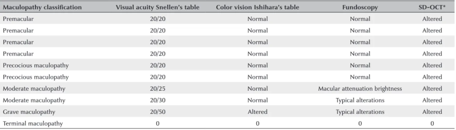
Table 3. Summary table with maculopathy classification and complete ophthalmological examination results for patients with toxic maculopathy asso-ciated with antimalarials
Maculopathy classification Visual acuity Snellen’s table Color vision Ishihara’s table Fundoscopy SD-OCT*
Premacular 20/20 Normal Normal Altered
Premacular 20/20 Normal Normal Altered
Premacular 20/20 Normal Normal Altered
Premacular 20/20 Normal Normal Altered
Precocious maculopathy 20/20 Normal Normal Altered
Precocious maculopathy 20/20 Normal Normal Altered
Moderate maculopathy 20/25 Normal Macular attenuation brightness Altered
Moderate maculopathy 20/30 Normal Typical alterations Altered
Grave maculopathy 20/50 Altered Typical alterations Altered
Terminal maculopathy 0 0 0 0
REFERENCES
1. Bernatsky S, Pineau C, Joseph L, Clarke A. Adherence to ophthalmolo-gic monitoring for antimalarial toxicity in a lupus cohort. J Rheumatol. 2003;30(8):1756-60.
2. Marmor MF, Kellner U, Lai TY, Lyons JS, Mieler WF; American Aca-demy of Ophthalmology. Revised recommendations on screening for chloroquine and hydroxychloroquine retinopathy. Ophthalmology. 2011;118(2):415-22.
3. Michaelides M, Stover NB, Francis PJ, Weleber RG. Retinal toxicity associated with hydroxychloroquine and chloroquine: risk factors, screening, and progression despite cessation of therapy. Arch Ophthalmol. 2011;129(1):30-9.
4. Marmor MF, Kellner U, Lai TY, Melles RB, Mieler WF; American Academy of Ophthalmology. Recommendations on Screening for Chloroquine and Hydroxychloroquine Retinopathy (2016 Revision). Ophthalmology. 2016;123(6):1386-94.
5. Missner S, Kellner U. Comparison of different screening methods for chloroquine/hydroxychloroquine retinopathy: multifocal elec-troretinography, color vision, perimetry, ophthalmoscopy, and fluorescein angiography. Graefes Arch Clin Exp Ophthalmol. 2012; 250(3):319-25.
6. Stepien KE, Han DP, Schell J, Godara P, Rha J, Carroll J. Spectral-domain optical coherence tomography and adaptive optics may detect hydroxychloroquine retinal toxicity before symptomatic vision loss. Trans Am Ophthalmol Soc. 2009;107:28-33.
7. Kiernan DF, Mieler WF, Hariprasad SM. Spectral-domain optical co-herence tomography: a comparison of modern high-resolution retinal imaging systems. Am J Ophthalmol. 2010;149(1):18-31.
8. Weinlander E, Ringeisen AL, Mititelu M. Retinopathy in the Era of Routine Hydroxychloroquine Monitoring. J Rheumatol. 2016; 43(6):1254.
9. Biccas Neto L, Mesquita AS. [Toxic maculopathy caused by antima-larial drugs: detection using spectral domain OCT: case reports]. Arq Bras Oftalmol. 2009;72(5):710-4. Portuguese.
10. Costedoat-Chalumeau N, Dunogué B, Leroux G, Morel N, Jallouli M, Le Guern V, et al. A critical review of the effects of hydroxychlo-roquine and chlohydroxychlo-roquine on the eye. Clin Rev Allergy Immunol. 2015;49(3):317-26.
11. Ruiz-Irastorza G, Ramos-Casals M, Brito-Zeron P, Khamashta MA. Clinical efficacy and side effects of antimalarials in systemic lupus erythematosus: a systematic review. Ann Rheum Dis. 2010; 69(1):20-8.
12. Bernstein HN. Ophthalmologic considerations and testing in patients receiving long-term antimalarial therapy. Am J Med. 1983;75(1 1A): 25-34.
13. Melles RB, Marmor MF. The risk of toxic retinopathy in patients on long-term hydroxychloroquine therapy. JAMA Ophthalmol. 2014; 132(12):1453-60.
14. Chen E, Brown DM, Benz MS, Fish RH, Wong TP, Kim RY, et al. Spectral domain optical coherence tomography as an effective screening test for hydroxychloroquine retinopathy (the “flying saucer” sign). Clin Ophthalmol. 2010;4:1151-8.
15. Wu L, Alpizar-Alvarez N. Choroidal imaging by spectral domain-opti-cal coherence tomography. Taiwan J Ophthalmol. 2013;3(1):3-13. 16. Laçava AC. Complicações oculares da terapêutica com a cloroquina
e derivados. Arq Bras Oftalmol. 2010;73(4):384-9.
17. Ponchet MR, Vilela MA, Sinahara KK, Dotto PF. Avaliação dos efeitos adversos desencadeados pelo uso de difosfato de cloroqui-na, com ênfase na retinotoxicidade, em 350 doentes com lúpus eritematoso. An Bras Dermatol. 2005;80 Suppl 3:S275-82. 18. Wolfe F, Marmor MF. Rates and predictors of hydroxychloroquine
retinal toxicity in patients with rheumatoid arthritis and systemic lupus erythematosus. Arthritis Care Res (Hoboken). 2010;62(6): 775-84.
19. Pasadhika S, Fishman GA. Effects of chronic exposure to hydro-xychloroquine or chloroquine on inner retinal structures. Eye (Lond). 2010;24(2):340-6.
20. Nebbioso M, Livani ML, Steigerwalt RD, Panetta V, Rispoli E. Retina in rheumatic diseases: standard full field and multifocal electro-retinography in hydroxychloroquine retinal dysfunction. Clin Exp Optom. 2011;94(3):276-83.
21. Kanski JJ. Doenças sistêmicas. In: Oftalmologia clínica: uma abordagem sistemática. 6a ed. Rio de Janeiro: Elsevier; 2008. p. 869-921.
22. Durcan L, Clarke WA, Magder LS, Petri M. Hydroxychloroqui-ne Blood Levels in Systemic Lupus Erythematosus: Clarifying Dosing Controversies and Improving Adherence. J Rheumatol. 2015;42(11):2092-7.
23. Oliveira-Santos M, Verani JF, Camacho LA, de Andrade CA, Fer-rante-Silva R, Klumb EM. Effectiveness of pharmaceutical care for drug treatment adherence in patients with systemic lupus erythe-matosus in Rio de Janeiro, Brazil: study protocol for a randomized controlled trial. Trials. 2016;17(1):181.
24. Prudente LR, Diniz JS, Ferreira TX, Lima DM, Silva NA, Saraiva G, et al. Medication adherence in patients in treatment for rheumatoid arthritis and systemic lupus erythematosus in a university hospital in Brazil. Patient Prefer Adherence. 2016;10(10):863-70.
25. Kanski JJ. Técnica de Exame ocular. In: Oftalmologia clínica: uma abordagem sistemática. 6a ed. Rio de Janeiro: Elsevier; 2008. p.1-24. 26. Costedoat-Chalumeau N, Dunogué B, Morel N, Le Guern V,
Guettrot-Imbert G. Hydroxychloroquine: a multifaceted treatment in lupus. Presse Med. 201443(6 Pt 2):e167-80.
27. Gass JD. Toxic disease affecting the pigment epithelium and
re-tina: chloroquine (Aralen®) and hydroxicloroquine (Plaquenil®)
retinopathy. In: Gass JD, editor. Stereoscopic atlas of macular diseases: diagnosis and treatment. 4th ed. St. Louis: Mosby; 1997. pp. 457-73.
28. Kanski JJ. Distúrbios Induzidos por medicamentos. In: Oftalmologia clínica: uma abordagem sistemática. 6a ed. Rio de Janeiro: Elsevier; 2008. p. 841-4.
29. Fonseca EC, Arruda GV, Rocha EM. Olho seco: etiopatogenia e tratamento. Arq Bras Oftalmol. 2010;73(2):197-203.