EDSON DA SILVA
EFEITOS DO DIABETES EXPERIMENTAL SOBRE A MORFOLOGIA E FUNÇÃO DO VENTRICULO ESQUERDO DE RATOS WISTAR PÚBERES:
IMPACTO DO TREINAMENTO DE NATAÇÃO
Tese apresentada à Universidade Federal de Viçosa, como parte das exigências do Programa de Pós-Graduação em Biologia Celular e Estrutural, para obtenção do título de Doctor Scientiae.
VIÇOSA
MINAS GERAIS – BRASIL 2013
i EDSON DA SILVA
EFEITOS DO DIABETES EXPERIMENTAL SOBRE A MORFOLOGIA E FUNÇÃO DO VENTRICULO ESQUERDO DE RATOS WISTAR PÚBERES:
IMPACTO DO TREINAMENTO DE NATAÇÃO
Tese apresentada à Universidade Federal de Viçosa, como parte das exigências do Programa de Pós-Graduação em Biologia Celular e Estrutural, para obtenção do título de Doctor Scientiae.
APROVADA: 08 de novembro de 2013.
_____________________________ ______________________________ Luciana Duarte Novais Silva Ita de Oliveira Silva
______________________________ _______________________________ Sirlene Souza Rodrigues Sartori Silvia Almeida Cardoso
ii O saber a gente aprende com os mestres e os livros. A sabedoria se aprende é com a vida e com os humildes.
iv AGRADECIMENTOS
A Deus pela vida e pela coragem de continuar a caminhada em todos os momentos.
À Marileila, minha esposa, amiga, companheira de todos os momentos, pelo exemplo de vida e pela força em superar tantas dificuldades e caminhar hoje ao meu lado.
À minha mãe Luíza, meus amados irmãos Noêmia, Luís, Eduardo e seus familiares, ao Salvador e sua família, por tudo que sempre fizeram por mim e por tudo que cada um representa ao longo da minha trajetória até aqui. Agradeço a todos pelo amor incondicional e pela representação de um modelo de esperança e fé.
Aos amigos do Laboratório de Biologia Estrutural: Ana Cláudia, Mariana, Carol, Suelem, Kyvia, Juliana, Kener, Marli, Wagner, Marta, Tatiana, Graziela, Suzana, Daiane, Camila, Stéphanie, Bruno e Bárbara, pela disposição em ajudar, pelas conversas e momentos de descontração, pela companhia na luta do dia-a-dia e pelo incentivo constante.
Aos companheiros de trabalho Mateus, Alex Bhering, Eliziária e Daise por iluminarem nossos caminhos em meio aos procedimentos e técnicas para as análises, pelo apoio nos experimentos, pela paciência, companhia e os adoráveis momentos de descontração.
Aos amigos do coração que fazem o dia-a-dia mais feliz: Claúdia, Eliziária, Kener, Rômulo, Gilton e Mírian.
À minha orientadora Profª. Izabel Regina dos Santos Costa Maldonado, por me acolher novamente na UFV. Obrigado pela parceria, confiança, paciência, amizade, pelos ensinamentos e apoio em questões acadêmicas, pessoais e profissionais. Uma pessoa exemplar e uma professora admirável.
Ao meu coorientador Prof. Antônio José Natali, pela orientação, disposição em ajudar, pelo apoio com o projeto junto à FAPEMIG, e incentivo para realizar os trabalhos e por ceder à infraestrutura do seu laboratório para realização de experimentos.
Ao meu coorientador Prof. Leandro Licursi de Oliveira, pelos conselhos em pesquisa, pela solicitude em ajudar e abrir as portas do seu laboratório para a realização de experimentos.
v Ao Departamento de Biologia Geral da UFV pela confiança em nosso trabalho de equipe, pelo apoio financeiro para os experimentos e solicitude em ajudar.
Ao Prof. José Eduardo Serrão pelos estímulos constantes para a busca de qualidade nas pesquisas, pelo apoio financeiro e de infraestrutura para a utilização dos laboratórios da UFV e realização de algumas etapas de experimentos.
Aos professores Gustavo Ferreira Martins e Jorge Abdalla Dergan por gentilmente permitirem a utilização de seus laboratórios para aquisição das imagens utilizadas.
À Universidade Federal de Viçosa e ao Programa de Pós-Graduação em Biologia Celular e Estrutural, pelo apoio em minha formação profissional e pessoal.
A todos os professores do Programa de Pós-Graduação em Biologia Celular e Estrutural, por todos os ensinamentos e incentivo.
Aos funcionários do Programa de Pós-Graduação em Biologia Celular e Estrutural, em especial Beth e Diana, pela ótima convivência, paciência, amizade e apoio sempre.
Às professoras Sirlene Souza Rodrigues Sartori, Ita de Oliveira Silva e Mariella Bontempo Duca de Freitas pelas orientações no ensino em anatomia humana e fisiologia. Pelo exemplo de simplicidade, profissionalismo, caráter e amizade. Sempre aprendo com vocês.
Aos colegas do Biotério Experimental do Departamento de Educação Física pela colaboração nos experimentos, apoio e convivência.
À Daise pela colaboração ao longo dos experimentos, pelos conselhos, dedicação, paciência, alegria, pelo exemplo de profissionalismo e de caráter e pela amizade.
À Marcinha pela parceria na pesquisa, pelos ensinamentos com o modelo experimental do diabetes, pelo exemplo de garra e disposição para o trabalho.
À FAPEMIG, pelo apoio financeiro.
À CAPES, pela concessão da bolsa de pesquisa durante parte do meu doutorado.
vi À Ieda Baracho dos Santos, funcionária do Departamento de Ciências Básicas da UFVJM pelas inúmeras orientações, informações e solicitude em ajudar a qualquer momento.
vii SUMÁRIO
LISTA DE ABREVIATURAS E SIGLAS...viii
RESUMO...xii
ABSTRACT ... xiii
INTRODUÇÃO GERAL ...1
REFERÊNCIAS BIBLIOGRÁFICAS... 6
ARTIGO 1...13
ARTIGO 2 ...38
viii LISTA DE ABREVIATURAS E SIGLAS
AMPK – Proteína quinase ativada por AMP ANOVA – Análise de variância
ATP – Adenosina trifosfato
ATPase – Enzima que catalisa a hidrólise do ATP BG – Blood glucose
bmp – Beats per minute BW – Body weight Ca2+ – Íon cálcio
CaCl2 – Calcium chloride
CaMKII – Calcium/calmodulin-dependent protein kinase II CE – Controle exercitado
CEUA – Ethics Committee on Animal Experimentation CRP – C-reactive protein
CS – Controle sedentário
DCM – Diabetic cardiomyophaty DE – Diabético exercitado
dL – Decilitro
DM – Diabetes mellitus DS – Diabético sedentário EC – Exercise control ECG – Electrocardiogram ECM – Extracellular matrix ED – Exercise diabetic
EGTA – Ethylene glycol-bis (ß-aminoethyl ether)-N, N, N’, N’-tetraacetic acid ELISA – Enzyme linked immunosorbent assay
eNOs – Endothelial nitric oxide synthase g – Gramas
GLUT 1 – Transportador de glicose 1 GLUT 4 – Transportador de glicose 4
ix HMW – High molecular weight
HR – Heart rate Hz – Hertz
ICAM-1 – Intercellular adhesion molecule-1 IL – Interleukin
IUR – Uniform random section KCl – Cloreto de potássio Kg – Quilograma
L – Litro
LMW – Low molecular weight LV – Left ventricle
M – Molar mg – Miligrama
MgCl2 – Cloreto de magnésio min – Minuto
mL – Mililitro mM – Milimolar
MMW – Middle molecular weight ms – Milissegundo
NaCl – Cloreto de sódio NCX – Na/Ca Exchange ng – Nanogramas NO – Nitric oxide ºC – Grau Celsius
PAS – Periodic acid-Schiff pH – Potencial hidrogeniônico PLB – Phospholamban
PN– Programa de natação
PPAR – Receptor ativado por proliferadores de peroxissoma gama
RyR2 – Canais receptores de rianodina S1P – Esfingosina-1 fosfato
x SEM – Standard error of the mean
SERCA2 – Sarcoplasmic reticulum Ca 2+ ATPase SphK1 – Esfingosina quinase 1
SR – Sarcoplasmic reticulum STZ – Estreptozotocina
T1DM – Type 1 diabetes mellitus T2DM – Type 2 diabetes mellitus TNF-α – Fator de necrose tumoral alfa
VCAM-1 – Vascular cell adhesion molecule-1 VE – Ventrículo esquerdo
vs – Versus VW – Weight gain WG – Ventricular weight
% r.c.l – Percentage of resting cell length µg – Microgramas
µm – Micrômetro µM – Micromolar
xi RESUMO
SILVA, Edson da, D.S., Universidade Federal de Viçosa, Novembro de 2013. Efeitos do diabetes experimental sobre a morfologia e função do ventrículo esquerdo de ratos Wistar púberes: impacto do treinamento de natação. Orientadora: Izabel Regina dos Santos Costa Maldonado. Coorientadores: Antônio José Natali e Leandro Licursi de Oliveira.
xiii ABSTRACT
1 INTRODUÇÃO GERAL
O Diabetes mellitus (DM) é uma desordem metabólica caracterizada pelo aumento da glicemia resultante de defeitos na secreção de insulina, ação da insulina, ou em ambos (AMERICAN DIABETES ASSOCIATION, 2013); com alta prevalência em adultos e adolescentes (MA, CHAN, 2009). O número de crianças, adolescentes e jovens adultos com DM aumenta a um ritmo alarmante (STEHNO-BITTEL, 2012; TRUONG et al., 2012). As doenças cardiovasculares representam a principal complicação do diabetes e podem ter início na infância (NADEAU et al., 2011) e influenciar de forma significativa a morbidade e a mortalidade na fase adulta (RAJESH et al., 2012; GIANNINI et al., 2011; GUO et al., 2007; LIBBY et al., 2005) .
A maioria dos casos de diabetes divide-se em duas categorias etiopatogenéticas: Diabetes Mellitus tipo 1 (DM1) e tipo 2 (DM2) (AMERICAN DIABETES ASSOCIATION, 2013). No DM1 a causa é uma deficiência absoluta da secreção de insulina, enquanto no DM2 a causa é uma combinação da resistência à ação da insulina e uma resposta secretora inadequada de insulina (AMERICAN DIABETES ASSOCIATION, 2013).
O DM1 é uma doença que ocorre principalmente por deficiência de insulina, a partir da destruição das células beta pancreáticas (AMERICAN DIABETES ASSOCIATION, 2013) e as doenças cardiovasculares podem estar associadas à resistência à insulina nesta forma de diabetes (PEREIRA et al., 2012; NADEAU et al., 2010; KILPATRICK et al., 2007; DEFRONZO et al., 1982). Além disto, o DM1 tem sido relacionado à inflamação de baixo grau e às complicações micro e macrovasculares em adultos (LLAURADÓ et al., 2012; GONZALEZ-CLEMENTE et al., 2007) e crianças (MANGGE et al., 2004). A inflamação sistêmica de baixo grau pode ser definida como a elevação, em duas a quatro vezes, das concentrações de citocinas que desencadeiam ações pró e anti-inflamatórias (BRUUNSGAARD, 2005).
2 qual aumenta os riscos para o desenvolvimento de insuficiência cardíaca de duas a cinco vezes (BELL, 1995).
Pessoas com diabetes podem desenvolver uma disfunção cardíaca denominada cardiomiopatia diabética (LAW et al., 2012). Esta é caracterizada por dilatação e hipertrofia do miocárdio, acompanhada por disfunção sistólica e/ou diastólica do ventrículo esquerdo (VE) e a sua presença é independente da coexistência de doença cardíaca isquêmica ou hipertensão (HAYAT et al., 2004). Apesar de a cardiomiopatia diabética ser uma condição subclínica crônica (VOULGARI et al., 2010), eventualmente leva à redução da elasticidade da matriz extracelular e diminuição da função contrátil do coração (FALCÃO-PIRES, LEITE-MOREIRA, 2012; RAJESH et al., 2012; ARAGNO et al., 2008; SEARLS et al., 2004). A associação entre o desenvolvimento da cardiomiopatia diabética e os elevados níveis de glicose têm efeitos significativos sobre a expressão, organização e modificação de componentes da matriz extracelular no coração (LAW et al., 2012).
A matriz extracelular cardíaca é uma rede dinâmica bem definida, constituída de proteínas estruturais, proteoglicanos, fatores de crescimento, citocinas e enzimas, essenciais para a organização e estrutura do tecido (LAW et al., 2012). É composta por colágeno (CAULFIELD, BORG, 1979), sendo colágeno fibrilar dos tipos I e III localizados no interstício do miocárdio, e colágeno não fibrilar dos tipos IV e VI e as glicoproteínas fibronectina e laminina, predominantes na membrana basal dos cardiomiócitos (BROWER et al., 2006; BISHOP, LAURENT, 1995; EGHBALI, WEBER, 1990). Elevações no estresse do miocárdio iniciam um remodelamento estrutural do coração, na tentativa de normalizar o estresse imposto. Este processo compreende a hipertrofia de cardiomiócitos com mudanças na quantidade e no fenótipo do colágeno, ocorrência de ligações cruzadas (cross-linking) entre moléculas de colágeno e remodelamento progressivo de componentes musculares, vasculares e da matriz extracelular do coração (BROWER et al., 2006). O aumento das concentrações intersticiais de colágeno e/ou de ligações cruzadas resulta em rigidez do miocárdio e disfunção diastólica do ventrículo esquerdo (BROWER et al., 2006).
3 et al., 2011). Um estado inflamatório crônico ocasiona alterações histopatológicas com lesão tecidual, disfunção endotelial e remodelamento cardíaco, os quais resultam em deterioração da contratilidade miocárdica e da função ventricular esquerda (LLAURADÓ ET AL., 2012; DUNCAN ET AL., 2007). A redução do transiente de Ca2+ (DUNCAN et al., 2007) e a redução da sensibilidade dos miofilamentos contrácteis ao Ca2+ são mecanismos possivelmente envolvidos neste processo (GOLDHABER et al., 1996).
Estudos confirmam que a adiponectina desempenha um papel importante no metabolismo de glicose e de lipídios (PEREIRA et al., 2012; GAREKANI et a.l, 2011; NUMAO et al., 2008), além de seu impacto relevante na patogênese do diabetes, da resistência à insulina e da lesão vascular (FENGER, 2013; GU et al., 2012). A adiponectina é uma proteína plasmática, secretada principalmente pelos adipócitos (BOBBERT et al., 2011; SUN, CHEN, 2010) e possui propriedades antidiabéticas, anti-inflamatórias, antiapoptóticas, antiaterogênicas (FORSBLOM et al., 2011; BOBBERT et al., 2011), imunomoduladoras e cardioprotetoras (JENKE et al., 2013; LETH et al., 2008). Dentre outras células, os cardiomiócitos são capazes de sintetizar adiponectina (PIÑEIRO et al., 2005; MAIA-FERNANDES et al., 2008), a qual tem função autócrina potencializando seu efeito cardioprotetor (HUI et al., 2012). As vias de sinalização dos efeitos cardioprotetores da adiponectina são mediadas principalmente pelo aumento de proteína quinase ativada por adenosina monofosfato (AMPK), receptor ativado por proliferadores de peroxissoma gama (PPAR ), esfingosina-1 fosfato (S1P) e esfingosina quinase 1 (SphK1) (HUI et al., 2012).
4 de IL-10 no coração (LO, MITSNEFES, 2012; GOLDSTEIN, SCALIA, MA, 2009; MAIA-FERANDES et al., 2008; HAN et al., 2007).
Por outro lado, o exercício físico regular é parte importante no manejo do DM1, devido aos efeitos benéficos à saúde, especialmente a prevenção de doenças cardiovasculares (GALASSET, RIDDELL, 2013). Assim, o treinamento físico tem sido recomendado como estratégia não farmacológica útil para reduzir a resistência à insulina, aumentar o metabolismo da glicose e de lipídios (KHAN, 2013), atenuar as alterações morfológicas e a disfunção contrátil em animais e humanos com cardiomiopatia diabética no DM1 (SILVA et al., 2013; CHIMEN et al, 2012; LOGANATHAN et al., 2012; RAJESH et al, 2012; SILVA et al., 2011; BIDASEE et al, 2008; SEARLS et al., 2004; DE ANGELIS et al., 2000). De modo geral, melhorias na função contrátil do miocárdio são observadas em seres humanos e animais, in vivo, em corações isolados e preparações multicelulares, submetidos a diferentes modelos de exercício (DI BELLO et al., 1996).
Além disto, a atividade física regular está associada à longevidade e à menor incidência de complicações decorrentes do diabetes (YARDLEY et al., 2012). Enquanto o treinamento físico de alta intensidade pode promover remodelamento cardíaco patológico em ratos (BENITO et al., 2011), o exercício aeróbico de baixa intensidade melhora a aptidão física e a força, reduz fatores de risco cardiovascular, melhora o bem-estar, reduz a necessidade de insulina e melhora a função autonômica cardíaca (CHIMEN et al, 2012; YARDLEY et al., 2012; DE ANGELIS et al., 2000). A literatura comprova que o exercício aeróbico de baixa a moderada intensidade é capaz de induzir adaptações cardiovasculares benéficas (por exemplo, melhora a função cardíaca, a estrutura do miocárdio, reduz a pressão arterial, e aumenta a sensibilidade baro e quimiorreflexa, o ritmo cardíaco intrínseco e o débito cardíaco), em ratos com diabetes induzido por estreptozotocina, um modelo que imita muito o DM1 humano (JORGE et al., 2012; LOGANATHAN et al., 2012; MOSTARDA et al., 2009; LOGANATHAN et al., 2007; DE ANGELIS et al., 2000). No entanto, os mecanismos subjacentes à histopatologia do DM1 e sua relação com o exercício físico, o remodelamento cardíaco e a disfunção cardíaca não são claros. Diante disto, torna-se importante conhecer os ajustes estruturais e morfológicos do miocárdio após o treinamento físico.
5 ANDO et al., 2009) principalmente com análises dos níveis circulantes de adiponectina total e HMW (ANDO et al., 2009). Até o momento, poucos estudos examinaram a relação de fatores que influenciam os níveis de adiponectina em
crianças e adolescentes com DM1, e os resultados destes estudos são inconsistentes
(KARAMIFAR et al, 2013; MENON, 2012; HABEEB et al., 2011; MESSAAOUI,
2012; HUERTA, 2006). Estudos recentes exibem redução na expressão de
adiponectina cardíaca em ratos com diabetes induzido por estreptozotocina (PEI et al, 2013), porém, a relação entre adiponectina cardíaca em ratos púberes com DM1 e
os efeitos de um programa de treinamento de natação nunca foi investigada.
Embora os relatos evidenciem de remodelamento cardíaco em humanos e animais adultos, dados semelhantes em ratos púberes com DM1 são escassos (SILVA et al., 2013; LAW et al., 2012; BROWER et al., 2006). Crianças e adolescentes com diabetes têm muitos dos benefícios que os adultos adquirem com a prática de exercícios, por isto devem praticar exercício físico com segurança, tanto para a saúde como para o lazer (ROBERTSON et al., 2009). Uma vez que os ratos são considerados um modelo animal válido para a compreensão do DM e são constantemente utilizados para este fim, acreditamos que ratos jovens podem servir como modelos para orientar o desenvolvimento de novas estratégias de prevenção e tratamento do DM.
Deste modo, o presente estudo foi realizado para verificar a influência do treinamento de natação de baixa intensidade sobre a estrutura dos cardiomiócitos, o remodelamento estrutural do miocárdio, níveis de citocinas e de óxido nítrico, e a densidade capilar do miocárdio, assim como a disfunção contrátil de cardiomiócitos do VE de ratos púberes com diabetes experimental não tratado com insulina.
6 REFERÊNCIAS BIBLIOGRÁFICAS
AMERICAN DIABETES ASSOCIATION. Diagnosis and classification of diabetes mellitus. Diabetes Care, v. 36, n. Suppl 1, p. S67-S74, 2013.
ANDO, D.; HOSAKA, Y.; SUZUKI, K.; YAMAGATA, Z. Effects of exercise training on circulating high molecular weight adiponectin and adiponectin oligomer composition: a randomized controlled trial. Journal of Atherosclerosis and Thrombosis, v. 16, n. 6, p. 733, 2009.
ARAGNO, M.; MASTROCOLA, R.; ALLOATTI, G.; VERCELLINATTO, I.; BARDINI, P.; GEUNA, S.; CATALANO, M.G.; DANNI, O.; BOCCUZZI, G. Oxidative stress triggers cardiac fibrosis in the heart of diabetic rats. Endocrinology, v. 149, n. 1, p. 380-388, 2008.
BELL, D.S.H. Diabetic Cardiomyopathy: A unique entity or a complication of coronary artery disease? Diabetes Care, v. 18, n. 5, p. 708-714, 1995.
BENITO, B.; GAY-JORDI, G.; SERRANO-MOLLAR, A.; YANFEN, E.G. Cardiac Arrhythmgenic Remodeling in a Rat Model of Long-Term Intensive Exercise TrainingClinical Perspective. Circulation, v. 123, n. 1, p. 13-22, 2011.
BIDASEE, K.R.; ZHENG, H.; SHAO, C.H.; PARBHU, P.K.; ROZANSKI, J.G.; PATEL, K.P. Exercise training initiated after the onset of diabetes preserves myocardial function: effects on expression of -adrenoceptors. Journal of Applied Physiology, v. 105, n. 3, p. 907-914, 2008.
BISHOP, J. E.; LAURENT, G. J. Collagen turnover and its regulation in the normal and hypertrophying heart. European Heart Journal, v. 16, n. suppl C, p. 38-44, 1995.
BOBBERT, P.; SCHEIBENBOGEN, C.; JENKE, A.; KANIA, G.; WILK, S.; KROHN, S.; STEHR, J.; KUEHL, U.; RAUCH, U.; ERIKSSON, U.; SCHULTHEISS, H.P.; POLLER, W.; SKURK, C. Adiponectin expression in patients with inflammatory cardiomyopathy indicates favorable outcome and inflammation control. European Heart Journal, v. 32, n. 9, p. 1134-1147, 2011.
BROWER, G.L.; GARDNER, J.D.; FORMAN, M.F.; MURRAY, D.B.; VOLOSHENYUK, T.V.; LEVICK, S.P.; JANICKI, J.S. The relationship between myocardial extracellular matrix remodeling and ventricular function. European Journal of Cardio-Thoracic Surgery, v. 30, n. 4, p. 604-610, 2006.
BRUUNSGAARD, H. Physical activity and modulation of systemic low-level inflammation. Journal of Leukocyte Biology, v. 78, n. 4, p. 819-835, 2005.
7 CHIMEN, M.; KENNEDY, A.; NIRANTHARAKUMAR, K.; PANG, T. T.; ANDREWS, R.; NARENDRAN, P. What are the health benefits of physical activity in type 1 diabetes mellitus? A literature review. Diabetologia, v. 55, n. 3, p. 542-551, 2012.
DE ANGELIS, K.L.; OLIVEIRA, A.R.; DALL'AGO, P.; PEIXOTO, L.R.; GADONSKI, G.; LACCHINI, S.; FERNANDES, T.G.; IRIGOYEN, M.C. Effects of exercise training on autonomic and myocardial dysfunction in streptozotocin-diabetic rats. Brazilian Journal of Medical and Biological Research, v. 33, n. 6, p. 635-641, 2000.
DEFRONZO, R. A.; HENDLER, R.; SIMONSON, D. Insulin resistance is a prominent feature of insulin-dependent diabetes. Diabetes, v. 31, n. 9, p. 795-801, 1982.
DI BELLO, V.; SANTORO, G.; TALARICO, L.; DI MURO, C.; CAPUTO, M. T.; GIORGI, D.; GIUSTI, C. Left ventricular function during exercise in athletes and in sedentary men. Medicine and Science in Sports and Exercise, v. 28, n. 2, p. 190-196, 1996.
DUNCAN, D. J.; HOPKINS, P. M.; HARRISON, S. M. Negative inotropic effects of tumour necrosis factor‐α and interleukin‐1 are ameliorated by alfentanil in rat ventricular myocytes. British Journal of Pharmacology, v. 150, n. 6, p. 720-726, 2007.
EGHBALI, M; WEBER, K. T. Collagen and the myocardium: fibrillar structure, biosynthesis and degradation in relation to hypertrophy and its regression. Molecular and Cellular Biochemistry, v. 96, n. 1, p. 1-14, 1990.
FALCÃO-PIRES I.; LEITE-MOREIRA, A.F. Diabetic cardiomyopathy: understanding the molecular and cellular basis to progress in diagnosis and treatment. Heart Failure Reviews, v. 17, n. 3, p. 325-344, 2012.
FENGER, M. The enigma of adiponectin. Atherosclerosis, v. 227, n.4, p.226-227, 2013.
FORSBLOM, C.; THOMAS, M.C.; MORAN, J.; SARAHEIMO, M.; THORN, L.; WADÉN, J.; GORDIN, D.; FRYSTYK, J.; FLYVBJERG, A.; GROOP, P.H. Serum adiponectin concentration is a positive predictor of all‐cause and cardiovascular mortality in type 1 diabetes. Journal of Internal Medicine, v. 270, n. 4, p. 346-355, 2011.
GALASSETTI, P.; RIDDELL, M.C. Exercise and Type 1 Diabetes (T1DM). Comprehensive Physiology, 2013.
8 GIANNINI, C.; MOHN, A.; CHIARELLI, F.; KELNAR, C.J. Macrovascular angiopathy in children and adolescents with type 1 diabetes. Diabetes/Metabolism Research and Reviews, v. 27, n. 5, p. 436-460, 2011.
GOLDHABER, J. I.; KIM, K. H.; NATTERSON, P. D.; LAWRENCE, T.; YANG, P.;WEISS, J. N. Effects of TNF-alpha on [Ca2+] i and contractility in isolated adult rabbit ventricular myocytes. American Journal of Physiology-Heart and Circulatory Physiology, v. 271, n. 4, p. H1449-H1455, 1996.
GOLDSTEIN, B.J.; SCALIA, R.G.; MA, X.L. Protective vascular and myocardial effects of adiponectin. Nature clinical practice. Cardiovascular medicine, v. 6, n. 1, p. 27-35, 2009.
GONZALEZ-CLEMENTE, J.M.; VILARDELL, C.; BROCH, M.; MEGIA, A.; CAIXAS, A.; GIMENEZ-PALOP, O.; RICHART, C.; SIMON, I.; MARTINEZ-RIQUELME, A.; ARROYO, J.; MAURICIO, D.; VENDRELL, J. Lower heart rate variability is associated with higher plasma concentrations of IL-6 in type 1 diabetes. European Journal of Endocrinology, v. 157, n. 1, p. 31-38, 2007.
GOTO, M.; GOTO, A.; MORITA, A.; DEURA, K.; SASAKI, S.; AIBA, N.; SHIMBO, T.; TERAUCHI, Y.; MIYACHI, M.; NODA, M.; WATANABE, S. Low‐molecular‐weight adiponectin and high‐molecular‐weight adiponectin levels in relation to diabetes. Obesity, 2013.
GU, W.; LI, Y.; GU, W.; LI, Y. The therapeutic potential of the adiponectin pathway. BioDrugs, v. 26, n. 1, p. 1-8, 2012.
GUO, Z.; XIA, Z.; YUEN, V. G.; MCNEILL, J.H. Cardiac expression of adiponectin and its receptors in streptozotocin-induced diabetic rats. Metabolism, v. 56, n. 10, p. 1363-1371, 2007.
HABEEB, N.M.M.; YOUSSEF, O.I.; SAAB, A.A.R.; EL HADIDI, E. Adiponectin as a Marker of Complications in Children with Type I Diabetes. Indian Pediatrics, v. 49, p. 277-280, 2011.
HAN, S.H.; QUON, M.J.; KIM, J.; KOH, K.K. Adiponectin and Cardiovascular Disease Response to Therapeutic Interventions. Journal of the American College of Cardiology, v. 49, n. 5, p. 531-538, 2007.
HAYAT, B.; PATEL, R.S.; KHATTAR, R.A.; MALIK, R.A. Diabetic cardiomyopathy: mechanisms, diagnosis and treatment. Clinical Science, v. 107, n. 6, p. 539-558, 2004.
HICKMAN, I.J.; WHITEHEAD, J.P. Structure, signalling and physiologic role of adiponectin-dietary and exercise-related variations. Current medicinal chemistry, v. 19, n. 32, p. 5427-5443, 2012.
9 HUI, X., LAM, K.S.; VANHOUTTE, P.M.; XU, A. Adiponectin and cardiovascular health: an update. British journal of pharmacology, v. 165, n. 3, p. 574-590, 2012. JENKE, A.; WILK, S.; POLLER, W.; ERIKSSON, U.; VALAPERTI, A.; RAUCH, B.H.; STROUX, A.; LIU, P.; SCHULTHEISS, H.P.; SCHEIBENBOGEN, C.; SKURK, C. Adiponectin protects against Toll-like receptor 4-mediated cardiac inflammation and injury. Cardiovascular Research, 2013.
JORGE , P.D.Y.; DA SILVA, D.D.; CONTI, F.F.; IRIGOYEN, M.C.; DE ANGELIS, K. Dynamic Aerobic Exercise Induces Baroreflex Improvement in Diabetic Rats. Experimental Diabetes Research, v. 2012, p 1-5, 2012.
KARAMIFAR, H.; HABIBIAN, N.; AMIRHAKIMI, G.; KARAMIZADEH, Z.; ALIPOUR, A. Adiponectin is a Good Marker for Metabolic State among Type 1 Diabetes Mellitus Patients. Iranian Journal of Pediatrics, v. 23, n. 3, p. 295, 2013.
KHAN, M.D. Exercise for the Management of Diabetes Mellitus: A Review of the Evidence. Journal of Enam Medical College, v. 3, n. 2, p. 99-108, 2013.
KILPATRICK, E.S.; RIGBY, A.S.; ATKIN, S.L. Insulin resistance, the metabolic syndrome, and complication risk in type 1 diabetes “Double diabetes” in the diabetes control and complications trial. Diabetes Care, v. 30, n. 3, p. 707-712, 2007.
LAW, B.; FOWLKES, F.; GOLDSMITH, J.G.; CARVER, W.; GOLDSMITH, E.C. Diabetes-induced alterations in the extracellular matrix and their impact on myocardial function. Microsc Microanal, v. 18, n. 1, p. 22-34, 2012.
LETH, H.; ANDERSEN, K. K.; FRYSTYK, J.; TARNOW, L.; ROSSING, P.; PARVING, H. H.; FLYVBJERG, A. Elevated levels of high-molecular-weight adiponectin in type 1 diabetes. Journal of Clinical Endocrinology & Metabolism, v. 93, n. 8, p. 3186-3191, 2008.
LI, C.J.; LV, L.; LI, H,; YU, D.M. LI. Cardiac fibrosis and dysfunction in experimental diabetic cardiomyopathy are ameliorated by alpha-lipoic acid. Cardiovascular Diabetology, v. 11, n. 1, p. 73, 2012.
LIBBY, P.; NATHAN, D. M.; ABRAHAM, K.; BRUNZELL, J.D.; FRADKIN, J. E.; HAFFNER, S.M.; RABADAN-DIEHL, C. Report of the National Heart, Lung, and Blood Institute-National Institute of Diabetes and Digestive and Kidney Diseases working group on cardiovascular complications of type 1 diabetes mellitus. Circulation, v. 111, n. 25, p. 3489-3493, 2005.
LLAURADÓ, G.; GALLART, L.; TIRADO, R.; MEGIA, A.; SIMÓN, I.; CAIXÀS, A.; GONZÁLEZ-CLEMENTE, J.M. Insulin resistance, low-grade inflammation and type 1 diabetes mellitus. Acta Diabetologica, v. 49, n. 1, p. 33-39, 2012.
10 LOGANATHAN, R.; BILGEN, M.; AL-HAFEZ, B.; ZHERO, S. V.; ALENEZY, M. D.; SMIRNOVA, I. V. Exercise training improves cardiac performance in diabetes: in vivo demonstration with quantitative cine-MRI analyses. Journal of Applied Physiology, v. 102, n. 2, p. 665-672, 2007.
LOGANATHAN, R.; NOVIKOVA, L.; BOULATNIKOV, I. G.; SMIRNOVA, I. V. Exercise-induced cardiac performance in autoimmune (Type 1) diabetes is associated with a decrease in myocardial diacylglycerol. Journal of Applied Physiology, v. 113, n. 5, p. 817-826, 2012.
MA, R.C.W.; CHAN, J.C.N. Diabetes: incidence of childhood type 1 diabetes: a worrying trend. Nature Reviews Endocrinology, v. 5, n. 10, p. 529-530, 2009.
MAIA-FERNANDES, T.; RONCON-ALBUQUERQUE JR, ROBERTO; LEITE-MOREIRA, A.F. Acções Cardiovasculares da Adiponectina: Implicações Fisiopatológicas [101]. Revista Portuguesa de Cardiologia, v. 27, n. 11, p. 1431-1450, 2008.
MANGGE, H.; SCHAUENSTEIN, K.; STROEDTER, L.; GRIESL, A.; MAERZ, W.; BORKENSTEIN, M. Low grade inflammation in juvenile obesity and type 1 diabetes associated with early signs of atherosclerosis. Experimental and Clinical Endocrinology and Diabetes, v. 112, n. 07, p. 378-382, 2004.
MENON, P.S.N. Adiponectin and Type 1 Diabetes Mellitus in Children. Indian Pediatrics, v. 49, p. 267-268, 2012.
MESSAAOUI, A.; WILLEMS, D.; MÉLOT, C.; DORCHY, H. Risk markers for cardiovascular disease in young type 1 diabetic patients: lipoproteins, high-sensitivity C-reactive protein and adiponectin. Acta Clinica Belgica, v. 67, n. 2, p. 79-82, 2012.
MOSTARDA, C.; ROGOW, A.; SILVA, I.C.M.; DE LA FUENTE, R.N.; JORGE, L.; RODRIGUES, B.; HEEREN, M.V.; CALDINI, E.G.; DE ANGELIS, K.; IRIGOYEN, I.C. Benefits of exercise training in diabetic rats persist after three weeks of detraining. Autonomic Neuroscience, v. 145, n. 1, p. 11-16, 2009.
NADEAU, K.J.; REGENSTEINER, J.G.; BAUER, T.A.; BROWN, M.S.; DOROSZ, J.L.; HULL, A.; ZEITLER, P.; DRAZNIN, B.; REUSCH, J.E. Insulin resistance in adolescents with type 1 diabetes and its relationship to cardiovascular function. Journal of Clinical Endocrinology and Metabolism, v. 95, n. 2, p. 513-521, 2010.
NADEAU, K.J.; REUSCH, J.E.B. Cardiovascular function/dysfunction in adolescents with type 1 diabetes. Current Diabetes Reports, v. 11, n. 3, p. 185-192, 2011.
11 NUMAO, S.; SUZUKI, M.; MATSUO, T.; NOMATA, Y.; NAKATA, Y.; TANAKA, K. Effects of acute aerobic exercise on high-molecular-weight adiponectin. Medicine and Science in Sports And Exercise, v. 40, n. 7, p. 1271, 2008.
PEI, H.; QU, Y.; LU, X.; YU, Q.; LIAN, K.; LIU, P.; YAN, W.; LIU, J.; MA, Y.; LIU, Y.; LI, C.; LI, W.; LAU, W.B.; ZHANG, H.; TAO, L. Cardiac-derived adiponectin induced by long-term insulin treatment ameliorates myocardial ischemia/reperfusion injury in type 1 diabetic mice via AMPK signaling. Basic Research in Cardiology, v. 108, n. 1, p. 1-11, 2013.
PEREIRA, R.I.; SNELL-BERGEON, J.K.; ERICKSON, C.; SCHAUER, I.E.; BERGMAN, B. C.; REWERS, M.; MAAHS, D. M. Adiponectin dysregulation and insulin resistance in type 1 diabetes. Journal of Clinical Endocrinology and Metabolism, v. 97, n. 4, p. E642-E647, 2012.
PIÑEIRO, R.; IGLESIAS, M.J.; GALLEGO, R.; RAGHAY, K.; EIRAS, S.; RUBIO, J.; LAGO, F. Adiponectin is synthesized and secreted by human and murine cardiomyocytes. FEBS Letters, v. 579, n. 23, p. 5163-5169, 2005.
RAJESH, M.; BÁTKAI, S.; KECHRID, M.; MUKHOPADHYAY, P.; LEE, W.; HORVÁTH, B.; CINAR, E.R.; LIAUDET, L.; MACKIE, K.; HASKÓ, G.; PACHER, P. Cannabinoid 1 receptor promotes cardiac dysfunction, oxidative stress, inflammation, and fibrosis in diabetic cardiomyopathy. Diabetes, v. 61, n. 3, p. 716-727, 2012.
ROBERTSON, K.; ADOLFSSON, P.; SCHEINER, G.; HANAS, R.; RIDDELL, M.C. Exercise in children and adolescents with diabetes. Pediatric Diabetes, v. 10, n. s12, p. 154-168, 2009.
RYBA, M.; MAREK, N.; HAK, Ł.; RYBARCZYK-KAPTURSKA, K.;
MY LIWIEC, M., TRZONKOWSKI, P., & MY LIWSKA, J.
Anti-TNF rescue CD4+ Foxp3+ regulatory T cells in patients with type 1 diabetes from effects mediated by TNF. Cytokine, v. 55, n. 3, p. 353-361, 2011.
SALEM, M.; EL BEHERY, S.; ADLY, A.; KHALIL, D.; EL HADIDI, E. Early predictors of myocardial disease in children and adolescents with type 1 diabetes mellitus. Pediatric Diabetes, v. 10, n. 8, p. 513-521, 2009.
SEARLS, Y.M.; SMIRNOVA, I.V.; FEGLEY, B.R.; STEHNO-BITTEL, L. Exercise attenuates diabetes-induced ultrastructural changes in rat cardiac tissue. Medicine and Science in Sports and Exercise, v. 36, n. 11, p. 1863-1870, 2004.
12 SILVA, M.F.; PELÚZIO, M.C.G.; AMORIM, P.R.S.; LAVORATO, V.N.; SANTOS, N.P.; BOZI, L.H.M.; PENITENTE, A.R.; FALKOSKI, D.L.; BERFORT, F.G.; NATALI, A.J. Treinamento em natação atenua a disfução contrátil de cardiomiócitos de ratos diabéticos; Swimming training attenuates contractile dysfunction in diabetic rat cardiomyocytes. Arq. bras. cardiol, v. 97, n. 1, p. 33-39, 2011.
STEHNO-BITTEL, L. Organ-Based Response to Exercise in Type 1 Diabetes. ISRN Endocrinology, v. 2012, 2012.
SUN, Y.; CHEN, X. Effect of adiponectin on apoptosis: Proapoptosis or antiapoptosis? Biofactors, v. 36, n. 3, p. 179-186, 2010.
TRUONG, U. T.; MAAHS, D.M.; DANIELS, S.R. Cardiovascular disease in children and adolescents with diabetes: where are we, and where are we going?. Diabetes Technology and Therapeutics, v. 14, n. S1, p. S-11-S-21, 2012.
VOULGARI, C.; PAPADOGIANNIS, D.; TENTOLOURIS, N. Diabetic cardiomyopathy: from the pathophysiology of the cardiac myocytes to current diagnosis and management strategies. Vascular Health and Risk Management, v. 6, p. 883, 2010.
WANG, T.; MAO, X.; LI, H.; QIAO, S.; XU, A.; WANG, J.; LEI, S.; LIU, Z.; NG, K.F.; WONG, G.T.; VANHOUTTE, P.M.; IRWIN, M.G.; XIA, Z. N-Acetylcysteine And Allopurinol Up-regulated the Jak/STAT3 and PI3K/Akt pathway via Adiponectin and Attenuated Myocardial Post-Ischemic Injury In Diabetes. Free Radical Biology and Medicine, 2013.
13 Artigo 1
Ventricular remodeling in growing rats with experimental diabetes: the impact of swimming training
Edson Silvaa,b, Antônio J. Natalic*, Márcia F. Silvaa, Gilton J. Gomesc, Daise N.Q. Cunhac, Regiane M.S. Ramosc, Marileila M. Toledod, Filipe R. Drummondc, Felipe G. Belfortc, Rômulo D. Novaesa,e, Izabel R.S.C. Maldonadoa*≠
*
The authors share senior authorship.
a
Department of General Biology, Federal University of Viçosa, Viçosa, MG, Brazil. bDepartment of Basic Sciences, Federal University of Jequitinhonha and Mucuri Valleys, Diamantina, MG, Brazil.
c
Department of Physical Education, Federal University of Viçosa, Viçosa, MG, Brazil.
d
Department of Medicine and Nursing, Federal University of Viçosa, Viçosa, MG, Brazil.
eDepartment of Biological Sciences and NUPEB, Federal University of Ouro Preto, MG, Brazil.
Keywords: Fibrosis; ventricular remodeling; collagen matrix; diabetes mellitus; exercise.
≠Address for correspondence: Izabel Regina dos Santos Costa Maldonado, Universidade Federal de Viçosa, Departamento de Biologia Geral. Edifício Chotaro Shimoya, Avenida PH Rolfs, s/nº, CEP: 35.570-000 Viçosa, Minas Gerais, MG, Brasil. Tel: +55 (031) 3899-3365. Email: irscosta@ufv.br
14 Abstract
15 Introduction
Diabetes mellitus (DM) and its long-term complications are considered one of the major health problems worldwide [33]. The number of children, adolescents, and young adults with DM continue to rise at a dramatic rate [54,55]. In patients with diabetes, cardiovascular complications represent the chief cause of morbidity and mortality in adults [46], children, and adolescents [20].
The pathways leading to DM-induced cardiac dysfunction and its histopathological changes are yet to be fully clarified [42,44,48]. However, this could be in part due to the complex and multifactorial nature of diabetes [31]. Probably the most important role has been attributed to persistent hyperglycemia [33]. This condition increases the risks for developing heart failure (HF) from two- to five fold [8].
Individuals with diabetes can develop cardiac dysfunction, termed diabetic cardiomyopathy (DCM) [31]. Diabetic cardiomyopathy is characterized by myocardial dilatation and hypertrophy, accompanied by systolic and diastolic left ventricle (LV) dysfunction, and its presence is independent of the coexistence of ischemic heart disease or hypertension [25]. Although DCM may be subclinical for a long time [58], eventually, it leads to decreased myocardial elasticity and impaired heart contractile function [3,18,46,49]. The link between elevated glucose levels and the development of DCM has significant effects on the expression, organization, and modification of extracellular matrix (ECM) components in the heart [31].
16 and/or cross-linking results in a stiffer myocardium and ventricular diastolic dysfunction [12].
Exercise training is of particular interest in both the prevention and treatment of DCM in type 1 DM [35]. Thus, exercise training has been suggested to be a useful nonpharmacological strategy to attenuate morphological changes and contractile dysfunction in both animals and human with DM [10,15,16,49]. Furthermore, regular physical activity is associated with greater longevity and lower frequency and severity of diabetes complications [59]. While long-term intensive exercise training is suggested to promote adverse remodeling (i.e., fibrosis) in a rat model [9], low-intensity aerobic exercises improves physical fitness and strength, reduces cardiovascular risk factors, improves well-being, reduces insulin requirements, and improves cardiac autonomic function [15,16,59]. Indeed, low- to moderate-intensity aerobic exercise has been shown to induce beneficial cardiovascular adaptations (e.g., improves cardiac function, myocardial structure; reduces blood pressure; increases baro- and chemoreflex sensitivity, intrinsic heart rate, and cardiac output) in STZ-induced diabetic rats, a model that imitates many of the human type 1 DM [16,29,34,35,41]. Nevertheless, the mechanisms underlying the histopathology of type 1 DM and their relationship to physical exercise, myocardial interstitial fibrosis, and cardiac functional impairment are unclear. Although reports show evidence of cardiac remodeling in adult humans and animals, similar data in growing rats with type 1 DM are lacking [12,31]. Children and adolescents with diabetes should have many of the same health and leisure benefits as adults and should be allowed to practice physical exercise with safety [47]. Since rats are considered a valid animal model for understanding the DM and are consistently used for this purpose, we believe that young rats could serve as models for guide the development of novel strategies for prevention and management of DM. Therefore, the present study was undertaken to verify the effects of low-intensity swimming training on the structural remodeling of the LV of growing rats with unmanaged experimental diabetes.
Materials and Methods
Animals and experimental groups
17 divided into four groups (n = 5, per group): sedentary control (SC), exercised control (EC), sedentary diabetic (SD), and exercised diabetic (ED). Rats were maintained on 12-h dark/light cycle at 22°C, housed in groups of five, and fed standard commercial rodent chow and water ad libitum. All procedures followed the Guidelines for Ethical Care of Experimental Animals and were approved by the Ethics Committee on Animal Experimentation (CEUA) from the Federal University of Viçosa (protocol number 46/2011).
Diabetes induction
Severe diabetes was induced in the animals by intraperitoneal injection of streptozotocin (STZ; Sigma-Aldrich, St. Louis, MO) dissolved in 0.1 M citrate buffer solution (0.1 M, pH 4.5) at the dose of 60 mg kg-1 body weight (BW) [28]. Equivalent volume (1 mL kg-1) of vehicle was injected into the rats assigned to the control groups. Animals were fasted overnight for 12 h prior to STZ administration. Water and food were available immediately after dosing. Development of diabetes was determined by observing hyperglycemia (> 300 mg dL-1) [51] as measured by an Accu-Chek Advantage glucometer (Boehringer Mannheim Corporation, Indianapolis, IN). Body weights and blood glucose levels were recorded once a week throughout the study. All animals were euthanatized 8 weeks after diabetes induction by intraperitoneal injection of sodium pentobarbital (120 mg kg-1).
Exercise protocol
18 increased by 1% of the animal’s BW from the fourth week on, such that at the eighth week, animals swam with a total load of 5% of their BW. During the swimming sessions, animals from the sedentary group (SD and SC) were placed in a polypropylene box containing warm water (28°C to 30°C) with a depth of 10 cm.
Electrocardiogram
Animals were anesthetized in an induction chamber with 2% isoflurane and 100% oxygen at a constant flow of 1 L/min. Once unconscious, they were placed on a platform in dorsal recumbency, with the four limbs fixed. Isoflurane was maintained at a concentration sufficient for restrain (0.5% to 1.0%), and animals were able to maintain spontaneous respiration during the electrocardiogram (ECG). A trichotomy of approximately 1 cm2 was performed in the forelimbs and left hindlimb for electrode insertion. Derivation II of the ECG was recorded using the data acquisition system PowerLab® (AD Instruments, São Paulo, Brazil), and data analysis was done with the program LabChart Pro® (ADInstruments LabChart 7, São Paulo, Brazil).
Electrocardiograms were performed at the end of the experiments by an experienced fellow blind to the study groups and treatments. Resting heart rate (HR) was derived from the ECG, following established guidelines [26].
Histological processing and histochemistry
19 Analysis of myocardial collagen content
To quantify interstitial fibrosis, LV was evaluated in histological sections 5 µm thick stained with Sirius Red dye (Sirius Red F3B, Mobay Chemical Co., Union, NJ, USA), which marks collagen fibers types I and III for further observation under a polarizing microscope [30]. The distribution of total collagen content was analyzed using the image analysis software Image Pro-Plus 4.5® (Media Cybernetics, Silver Spring, MD, USA) based on the birefringence properties of the collagen fibrils under polarized light. In this analysis, 12 microscopic fields from five rats per group were investigated (magnification ×200) randomly. A 208-point grid was superimposed on each image, and the total of 2,496 points was assigned for each rat. The area was determined by adding up these points, then dividing the result by the total points for the cross-section. Results were expressed as mean ± SD. Samples were observed under an Olympus BX53 microscope equipped with a DP-73 digital camera and imaging system (CellSens, OlympusCorporation, Tokyo, Japan).
Periodic acid-Schiff and Gomori’s reticulin staining
A qualitative analysis based on the intensity of the staining reaction with the ventricular muscle was performed using PAS stain and Gomori’s reticulin stain. In this analysis, 12 microscopic fields from five rats per group were investigated (magnification ×1,000) randomly. Tissue sections of LV were stained with PAS for detection of polysaccharides [53], and amylase-treated sections served as negative controls. Other sections of LV were stained by Gomori’s reticulin. The level of polysaccharides/collagen staining received a score that varied from 1 cross (+), representing weak reaction for polysaccharides/collagen fibers, to 4 crosses (++++), representing the strongest staining for polysaccharides/collagen (adapted from Spillman et al. [53]).
Statistical analysis
20 Results
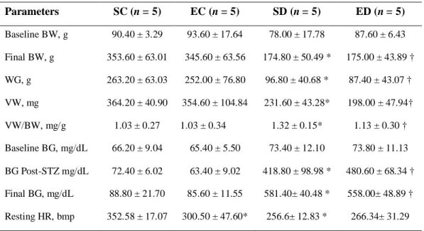
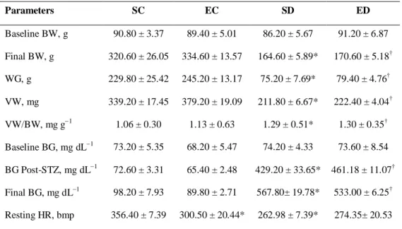
The animals of the groups SD and ED developed the expected hyperglycemia of severe diabetes compared with control groups. Initially, on the baseline, there were no differences in blood glucose levels between diabetic (SD vs. ED) and control rats (SC vs. EC) (Table 1). Blood glucose increased throughout the experiment in both SD and ED rats. Seven days after the application of STZ and at the end of the study, the glycemic levels for the diabetic groups (SD and ED) were significantly greater compared with their controls (Table 1). Swimming training reduced BG level by 23 mg/dL in the diabetic groups, but this did not reach statistical difference.
The analyses of the baseline body weights were not different among the four groups (Table 1). Diabetic rats had lower BW gain compared with normal rats (SC and EC) (p < 0.05). Similarly, the LV weighted significantly less in the diabetic rats (SD and ED).
Table 1. Biometrical and functional parameters
Parameters SC (n = 5) EC (n = 5) SD (n = 5) ED (n = 5)
Baseline BW, g 90.40 ± 3.29 93.60 ± 17.64 78.00 ± 17.78 87.60 ± 6.43
Final BW, g 353.60 ± 63.01 345.60 ± 63.56 174.80 ± 50.49 * 175.00 ± 43.89 †
WG, g 263.20 ± 63.03 252.00 ± 76.80 96.80 ± 40.68 * 87.40 ± 43.07 †
VW, mg 364.20 ± 40.90 354.60 ± 104.84 231.60 ± 43.28* 198.00 ± 47.94†
VW/BW, mg/g 1.03 ± 0.27 1.03 ± 0.34 1.32 ± 0.15* 1.13 ± 0.30 †
Baseline BG, mg/dL 66.20 ± 9.04 65.40 ± 5.50 73.40 ± 12.10 73.80 ± 11.13
BG Post-STZ mg/dL 72.40 ± 6.02 63.40 ± 9.02 418.80 ± 98.98 * 480.60 ± 68.34 †
Final BG, mg/dL 88.80 ± 21.70 85.60 ± 11.55 581.40± 40.48 * 558.00± 48.89 †
Resting HR, bmp 352.58 ± 17.07 300.50 ± 47.60* 256.6± 12.83 * 266.34± 31.29
21 As expected, STZ-induced diabetes resulted in decreased resting HR in SD animals compared with SC (Table 1). Resting HR did not differ between ED and EC rats. The swimming training program was capable of reducing the resting HR for the EC group when baseline and end-study comparisons were made.
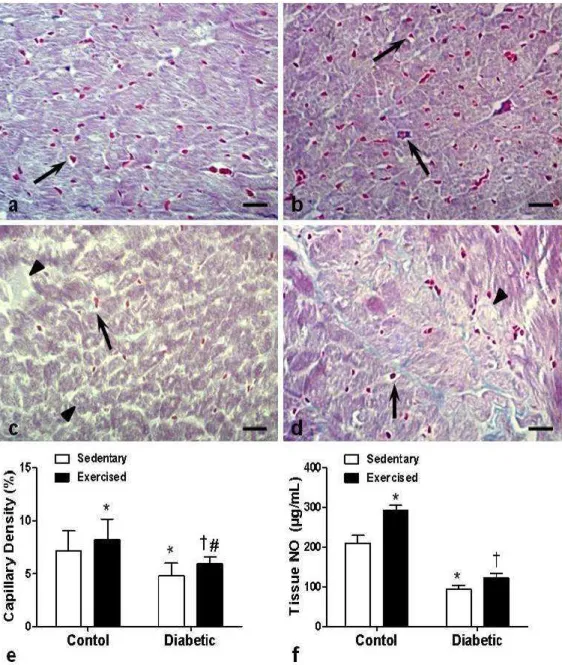
Diabetes was associated with interstitial fibrosis by the accumulation of myocardial collagen (Fig. 2 and 3). Collagen content was significantly greater in SD rats compared with SC (7.41 ± 2.08% vs. 3.04±0.26%, respectively; p < 0.05) and in ED compared with EC rats (4.29 ± 0.36 vs. 2.82±0.38, respectively; p < 0.047) (Fig. 1 and 2). Interestingly, the exercise training reduced collagen accumulation in the LV of diabetic animals (SD vs. ED, p < 0.05). However, this was not true in the nondiabetic animals (Fig. 1, 2 and 3).
Contol Diabetic 0 2 4 6 8 10 Exercised Sedentary
*
# †
L V T o ta l c o ll a g e n ( % )23 Figure 3. Diabetes-induced cardiac fibrosis. The red color of Sirius Red staining under light microscopy indicates total collagen deposition. (A) Sedentary control, (B) exercised control, (C) sedentary diabetic, and (D) exercised diabetic groups. Arrows = collagen fibers. Observe the increased amount of collagen fibers in panel C and its reduction (near normality) in panel D. Asterisks = interstitial fibrosis; magnification, ×400; bar: 30 µm.
24 Table 2. Qualitative analysis of the LV of rats by Periodic acid-Schiff (PAS) and Gomori’s reticulin histochemical techniques
Histochemistry techniques SC (n = 5) EC (n = 5) SD (n = 5) ED (n = 5)
PAS-sarcoplasm + + ++++ +++
PAS-endomysium + + ++++ ++
Gomori’s reticulin + + +++ ++
PAS staining and Gomori’s reticulin staining score: (+) weak reaction with the staining technique, (++) moderate reaction with the staining technique, (+++) strong reaction with the staining technique, (++++) intense reaction with the staining technique. (A) Sedentary control, (B) exercised control, (C) sedentary diabetic, and (D) exercised diabetic groups.
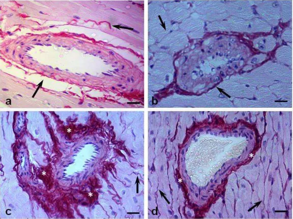
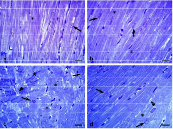
26 Figure 5. Silver-impregnated section of myocardium subjected to the Gomori’s reticulin technique. (A) Sedentary control, (B) exercised control, (C) sedentary diabetic, and (D) exercised diabetic groups. Arrows = reticular fibers consisting mainly of collagen type III. Observe the increase of reticular fibers in panel C and its reduction in panel D. Note in panel C the reticular fibers associated with cardiomyocyte degeneration and possibly a process of recovery from injury. Asterisks = thick collagen fibers; magnification, ×1,000; bar: 20 µm.
Discussion
27 chronic exercise, as observed by reduced collagen deposition and the accumulation of glycogen.
We found increased LV collagen deposition on STZ-induced diabetic rats, which is in agreement with previous studies [2,4,13,32,33]. Our results showed that the degree of total collagen deposition in SD rats was significantly more intense, and consequently, fibrosis was more evident compared with normal rats. These data substantiates a histomorphological remodeling process, which is able to endanger the heart function. In fact, collagen deposition leads to a stiffer myocardium, and consequently, diastolic dysfunction is observed [12]. Even though this was not directly evaluated in this study, based on collagen findings, it is possible that diabetic animals have developed diastolic dysfunction as observed by Loganathan et al. [35].
Additionally, the degree of interstitial fibrosis in ED rats was reduced compared with the SD group. Cardiac fibrosis is characterized by the proliferation of cardiac fibroblasts and the excessive accumulation of matrix proteins, mainly collagen types I and III, in the extracellular space [31,56,60]. This occurs due to imbalanced ECM metabolism, characterized by increased collagen synthesis and decreased collagen degradation [56]. Moreover, glycated proteins can undergo a series of chemical rearrangements to form complex compounds and cross-links known as advanced glycation end products (AGEs) [40]. The accumulation of AGE in collagen was associated with reduced collagen turnover, increased cross-linking of collagen, and stiffness of arteries and myocardium [40]. Furthermore, collagen glycation increases the formation and migration of myofibroblasts in the heart, a critical event during fibrosis development in diabetes [60].
28 child and adolescent can produce myocardial dysfunction that may be a threat to the development of severe cardiomyopathy at adulthood [7].
Our qualitative analysis of PAS staining demonstrated a greater tissue distribution of polysaccharides in the LV myocardium of diabetics compared with control animals. The accumulation of PAS-positive material was also observed in the sarcoplasm of cardiomyocyte of the SD rats, which is in concordance with previous studies [13,36,43].
The effects of diabetes on myocardial glycogen metabolism are directly related to decreased glucose transport, which might result in increased myocardial glycogen [43]. However, the excess of glycogen brings about cardiac structural and physiological impairments, including changes in pH, ionic imbalances, and stimulations of pathways leading to hypertrophic signaling [45]. It is known that energy metabolism is rapidly shifted in the diabetic metabolism, resulting in augmented fatty acid and decreased glucose consumption [21]. The switch of cardiac energy substrate utilization from carbohydrate to lipids increases intracellular glycogen, probably through increased glycogen synthesis or impaired glycogenolysis or a combination of both [24]. Therefore, the greatest intensity in the PAS-positive reaction observed in the LV of sedentary diabetic rats in the present study may be related to glycogen stored and spared in the myocardium [13]. In addition, a higher reaction found in the endomysium by PAS staining may be related to morphological changes involved in the mechanisms of type III and type IV collagen deposition, both positive to PAS technique [13,19].
Although exercise seemed to improve accumulation of glycogen in our trained animals, diabetes played its role. This was demonstrated by a slightly greater reaction of PAS-positive material in the sarcoplasm of the exercised diabetic rats when compared with the nondiabetic animals. These results highlight the benefits that the exercise training exerted on these animals by partially recovering the polysaccharides tissue distribution. According to Castellar et al. [13], this recovery might be attributed to an improved metabolic status provided by physical exercise, which apparently reduces the necessity for glycogen production.
29 total collagen deposition in the ECM of diabetic animals reported previously [31]. This adaptation was reduced by exercise training (ED animals) and was unchanged in nondiabetic animals (SC and EC). However, the reduction of reticular fiber in ED animals is partially different from previous reports [13]. Castellar and colleagues [13] found only a slight increased stain reaction to silver impregnation in sedentary diabetic rats compared with exercised diabetic and control animals. These controversial results could be attributed to differences in exercise training protocols or even to age differences among these studies. In this study, the duration of the training sessions was 30 min longer, and the animals were 40 days younger than those of Castellar’s protocol [13]. A higher reaction of the endomysium by Gomori’s reticulin staining in our study also may be involved in the mechanisms of type III collagen deposition, corroborating with our data of PAS staining [13]. Though the interactions of ECM components with silver may not be specific, they can nevertheless provide important insights into the mechanisms of the chemical reactions involved in ECM remodeling [57]. However, the precise mechanism for the protection associated with decreased reticular fiber by exercise in our study is unknown.
30 In the current study, we found that diabetes in rats resulted in decreased HR at rest. It was surprising that we found no statistical differences in the HR between exercised and sedentary diabetic animals. Bradycardia has been reported previously in adult rats [16,52] and adolescent [37] with type 1 DM. Thus, the decreased HR observed in our study might be associated with autonomic dysfunction. Bradycardia is a very early indication of DCM [52]. Furthermore, cardiovascular autonomic neuropathy in diabetes commonly leads to abnormalities in HR control and vascular dynamics [39]. Additionally, in rats, degenerative changes in autonomic neurons can be observed from 3 days to several weeks after STZ injection [16].
Conversely, studies have reported that exercise training prevents cardiac autonomic nervous dysfunction in diabetics [15,59] and reverses bradycardia [16]. Diabetic rats were exercised at a low intensity during 7 weeks, and there was a significant decrease in resting and post-stress test HR [52]. However, further investigation is needed to clarify the causes of the lower heart rate in young rats with type 1 DM.
Conclusion
We concluded that low-intensity swimming training attenuated total collagen deposition on the ECM and the accumulation of glycogen in the LV of growing rats with untreated severe experimental diabetes. Our results support the idea that this type of regular physical activity plays a beneficial role in the adverse remodeling of the myocardium in rats with type 1 DM. Further studies focusing on the metabolic disorders associated with diabetes in the youth are required.
Acknowledgements
31 References
1. D. An, B. Rodrigues, Role of changes in cardiac metabolism in development of diabetic cardiomyopathy. Am J Physiol Heart Circ Physiol 291 (2006) H1489-H1506.
2. A. Aneja, W.H. Tang, S. Bansilal, M.J. Garcia, M.E. Farkouh, Diabetic cardiomyopathy: Insights into pathogenesis, diagnostic challenges and therapeutic options. Am J Med 121 (2008) 748-757.
3. M. Aragno, R. Mastrocola, G. Alloatti, I. Vercellinatto, P. Bardini, S. Geuna, M.G. Catalano, O. Danni, G. Boccuzzi, Oxidative stress triggers cardiac fibrosis in the heart of diabetic rats. Endocrinology 149 (2008) 380-388.
4. S. Ares-Carrasco, B. Picatoste, A. Benito-Martín, I. Zubiri, A.B. Sanz, M.B. Sánchez-Niño, A. Ortiz, J. Egido, J. Tuñón, O. Lorenzo, Myocardial fibrosis and apoptosis, but not inflammation, are present in long-term experimental diabetes. Am J Physiol Heart Circ Physiol 297 (2009) H2109-H2119.
5. D. Aronson, M.A. Violan, S.D. Dufresne, D. Zangen, R.A. Fielding, L.J. Goodyear, Exercise stimulates the mitogen-activated protein kinase pathway in human skeletal muscle. J Clin Invest 99 (1997) 1251-1257.
6. J. Asbun, F.J. Villarreal, The pathogenesis of myocardial fibrosis in the setting of diabetic cardiomyopathy. J Am Coll Cardiol 47 (2006) 693-700.
7. V.C. Baum, L.L. Levitsky, R.M. Englander, Abnormal cardiac function after exercise in insulin-dependent diabetic children and adolescents. Diabetes Care 10 (1987) 319-323.
8. D.S. Bell, Diabetic cardiomyopathy. A unique entity or a complication of coronary artery disease? Diabetes Care 18 (1995) 708-714.
32 10. K.R. Bidasee, H. Zheng, C.H. Shao, P.K. Parbhu, J.G. Rozanski, K.P. Patel, Exercise training initiated after the onset of diabetes preserves myocardial function: effects on expression of -adrenoceptors. J Appl Physiol 105 (2008) 907-914.
11. J.E. Bishop, G.J. Laurent, Collagen turnover and its regulation in the normal and hypertrophying heart. Eur Heart J 16 (1995) 38-44.
12. G.L. Brower, J.D. Gardner, M.F. Forman, D.B. Murray, T.V. Voloshenyuk, S.P. Levick, J.S. Janicki, The relationship between myocardial extracellular matrix remodeling and ventricular function. Eur J Cardiothorac Surg 30 (2006) 604-610.
13. A. Castellar, R.N. Remedio, R.A. Barbosa, R.J. Gomes, F.H. Caetano, Collagen and reticular fibers in left ventricular muscle in diabetic rats: Physical exercise prevents its changes? Tissue Cell 43 (2011) 24-28.
14. J.B. Caulfield, T.K. Borg, The collagen network of the heart. Lab Invest 40 (1979) 364-372.
15. M. Chimen, A. Kennedy, K. Nirantharakumar, T.T. Pang., R. Andrews, P. Narendran, What are the health benefits of physical activity in type 1 diabetes mellitus? A literature review. Diabetologia 55 (2012) 542-551.
16. K.L. De Angelis, A.R. Oliveira, P. Dall'Ago, L.R. Peixoto, G. Gadonski, S. Lacchini, T.G. Fernandes, M.C. Irigoyen, Effects of exercise training on autonomic and myocardial dysfunction in 546 streptozotocin-diabetic rats. Braz J Med Biol Res 33 (2000) 635-641.
17. M. Eghbali, K.T. Weber, Collagen and the myocardium: fibrillar structure, biosynthesis and degradation in relation to hypertrophy and its regression. Mol Cell Biochem 96 (1990) 1-14.
33 19. E.P. Feener, G.L. King, Vascular dysfunction in diabetes mellitus. Lancet 350
(1997) SI9-13.
20. C. Giannini, A. Mohn, F. Chiarelli, C.J. Kelnar, Macrovascular angiopathy in children and adolescents with type 1 diabetes. Diabetes Metab Res Rev 27 (2011) 436-460.
21. R.J. Gomes, J.A.C.A. Leme, A.R.M. Mello, E. Luciano, F.H. Caetano, Efeitos do treinamento de natação em aspectos metabólicos e morfológicos de ratos diabéticos. Motriz 14 (2008) 320-328.
22. R.J. Gomes, M.A.R. de Mello, F.H. Caetano, C.Y. Cybuia, C.A. Anaruma, G.P. Rogatto, J.R. Pauli, E. Luciano, Effects of swimming training on bone mass and the GH/IGF-1 axis in diabetic rats. Growth Horm IGF Res 16 (2006) 326-331.
23. R.J. Gomes, J.A.C.A. Leme, L.P. de Moura, M.B. de Araujo, G.P. Rogatto, R.F. de Moura, E. Luciano, M.A.R. Mello, Growth factors and glucose homeostasis in diabetic rats: effects of exercise training. Cell Biochem Funct 27 (2009) 199-204.
24. G.W. Goodwin, H. Taegtmeyer, Improved energy homeostasis of the heart in the metabolic state of exercise. Am J Physiol Heart Circ Physiol 279 (2000) H1490-1501.
25. S.A. Hayat, B. Patel, R.S. Khattar, R.A. Malik, Diabetic cardiomyopathy: mechanisms, diagnosis and treatment. Clin Sci 107 (2004) 539-557.
26. K.S. Heffernan, S.Y. Jae, B. Fernhall, Heart rate recovery after exercise is associated with resting QTc interval in young men. Clin Auton Res 17 (2007) 356-363.
34 28. F.C. Howarth, F.A. Almugaddum, M.A. Qureshi, M. Ljubisavljevic, The effects of heavy long-term exercise on ventricular myocyte shortening and intracellular Ca2+ in streptozotocin-induced diabetic rat. J Diabetes Complications 24 (2010) 278-285.
29. L. Jorge, D.Y. da Pureza, D. da Silva Dias, F.F. Conti, M.C. Irigoyen, K. De Angelis, Dynamic aerobic exercise induces baroreflex improvement in diabetic rats. Exp Diabetes Res (2012) 2012:108680.
30. L.C.U. Junqueira, G. Bignolas, R.R. Brentani, Picrosirius staining plus polarization microscopy, a specific method for collagen detection in tissue sections. Histochem J 11 (1979) 447-455.
31. B Law, V. Fowlkes, J.G. Goldsmith, W. Carver, E.C. Goldsmith, Diabetes-induced alterations in the extracellular matrix and their impact on myocardial function. Microsc Microanal 18 (2012) 22-34.
32. J. Li, H. Zhu, E. Shen, L. Wan, J.M. Arnold, T. Peng, Deficiency of Rac1 Blocks NADPH Oxidase Activation, Inhibits Endoplasmic Reticulum Stress, and Reduces Myocardial Remodeling in a Mouse Model of Type 1 Diabetes. Diabetes 59 (2010) 2033-2042.
33. C.J. Li, L. Lv, H. Li, D.M. Yu, Cardiac fibrosis and dysfunction in experimental diabetic cardiomyopathy are ameliorated by alpha-lipoic acid. Cardiovasc Diabetol 19 (2012) 11-73.
34. R. Loganathan, M. Bilgen, B. Al-Hafez, S.V. Zhero, M.D. Alenezy, I.V. Smirnova, Exercise training improves cardiac performance in diabetes: in vivo demonstration with quantitative cine-MRI analyses. J Appl Physiol 102 (2007) 665-672.
35 36. E. Luciano, F.B. Lima, Metabolismo de ratos diabéticos treinados submetidos ao
jejum e ao exercício agudo. Rev Cien Biomed 18 (1997) 47-60.
37. D. Lucini, G.V. Zuccotti, A. Scaramuzza, M. Malacarne, F. Gervasi, M. Pagani, Exercise might improve cardiovascular autonomic regulation in adolescents with type 1 diabetes. Acta Diabetol Sep 1 (2012) [Epub ahead of print].
38. C.A. Mandarim-de-Lacerda, Stereological tools in biomedical research. An Acad Bras Cienc 75 (2003) 469-486.
39. R.E. Maser, B.D. Mitchell, A.L. Vinik, R. Freeman, The association between cardiovascular autonomic neuropathy and mortality in individuals with diabetes: a meta-analysis. Diabetes Care 26 (2003) 1895-1901.
40. T. Miki, S. Yuda, H. Kouzu, T. Miura, Diabetic cardiomyopathy: pathophysiology and clinical features. Heart Fail Rev 18 (2013) 149-166.
41. C. Mostarda, A. Rogow, I.C.M. Silva, R.N. De La Fuente, L. Jorge, B. Rodrigues, M.V. Heeren, E.G. Caldini, K. De Angelis, M.C. Irigoyen, Benefits of exercise training in diabetic rats persist after three weeks of detraining. Auton Neurosci 145 (2009) 11-16.
42. K.J. Nadeau, J.G. Regensteiner, T.A. Bauer, M.S. Brown, J.L. Dorosz, A. Hull, P. Zeitler, B. Draznin, J.E. Reusch, Insulin resistance in adolescents with type 1 diabetes and its relationship to cardiovascular function. J Clin Endocrinol Metab 95 (2010) 513-521.
43. M. Nakao, T. Matsubara, N. Sakamoto, Effects of diabetes on cardiac glycogen metabolism in rats. Heart Vessels 8 (1993) 171-175.
44. O. Nemoto, M. Kawaguchi, H. Yaoita, K. Miyake, K. Maehara, Y. Maruyama, Left Ventricular Dysfunction and Remodeling in Streptozotocin-Induced Diabetic Rats. Circ J 70 (2006) 327-334.
36 Cardiac glycogen accumulation after dexamethasone is regulated by AMPK. Am J Physiol Heart Circ Physiol 295 (2008) H1753-H1762.
46. M. Rajesh, S. Bátkai, M. Kechrid, P. Mukhopadhyay, W. Lee, B. Horváth, E.R. Cinar, L. Liaudet, K. Mackie, G. Haskó, P. Pacher, Cannabinoid 1 receptor promotes cardiac dysfunction, oxidative stress, inflammation, and fibrosis in diabetic cardiomyopathy. Diabetes 61 (2012) 716-727.
47. K. Robertson, P. Adolfsson, G. Scheiner, R. Hanas, M.C. Riddell, Exercise in children and adolescents with diabetes. Pediatr Diabetes 10 (2009) 154-168.
48. M. Salem, S. El Behery, A. Adly, D. Khalil, E. El Hadidi, Early predictors of myocardial disease in children and adolescents with type 1 diabetes mellitus. Pediatr Diabetes 10 (2009) 513-521.
49. Y.M. Searls, I.V. Smirnova, B.R. Fegley, L. Stehno-Bittel, Exercise attenuates diabetes-induced ultrastructural changes in rat cardiac tissue. Med Sci Sports Exerc 36 (2004) 1863-1870.
50. M.F. Silva, M.C.G. Pelúzio, P.R.S. Amorim, V.N. Lavorato, N.P Santos, L.H.M. Bozi, A.R. Penitente, D.L. Falkoski, F.G. Berfort, A.J. Natali, Swimming Training Attenuates Contractile Dysfunction in Diabetic Rat Cardiomyocytes. Arq Bras Cardiol 97 (2011) 33-39.
51. B. Siu, J. Saha, W.E. Smoyer, K.A. Sullivan, F.C. Brosius, Reduction in podocyte density as a pathologic feature in early diabetic nephropathy in rodents: prevention by lipoic acid treatment, BMC Nephrol 15 (2006) 7-6.
52. I.V. Smirnova, N. Kibiryeva, E. Vidoni, R. Bunag, S. Stehno-Bittel, Abnormal EKG stress test in rats with type 1 diabetes is deterred with low-intensity exercise programme, Acta Diabetologica 43 (2006) 66-74.
37 54. L. Stehno-Bittel, Organ-based response to exercise in type 1 diabetes. ISRN
Endocrinol 2012 (2012) 1-14.
55. U.T. Truong, D.M. Maahs, S.R. Daniels, Cardiovascular disease in children and adolescents with diabetes: where are we, and where are we going? Diabetes Technol Ther 14 (2012) S11-21.
56. G.P. Vadla, E. Vellaichamy, Anti-fibrotic cardio protective efficacy of aminoguanidine against streptozotocin induced cardiac fibrosis and high glucose induced collagen up regulation in cardiac fibroblasts. Chem Biol Interact 197 (2012) 119-128.
57. B.C. Vidal, Histochemical and anisotropical properties characteristics of silver impregnation: The differentiation of reticulin fibers from the other interstitial collagens. Zool Jb Anat 117 (1988) 485-494.
58. C. Voulgari, D. Papadogiannis, N. Tentolouris, Diabetic cardiomyopathy: from the pathophysiology of the cardiac myocytes to current diagnosis and management strategies. Vasc Health Risk Manag 6 (2010) 883-903.
59. J.E. Yardley, G.P. Kenny, B.A. Perkins, M.C. Riddell, J. Malcolm, P. Boulay, F. Khandwala, R.J. Sigal, Effects of performing resistance exercise before versus after aerobic exercise on glycemia in type 1 diabetes. Diabetes Care 35 (2012) 669-675.