RESEARCH ARTICLE
Cardiac Dysfunction in a Porcine Model of
Pediatric Malnutrition
Christian Fabiansen1, Mikkel Lykke1, Anne-Louise Hother1, Jørgen Koch2, Ole
Bækgaard Nielsen3, Ingrid Hunter4, Jens P. Goetze4, Henrik Friis1, Thomas Thymann1,2*
1Department of Nutrition, Exercise and Sports, University of Copenhagen, Frederiksberg, Denmark, 2Department of Clinical Veterinary and Animal Sciences, University of Copenhagen, Frederiksberg, Denmark,3Department of Biomedicine, Aarhus University, Aarhus, Denmark,4Department of Clinical Biochemistry, Rigshospitalet Copenhagen, Copenhagen, Denmark
Abstract
Background
Half a million children die annually of severe acute malnutrition and cardiac dysfunction may contribute to the mortality. However, cardiac function remains poorly examined in cases of severe acute malnutrition.
Objective
To determine malnutrition-induced echocardiographic disturbances and longitudinal changes in plasma pro-atrial natriuretic peptide and cardiac troponin-T in a pediatric porcine model.
Methods and Results
Five-week old piglets (Duroc-x-Danish Landrace-x-Yorkshire) were fed a nutritionally inade-quate maize-flour diet to induce malnutrition (MAIZE, n = 12) or a reference diet (AGE-REF, n = 12) for 7 weeks. Outcomes were compared to a weight-matched reference group (WEIGHT-REF, n = 8). Pro-atrial natriuretic peptide and cardiac troponin-T were measured weekly. Plasma pro-atrial natriuretic peptide decreased in both MAIZE and AGE-REF dur-ing the first 3 weeks but increased markedly in MAIZE relative to AGE-REF durdur-ing week 5–7 (p0.001). There was overall no difference in plasma cardiac troponin-T between
groups. However, further analysis revealed that release of cardiac troponin-T in plasma was more frequent in AGE-REF compared with MAIZE (OR: 4.8; 95%CI: 1.2–19.7; p = 0.03). However, when release occurred, cardiac troponin-T concentration was 6.9-fold higher (95%CI: 3.0–15.9; p<0.001) in MAIZE compared to AGE-REF. At week 7, the mean body weight in MAIZE was lower than AGE-REF (8.3 vs 32.4 kg, p<0.001), whereas heart-weight relative to body-heart-weight was similar across the three groups. The myocardial perfor-mance index was 86% higher in MAIZE vs AGE-REF (p<0.001) and 27% higher in MAIZE vs WEIGHT-REF (p = 0.025).
a11111
OPEN ACCESS
Citation:Fabiansen C, Lykke M, Hother A-L, Koch J, Nielsen OB, Hunter I, et al. (2015) Cardiac Dysfunction in a Porcine Model of Pediatric Malnutrition. PLoS ONE 10(10): e0140472. doi:10.1371/journal.pone.0140472
Editor:Marià Alemany, University of Barcelona, Faculty of Biology, SPAIN
Received:May 13, 2015
Accepted:September 25, 2015
Published:October 16, 2015
Copyright:© 2015 Fabiansen et al. This is an open access article distributed under the terms of the
Creative Commons Attribution License, which permits unrestricted use, distribution, and reproduction in any medium, provided the original author and source are credited.
Data Availability Statement:All relevant data are within the paper and its Supporting Information files.
Funding:TT received grants from Nutriset (www. nutriset.fr) and the University of Copenhagen (www. ku.dk).
Conclusions
Malnutrition associates with cardiac dysfunction in a pediatric porcine model by increased myocardial performance index and pro-atrial natriuretic peptide and it associates with car-diac injury by elevated carcar-diac troponin-T. Clinical studies are needed to see if the same applies for children suffering from malnutrition.
Introduction
Half a million children die every year of severe acute malnutrition (SAM) [1,2], and impaired cardiac function may contribute to the mortality. Cardiac function is central for hemodynamic and concomitant fluid and sodium homeostasis, yet it is poorly examined in relation to SAM. Measurement of plasma cardiac natriuretic peptides and troponins together with Tissue Dopp-ler Imaging (TDI) with cardiac event timing are routinely used in high-income countries, but unavailable in low-income countries.
Children with SAM suffer from organ atrophy and altered physiology and metabolism, including changes in fluid and electrolytes [3]. Concerns of sodium and water retention in addition to impaired cardiac function have led the World Health Organization (WHO) to develop clinical guidelines for shock, dehydration and blood transfusions in SAM that differ from standard treatment in well-nourished children. The recommendations include small vol-ume therapy and low-sodium fluids to prevent overload and heart failure [4,5]. However, the evidence behind these recommendations [6–8] is controversial and WHO has called for further research to produce evidence-based recommendations [9].
It is not yet clear whether cardiac function is impaired in SAM or rather adapted to different metabolic demand. There is some evidence of heart failure in children with SAM, but mainly when inappropriately treated with fluids [10–12]. Moreover, sudden unexpected death, as described in SAM, has led authors to suggest cardiac involvement [13,14]. Some echocardiography studies in malnourished children report overall preserved pumping function [15–17] whereas others report systolic dysfunction [18–21]. Reports on diastolic dysfunction are found in rats [22] and dogs [23].
The Myocardial Performance Index (MPI) and the natriuretic peptides have only recently been investigated in conjunction with malnutrition, and only preliminary results are available [24]. Cardiac troponins indicating cardiac injury have only been investigated twice in malnour-ished children and with conflicting results [15,18].
At present, no large animal model exists that uses growing animals with long-term malnu-trition. An animal malnutrition model would enable characterization of changes in cardiac function during development of pronounced malnutrition and allow testing of novel diagnostic approaches prior to pediatric application. Piglets are considered relevant models as they are similar to humans in terms of genome, diet and cardiac function [25,26].
We hypothesized that malnutrition in our novel piglet model associates with cardiac dys-function and injury as determined by echocardiographic assessment and the circulating cardiac biomarkers pro-atrial natriuretic peptide (proANP) and cardiac troponin-T (cTnT).
Materials and Methods
Animals and diets
pig growth. The pigs were group-housed and kept under hygienic conditions in rooms with controlled ventilation and thermoregulation. The pigs were monitored twice daily for health and welfare. After 5 days, 12 pigs were switched toad libitumaccess to maize-flour (MAIZE, n = 12) while the other 12 remained on the reference diet (AGE-REF, n = 12). Maize represents a human diet with a high content of starch, low protein quality and low bioavailability of min-erals and vitamins, as encountered in diets in rural, low-income settings. Diet composition is listed inTable 1. To reduce aggressiveness and avoid major electrolyte disturbances, the maize diet was supplemented with 0.2% magnesium oxide for 2 days during week 5 and pigs were given access to a mineral lick stone for 5 days.
Modern pigs have been selected for fast growth, and have a total body-weight accretion of approximately 100 kg in less than six months, i.e. the age they reach puberty. However, this high growth potential can only be realized if all nutrients are supplied in adequate amounts to fulfill their requirements. Although this is a higher growth velocity than in children, it is never-theless a useful model of malnutrition [28].
Growth and blood measurement
Body weight and crown-rump length (CRL) were measured weekly and blood samples were collected at the same time by puncture ofV.jugularis externa. Hemoglobin was determined on an Advia 120 Hematology System (Siemens Healthcare Diagnostics, Tarrytown, NY, USA) in blood collected in EDTA vacutainers. Blood collected in vacutainers was left to clot at room temperature. Serum was isolated following centrifugation (2500xG, 4°C, 10 min), and electro-lytes, creatinine and urea concentrations were determined on an Advia 1800 Chemistry System (Siemens Healthcare Diagnostics). The final blood samples were collected in heparinized vacu-tainers and plasma isolated following centrifugation (1300xG, 4°C, 10 min) and stored at -80°C until analysis. ProANP was determined using a species-independent and processing-indepen-dent assay [29,30]. cTnT was determined using a high-sensitivity assay on the automated plat-form, Elecsys-2010 (Roche Diagnostics, Basel, Switzerland). The lowest detection limit was 3 ng/l. In healthy adults, the 99thpercentile was 13 ng/l [31].
Echocardiographic examination
Following 7 weeks of dietary intervention, animals were anesthetized with 0.1 ml/kg of the solution: zolazepam/tiletamin (Zoletil 50 mg/ml, Virbac, Kolding, Denmark), xylazine (Nar-coxyl 20 mg/ml, MSD Animal Health, Ballerup Denmark), ketamine (Ketaminol 100 mg/ml, MSD Animal Health) and butorphanol (Torbugesic 10mg/mL, ScanVet, Fredensborg, Den-mark). Transthoracic echocardiography was performed on eight randomly selected animals from MAIZE and AGE-REF with a Vivid 7 Dimension ultrasonographic system (GE Health-care, Brøndby, Denmark) with a 5S MHz phased array transducer. The echocardiograms were
Table 1. Macronutrient composition of experimental diets*.
Reference Maize
Energy, MJ/kg 8.71 9.37
Protein, g/kg 219 90
Carbohydrate, g/kg 526 715
Fat, g/kg 61.2 43
Sodium g/kg 1.88 0.1
*Diet composition is listed in detail elsewhere [27].
doi:10.1371/journal.pone.0140472.t001
digitally stored for later analysis using Echo Pac for PC, 7.0 (GE Healthcare). The anesthetized animals were examined from below in right and left lateral recumbent position. All pigs under-went complete echocardiographic examination from the right parasternal long-axis and short-axis views and left apical 4- and 5-chamber views, which included 2D, M-mode, spectral and colour flow Doppler imaging with continuous ECG monitoring. All cardiac dimensions were measured according to current recommendations [32].
Left ventricular and atrial volumes were derived from a single plane using left apical 4-chamber view due to difficulties in obtaining a left apical 2-chamber view. Trivial-to-mild mitral regurgitation (MR) was graded semi-quantitatively with the jet area % method. Moder-ate-to-severe regurgitations were estimated by the proximal convergence method (PISA) [33]. Tissue Doppler imaging (TDI) was performed with a narrow sector view on the left apical 4-chamber view with a sampling frequency>150 Hz. All measurements were averaged from
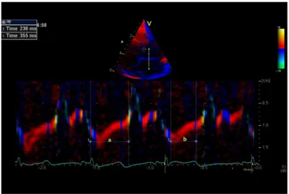
three consecutive cardiac cycles. TDI-derived MPI was measured with cardiac event timing using curved anatomical m-mode modality, which has a good temporal resolution [34]. A 2 cm line was drawn manually perpendicular through the anterior mitral leaflet. The positions of mitral valve opening and closure together with aortic valve opening were identified on the curved anatomical m-mode (Fig 1), and aorta valve closure was identified as the end of the thin blue line after ejection (end systole) [32,35]. MPI is the sum of isovolumic contraction time (IVCT) and isovolumic relaxation time (IVRT), divided by ejection time (ET). TDI-MPI was derived as (a-b)/b, where‘a’is the measured duration from mitral-closure-to-opening and‘b’is the aortic flow ejection time.
Sample collection
Anesthetized animals were euthanized with an intracardiac injection of sodium pentobarbital (60 mg/kg). Hearts were excised and weighed, and apex samples were snap-frozen in liquid nitrogen and stored at–80°C.
Cardiac dysfunction has been associated with alterations in myocardial Na/K-ATPase and other key Ca2+handling proteins [36,37], leading to diminished sarcoplasmic reticulum Ca2+ sequestration, decreased cytosolic Ca2+transients during contractions and increased cytosolic Ca2+levels during diastole, with ensuing impairment of contractility and relaxation [38,39]. Based on this, the myocardial content of Na+/K+-ATPase was quantified to provide an addi-tional marker for the effects of malnutrition on cardiac function by binding a radioactively labelled ligand (ouabain) to functional Na+/K+-ATPases with a stoichiometry of 1:1 according to pre-established techniques [40].
Weight-matched reference group
A third group of pigs (WEIGHT-REF, n = 8) (Duroc-x-Danish Landrace-x-Yorkshire, Ros-kilde, Denmark) were weaned at 4 weeks of age and givenad libitumaccess to the optimized reference diet and water. Upon reaching a bodyweight similar to the final bodyweight in MAIZE, these pigs underwent echocardiographic evaluation and were hereafter euthanized. Tissue samples were collected as previously described.
Ethics statement
Statistics
Analyses were carried out using Stata version 12 (Stata/IC) (StataCorp LP, College Station, Texas, USA) and R (R Core Team, 2013, Vienna, Austria). Except blood-derived outcomes, dif-ferences between MAIZE, AGE-REF, and WEIGHT-REF were evaluated by one-way ANOVA. In the event of variance heterogeneity between groups, data was either log-transformed or the variation explicitly modelled by assuming different variances for the different groups. However, outcomes on valve regurgitation were evaluated using chi-square tests. For blood-derived out-comes, the difference in MAIZE vs. AGE-REF was evaluated using a linear mixed-effects model including time and treatment, and their interaction as fixed effects and pig-specific ran-dom effects to account for repeated measurements, but cTnT was analysed in a two-step analy-sis. The analysis of cTnT, defined as above/below the detection limit (cTnT<3 ng/l), included
main effects of time and treatment. If they were above the detection limit, log-transformed cTnT values were analysed using a linear mixed-effects model including main effects of time and treatment as fixed effects. Both analyses included pig-specific random effects. P-values from post-hoc pairwise comparisons were Bonferroni adjusted. A linear regression model was used to explore associations. P-values below 0.05 were considered significant.
Results
After 7 weeks, MAIZE weighed 26% and CRL was 71% of AGE-REF, as explained previously [27]. As intended, the average weight of WEIGHT-REF was similar to MAIZE (Table 2). Two MAIZE pigs were prematurely euthanized due to ear biting by littermates. A third MAIZE pig died for unknown reasons in the final week.
Echocardiography
TDI-derived MPI was 86% higher in MAIZE than AGE-REF (0.59 vs. 0.32, p<0.001) and 27%
higher than WEIGHT-REF (0.59 vs. 0.46, p = 0.025). Also MPI was 46% higher in WEIGH-T-REF than AGE-REF (0.46 vs. 0.32, p = 0.001). There were negative associations between Fig 1. Myocardial Performance Index (MPI) derived by color tissue Doppler echocardiography.Top:
Apical left 4-chamber view in end-systole showing the position of M-mode line used for measurement of cardiac intervals.Bottom: Colour diagram of the tissue Doppler imaging M-mode of the anterior mitral leaflet. a = interval from mitral valve closing to opening; b = ejection time. Myocardial performance index = (a—b)/ b = 355 ms–236 ms/236 ms = 0.50.
doi:10.1371/journal.pone.0140472.g001
heart rate and MPI and its components (MPI, r2= 0.42; IVCT, r2= 0.34; IVRT, r2= 0.64; ET, r2= 0.46, all p<0.003,Fig 2). No changes in systolic indices, i.e. fractional shortening and
ejec-tion fracejec-tion, were observed between the groups. Left ventricular dimensions, wall thickness, peak aorta flow and mitral inflow velocities were lower in MAIZE relative to AGE-REF, but not to WEIGHT-REF (Table 3). Mitral regurgitation was more frequent in MAIZE (MAIZE 7/ 8, AGE-REF 3/8, WEIGHT-REF 2/8, p = 0.03), four MAIZE pigs had severe MR (RF>50%)
while all remaining MR were trivial-to-mild (mean jet area/LA ratio = 22%). All MR velocities were approximately 5 m/s. Tricuspid regurgitation (TR) was similar between groups (MAIZE 7/8, AGE-REF 5/8, WEIGHT-REF 7/8, p = 0.36). All TR velocities were<2.5 m/s.
Cardiac weight and myocardial Na
+/K
+-ATPase density
During necropsy, immediately after echocardiography, hearts in MAIZE presented with a pale and soft myocardium, which tended to collapse. The absolute heart weight in MAIZE was markedly lower than in AGE-REF and similar to WEIGHT-REF, but the heart weight relative to body weight was similar (Table 2). Myocardial Na+/K+-ATPase density was 51% higher in MAIZE vs. AGE-REF (1650 vs. 1095 pmol/g wet weight (ww), p<0.001) and 26% lower in
MAIZE vs. WEIGHT-REF (1650 vs. 2242 pmol/g ww, p<0.001) (Fig 3).
Biochemistry and haematology
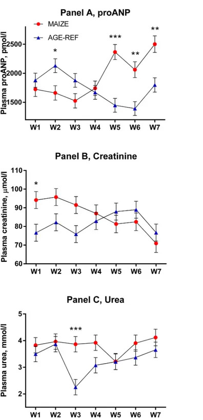
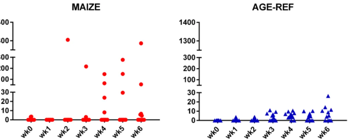
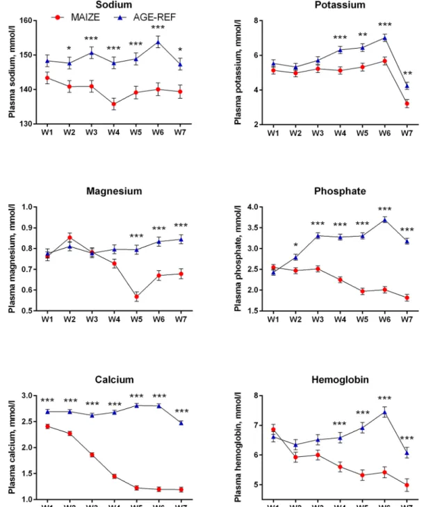
Plasma proANP decreased during the first 3 weeks for both MAIZE and AGE-REF, followed by a marked increase in MAIZE relative to AGE-REF during weeks 5–7 (Fig 4,Panel A, p0.001). Moreover, there was a positive association between MPI and proANP at the time of killing (r2= 0.34, p = 0.017). As circulating proANP may be influenced by kidney function, we measured creatinine and urea and found no consistent differences between MAIZE and AGE-REF (Fig 4,Panels B and C).There was overall no difference in cTnT between groups. Release of cTnT in plasma was more frequent in AGE-REF than MAIZE (OR: 4.8; 95%CI: 1.2– 19.7; p = 0.03). However, when release occurred, cTnT concentration was 6.9-fold higher (95% CI: 3.0–15.9; p<0.001) in MAIZE (Fig 5). Finally, electrolytes and hemoglobin declined
pro-gressively in MAIZE relative to AGE-REF (Fig 6). Serum albumin followed a similar pattern, with markedly lower levels in MAIZE vs AGE-REF at week 7 (20.7±0.7 g/L vs. 35.6±0.7 g/L, p<0.001).
Discussion
We found cardiac dysfunction in piglets with clinical signs of malnutrition, disturbed electro-lytes and anaemia, based on MPI and the cardiac biomarkers proANP and cTnT. These Table 2. Body and heart dimensions at the end of the experiment*.
MAIZE AGE-REF WEIGHT-REF
(n = 9) (n = 12) (n = 8)
Body weight, kg 8.3±1.8a 32.4±4.3b 6.8±1.5a
Crown-rump length, cm 55.1±4.4a 77.7±5.6b 48±2c
Heart weight, g 49.8±7.1a 178±23b 43.8±11.4a
Heart weight/body weight, g/kg 6.2±1.2 5.5±0.6 6.7±2.2
*Differences reported here for all pigs are also seen among the subsample of pigs used in echocardiography. Data are (means±sd).
a,b,cMeans not sharing a superscript are different (p<0.05).
findings were obtained even with a similar heart-weight relative to body-weight across the mal-nourished, the age-matched and the weight-matched reference group. Normalisation of cardiac tissue to body weight/surface area during malnutrition has previously been described
[20,22,41], although both decreased [15] and increased ratios [16,42] have been reported. We induced malnutrition by feeding a pure maize-diet which is low in protein, phosphate and other nutrients. Here we focus on cardiac effects, however, we also studied other pathologi-cal changes associated with malnutrition, including hepatic fat infiltration, gut atrophy and altered body composition (data presented elsewhere [27]). Insufficient protein intake alters metabolism and there may be discrepancies in amino acid metabolism not only between healthy and severely malnourished infants, but also between different manifestations of malnu-trition [43]. Amino acid availability and metabolism is likely to be important for heart function and warrants future investigations.
Echocardiography
MPI was markedly increased in MAIZE relative to both AGE-REF and WEIGHT-REF, indicat-ing that malnutritionper seinfluenced cardiac function independently of body weight. Several studies document that elevated MPI is reliable for global evaluation of left-ventricular
Fig 2. Comparison of Myocardial performance index (MPI), Isovolumic relaxation time (IVRT) Isovolumic contraction time (IRCT) and Ejection time (ET) (means±SEM).Measurements from individual pigs: MAIZE (n = 8), AGE-REF (n = 8), WEIGHT-REF (n = 8).a,b,cMeans not sharing a letter in their superscript are significantly different (p<0.05).
doi:10.1371/journal.pone.0140472.g002
Table 3. Echocardiography results*.
MAIZE AGE-REF WEIGHT-REF
(n = 8) (n = 8) (n = 8)
Heart rate, beats/min 83±17a 118±6b 99±19a
M-mode
Inter-ventricular septum thickness in diastole, mm 5.9±0.5a 8.8±0.6b 5.5±0.8a
Left ventricular internal diameter in diastole, mm 26.6±1.5a 40.1±2.7b 22.3±1.7c
Left ventricular free wall thickness in diastole, mm 4.6±0.4a 7.1±0.7b 4.2±0.6a
Inter-ventricular septum thickness in systole, mm 9.4±1.4a 13.7±1.3b 8.7±1.0a
Left ventricular internal diameter in systole, mm 17.7±2.2a 27.0±1.5b 16.1±1.6a
Left ventricular free wall thickness in systole, mm 7.4±0.6a 12.1±1.2b 6.5±1.0a
Fractional shortening, % 32.3±6.1 32.3±4.4 27.5±4.0
E-point septal separation, mm 0.6±0.9 1.8±0.9 1.4±0.9
2-dimensional echocardiogram
Left atrium diameter, mm 17.6±2.3a 22.0±2.3b 15.7±1.2a
Aortic diameter, mm 12.2±1.1a 17.2±1.0b 11.0±0.7a
Left ventricular end-diastolic volume, ml 13.5±3.6a 42±7.5b 9.6±2.0a
Left ventricular end-systolic volume, ml 4.4±1.8a 15.6±4.2b 3.8±1.0a
Stroke volume, ml 9.2±2.8a 26.4±4b 5.9±1.4a
Ejection fraction, % 67.1±9.4 63.6±5.8 63.5±4.9
Doppler
Peak velocity of pulmonaryflow, m/s 0.6±0.1a 1.0±0.1b 0.6±0.1a
Peak velocity of aorticflow, m/s 0.9±0.1a 1.3±0.1b 0.79±0.9a
Mitral E, peak earlyfilling velocity in diastole, m/s 0.8±0.1a 1.0±0.1b 0.7±0.1a
Mitral A, peak activefilling velocity in diastole, m/s 0.6±0.2ab 0.8±0.1a 0.4±0.2b
E/A ratio 1.5±0.5 1.3±0.2 1.5±0.4
*Data are (means±sd).
a,b,cMeans not sharing a superscript are different (p<0.05).
doi:10.1371/journal.pone.0140472.t003
Fig 3. Comparison of myocardial Na+/K+-ATPase density (means±SEM).Measurements of Na+/K+ -ATPase density expressed as ouabain-binding sites in myocardial tissue of individual pigs: MAIZE (n = 9), AGE-REF (n = 11), WEIGHT-REF (n = 8).a,b,cMeans not sharing a letter in their superscript are significantly different (p<0.001). ww = wet weight.
Fig 4. Comparison of temporal changes in plasma proANP, creatinine and urea (means±SEM). Weekly measurements from pig groups: MAIZE (n = 12), AGE-REF (n = 12). Differences in means*(p<0.05),
**(p<0.01),***(p<0.001).
doi:10.1371/journal.pone.0140472.g004
dysfunction in a variety of heart diseases [44–47], and has prognostic value for heart failure and death [47–50]. MPI is considered independent of geometric assumptions and unaffected by heart rate [46,51,52], whereas loading conditions may affect the index [53,54]. Furthermore, in a pig model, MPI has been shown to correlate with invasive measurements of ventricular function [55]. Surprisingly, we found that heart rate was negatively associated with MPI and its components, which may be a limitation.
Additionally, IVRT was markedly elevated in MAIZE relative to both reference groups and appeared to be a main determinant of the MPI index. IVCT was elevated in both MAIZE and WEIGHT-REF relative to AGE-REF, indicating that bodyweight alone may affect this compo-nent. The marked influence of the extended isovolumic relaxation time on the MPI index is further substantiated by an elevated ET in MAIZE relative to both reference groups, even though thisper sewould tend to lower the MPI index. The elevated MPI in MAIZE suggests a subclinical global left ventricular dysfunction as a result of malnutrition, and the markedly increased IVRT indicates an impaired left ventricular relaxation that was not detectable by mitral inflow E/A-ratio derived from conventional Doppler echocardiography.
Theα2-agonist effect (xylazine) has a known bradycardic effect and the use of ketamine/ xylaxine has been associated with MR in normal rats [56]. It is possible that the anaesthetic reg-imen served as a stress test, unmasking cardiac dysfunction due to malnutrition. However, since pigs in all groups received the same anaesthetic per kg body weight, the differences between groups can only be explained by underlying malnutrition.
Previous studies show different effects of malnutrition on cardiac pump function. In line with our results, no diastolic dysfunction has been found in malnutrition as evaluated by the E/ A ratio [15,18]. However, diastolic dysfunction has been reported in malnourished rats as increased passive stiffness with preserved LV compliance [22] and in malnourished dogs as decreased LV compliance [23]. We identified no systolic dysfunction in malnourished pigs, which is in line with other animal studies [22,42,57]. Likewise, preserved systolic function is described in several studies with children with SAM [15–17], whereas other studies docu-mented reduced ejection fraction [18,21], reduced fractional shortening [18–21] and reduced velocity of circumferential shortening [20,21].
Fig 5. Comparison of cardiac Troponin T release.Weekly measurements of cardiac Troponin T release in individual pigs: MAIZE (n = 12), AGE-REF (n = 12). Blood samples at killing week 7 drawn by cardiac puncturing and not shown.
proANP
The initial decrease in proANP concentrations for both MAIZE and AGE-REF presumably reflects normal development. As MAIZE pigs became increasingly malnourished, proANP increased from week 3 and onwards, whereas it continued to decline in AGE-REF. This indi-cates that cardiac function or fluid and salt homeostasis, or both, are affected by the monoto-nous maize diet.
Fig 6. Comparison of temporal changes in electrolytes and hemoglobin (means±SEM).Weekly measurements of electrolytes and hemoglobin levels in pig groups: MAIZE (n = 12), AGE-REF (n = 12). Differences in means*(p<0.05),**(p<0.01),***(p<0.001).
doi:10.1371/journal.pone.0140472.g006
The natriuretic peptides, atrial natriuretic peptide (ANP) and brain natriuretic peptide (BNP) are released from the heart in response to cardiac pressure and volume overload [58]. The instability of circulating natriuretic peptides has led to the development of assays for more stable fragments including proANP [30]. The ANP gene is structurally better conserved than BNP during evolution, and proANP immune assays can be used across mammalian species, allowing comparative studies between pigs and humans [29].
Natriuretic peptides and their proforms are increased in various cardiac diseases [59,60], with a clear focus on systolic heart failure. Additionally, natriuretic peptide measurement pre-dicts mortality in a variety of acute disease states, and there is a growing interest in pediatric application as both a diagnostic and prognostic tool [60]. Therefore, the rise in proANP levels in the MAIZE group and the positive correlation between proANP and MPI may be clinically useful and also indicate that malnourished pigs are at risk of cardiac failure.
Troponin
There was overall no difference in plasma cardiac troponin-T between groups. Detailed analy-sis revealed that MAIZE had either no or high cTnT release, whereas AGE-REF had frequent but smaller cTnT releases, which may reflect normal physiology or a stress-response to blood sampling.
Two MAIZE pigs showed single extremely high cTnT values at 100-fold the 99thpercentile of upper reference value. One of these was the last sample taken from the only MAIZE pig found dead; this indicates that the death may be related to antecedent myocardial muscle necrosis. However, with no data on ischemia or immediate cTnT kinetics, no definite conclu-sion can be drawn as to the aetiology behind the elevated cTnT values. Nevertheless, high cTnT values in MAIZE showed fluctuations from week to week, which may indicate an acute pathogenesis. When first studied in malnourished children, cTnI was detected in only two of 30 children [15], while a later study found that elevated cTnT predicted mortality and corre-lated to the severity of malnutrition, anaemia, electrolyte deficiency, and sepsis [18].
Myocardial Na
+/K
+-ATPase concentration
In humans, the myocardial Na+/K+-ATPase concentration decreases markedly during the first few years of life, to reach a more constant level of approximately 700 pmol/g ww in early adult-hood [36]. Hence, the higher myocardial content of Na+/K+-ATPase in MAIZE when com-pared to AGE-REF was most likely related to the reduced growth in MAIZE. However, compared to the WEIGHT-REF group, the myocardial Na+/K+-ATPase content in MAIZE was significantly reduced. Similar reductions in myocardial Na+/K+-ATPase content are gener-ally observed in heart failure associated with myocardial hypertrophy, ischaemic heart disease or heart dilatation in humans [36], and the observation therefore provides further evidence for cardiac dysfunction in MAIZE. At a mechanistic level, the ensuing decrease in the capacity for active extrusion of cell Na+can cause cytosolic Na+to increase [61], which reduces the driving force for active Ca2+extrusion via the Na/Ca exchanger [38], leading to increased accumulation of Ca2+in the cytosol and the sarcoplasmatic reticulum. Whereas this may support the contrac-tility of the heart, the increased basal level of cytosolic Ca2+may also lead to increased cell deg-radation and a general decrease in cardiac performance [37,39].
Conclusions
cardiac development between humans and pigs, including also cardiac responses to malnutri-tion. Yet future studies in human cohorts are required to fully delineate similarities and dis-crepancies to this porcine malnutrition model.
Supporting Information
S1 Data.
(XLS)
S2 Data.
(XLS)
S3 Data.
(XLS)
S4 Data.
(XLS)
S5 Data.
(XLS)
S6 Data.
(XLS)
S7 Data.
(XLS)
Acknowledgments
The authors thank André Briend, Jesper Reimers and Michael Golden for constructive com-ments on the manuscript.
Author Contributions
Conceived and designed the experiments: ML ALH TT. Performed the experiments: ML ALH TT. Analyzed the data: CF. Contributed reagents/materials/analysis tools: JK OBN IH JPG. Wrote the paper: CF ML ALH JK OBN IH JPG HF TT.
References
1. Black RE, Victora CG, Walker SP, Bhutta ZA, Christian P, de Onis M, et al. Maternal and child undernu-trition and overweight in low-income and middle-income countries. Lancet. 2013; 382: 427–451. doi: 10.1016/S0140-6736(13)60937-XPMID:23746772
2. WHO, UNICEF. WHO Child Growth Standards and the Identification of Severe Acute Malnutrition in Infants and Children: A Joint Statement by the World Health Organization and the United Nations Chil-dren’s Fund. 2009.
3. Waterlow JC, Tomkins A, Grantham-McGregor SM. Protein-energy malnutrition. Edward Arnold. 1992: 26–111.
4. WHO. Management of severe malnutrition: a manual for physicians and other senior health workers. 1999.
5. WHO. Pocket book of hospital care for children: guidelines for the management of common childhood illnesses. 2013.
6. Maitland K, Berkley JA, Shebbe M, Peshu N, English M, Newton CRJC. Children with Severe Malnutri-tion: Can Those at Highest Risk of Death Be Identified with the WHO Protocol? PLoS Med. 2006; 3: e500. doi:10.1371/journal.pmed.0030500PMID:17194194
7. Brent B, Obonyo N, Maitland K. Tailoring management of severe and complicated malnutrition: more research is required first. Pathog Glob Health. 2012; 106: 197–199. doi:10.1179/2047772412Z.
00000000061PMID:23265419
8. Maitland K. Joint BAPEN and Nutrition Society Symposium on“Feeding size 0: the science of starva-tion”. Severe malnutrition: therapeutic challenges and treatment of hypovolaemic shock. Proc Nutr Soc. 2009; 68: 274–280. doi:10.1017/S0029665109001359PMID:19490738
9. WHO. Guideline: updates on the management of severe acute malnutrition in infants and children. 2013.
10. Edozien JC, Rahim-Khan MA. Anaemia in protein malnutrition. Clin Sci. 1968; 34: 315–326. PMID: 5653689
11. Wharton BA, Howells GR, McCance RA. Cardiac failure in kwashiorkor. Lancet. 1967; 2: 384–387. PMID:4166331
12. Grellety Y. Management of Severe Malnutrition in Africa. PhD Thesis. University of Aberdeen; 2000. 13. Smythe PM, Swanepoel A, Campbell J a. H. The Heart in Kwashiorkor. BMJ. 1962; 1: 67. PMID:
13914550
14. Piza J, Troper L, Cespedes R, Miller JH, Berenson GS. Myocardial Lesions and Heart Failure in Infan-tile Malnutrition. Am J Trop Med Hyg. 1971; 20: 343–355. PMID:5553269
15. El-Sayed HL, Nassar MF, Habib NM, Elmasry OA, Gomaa SM. Structural and functional affection of the heart in protein energy malnutrition patients on admission and after nutritional recovery. Eur J Clin Nutr. 2005; 60: 502–510.
16. Kothari SS, Patel TM, Shetalwad AN, Patel TK. Left ventricular mass and function in children with severe protein energy malnutrition. Int J Cardiol. 1992; 35: 19–25. doi:10.1016/0167-5273(92)90050-D PMID:1563875
17. Shoukry I, Shoukry AS, Ibrahim MM, Fahmy N, Madkour MA, Said GE. Cardiac Atrophy and Ventricular Function in Infants with Severe Protein Calorie Malnutrition (Kwashiorkor Disease). In: EFD M.D, MAE M.D, WMG M.D, WJR M.D, NST M.D, editors. Pediatric Cardiology. Springer New York; 1986. pp. 1169–1171.
18. Faddan NHA, Sayh KIE, Shams H, Badrawy H. Myocardial dysfunction in malnourished children. Ann Pediatr Cardiol. 2010; 3: 113–118. doi:10.4103/0974-2069.74036PMID:21234188
19. Olowonyo MT, Ogunkunle OO, Akinbami FO, Jaiyesimi F. The Echocardiographic Findings in Kwashi-orkor. J Trop Pediatr. 1995; 41: 74–76. doi:10.1093/tropej/41.2.74PMID:7776400
20. Phornphatkul C, Pongprot Y, Suskind R, George V, Fuchs G. Cardiac function in malnourished chil-dren. Clin Pediatr (Phila). 1994; 33: 147–154.
21. Singh GR, Malathi KE, Kasliwal RR, Ommar A, Padmavati S, Ramji S. An evaluation of cardiac function in malnourished children by non-invasive methods. Indian Pediatr. 1989; 26: 875–881. PMID:2517425 22. Fioretto JR, Querioz SS, Padovani CR, Matsubara LS, Okoshi K, Matsubara BB. Ventricular
remodel-ing and diastolic myocardial dysfunction in rats submitted to protein-calorie malnutrition. Am J Physiol Heart Circ Physiol. 2002; 282: H1327–H1333. doi:10.1152/ajpheart.00431.2001PMID:11893568 23. Abel RM, Grimes JB, Alonso D, Alonso M, Gay WA Jr.. Adverse hemodynamic and ultrastructural
changes in dog hearts subjected to protein-calorie malnutrition. Am Heart J. 1979; 97: 733–744. doi: 10.1016/0002-8703(79)90008-5PMID:107775
24. Brent B, Obonyo N, Maitland K, Tulloh R. Long axis dysfunction and stiff arteries in children with severe acute malnutrition in Kenya. Cardiology in the Young. 2014; 24: S46.
25. Nielsen KL, Hartvigsen ML, Hedemann MS, Lærke HN, Hermansen K, Knudsen KEB. Similar meta-bolic responses in pigs and humans to breads with different contents and compositions of dietary fibers: a metabolomics study. Am J Clin Nutr. 2014; 99: 941–949. doi:10.3945/ajcn.113.074724PMID: 24477039
26. Suzuki Y, Yeung AC, Ikeno F. The Representative Porcine Model for Human Cardiovascular Disease. J Biomed Biotechnol. 2011; 2011: 1–10. doi:10.1155/2011/195483
27. Lykke M, Hother A-L, Hansen CF, Friis H, Mølgaard C, Michaelsen KF, et al. Malnutrition induces gut
atrophy and increases hepatic fat infiltration: studies in a pig model of childhood malnutrition. Am J Transl Res. 2013; 5: 543–554. PMID:23977413
28. Yang TS, Lin JH. Variation of heart size and its correlation with growth performance and vascular space in domestic pigs. Anim Sci. 1997; 64: 523–528. doi:10.1017/S1357729800016155
30. Hunter I, Alehagen U, Dahlström U, Rehfeld JF, Crimmins DL, Goetze JP. N-Terminal Pro—Atrial Natri-uretic Peptide Measurement in Plasma Suggests Covalent Modification. Clin Chem. 2011; 57: 1327–
1330. doi:10.1373/clinchem.2011.166330PMID:21715695
31. Giannitsis E, Kurz K, Hallermayer K, Jarausch J, Jaffe AS, Katus HA. Analytical Validation of a High-Sensitivity Cardiac Troponin T Assay. Clin Chem. 2010; 56: 254–261. doi:10.1373/clinchem.2009. 132654PMID:19959623
32. Lang RM, Bierig M, Devereux RB, Flachskampf FA, Foster E, Pellikka PA, et al. Recommendations for chamber quantification. Eur J Echocardiogr. 2006; 7: 79–108. doi:10.1016/j.euje.2005.12.014PMID: 16458610
33. Zoghbi W. Recommendations for evaluation of the severity of native valvular regurgitation with two-dimensional and doppler echocardiography. J Am Soc Echocardiogr. 2003; 16: 777–802. doi:10.1016/ S0894-7317(03)00335-3PMID:12835667
34. Kjaergaard J, Hassager C, Oh JK, Kristensen JH, Berning J, Sogaard P. Measurement of Cardiac Time Intervals by Doppler Tissue M-Mode Imaging of the Anterior Mitral Leaflet. J Am Soc Echocardiogr. 2005; 18: 1058–1065. doi:10.1016/j.echo.2005.03.043PMID:16198883
35. Aase SA, Torp H, Støylen A. Aortic valve closure: relation to tissue velocities by Doppler and speckle
tracking in normal subjects. Eur J Echocardiogr. 2008; 9: 555–559. doi:10.1093/ejechocard/jen120 PMID:18490310
36. Bundgaard H, Kjeldsen K. Human myocardial Na,K-ATPase concentration in heart failure. Mol Cell Bio-chem. 1996; 163–164: 277–283. PMID:8974067
37. Schmidt TA, Kjeldsen K. Human myocardial Na,K-ATPase—quantification, regulation and relation to Ca. Cardiovasc Res. 1998; 37: 335–345. PMID:9614490
38. Piacentino V, Weber CR, Chen X, Weisser-Thomas J, Margulies KB, Bers DM, et al. Cellular Basis of Abnormal Calcium Transients of Failing Human Ventricular Myocytes. Circ Res. 2003; 92: 651–658. doi:10.1161/01.RES.0000062469.83985.9BPMID:12600875
39. Luo M, Anderson ME. Mechanisms of Altered Ca2+Handling in Heart Failure. Circ Res. 2013; 113: 690–708. doi:10.1161/CIRCRESAHA.113.301651PMID:23989713
40. Nørgaard A, Kjeldsen K, Hansen O, Clausen T, Larsen CG, Larsen FG. Quantification of the 3H
-oua-bain binding site concentration in human myocardium: a postmortem study. Cardiovasc Res. 1986; 20: 428–435. PMID:2430708
41. Gruber C, Nink N, Nikam S, Magdowski G, Kripp G, Voswinckel R, et al. Myocardial remodelling in left ventricular atrophy induced by caloric restriction: Caloric restriction and myocardial remodelling. J Anat. 2012; 220: 179–185. doi:10.1111/j.1469-7580.2011.01453.xPMID:22077432
42. Nutter DO, Murray TG, Heymsfield SB, Fuller EO. The effect of chronic protein-calorie undernutrition in the rat on myocardial function and cardiac function. Circ Res. 1979; 45: 144–152. doi:10.1161/01.RES. 45.1.144PMID:109229
43. Manary MJ, Broadhead RL, Yarasheski KE. Whole-body protein kinetics in marasmus and kwashiorkor during acute infection. Am J Clin Nutr. 1998; 67: 1205–1209. PMID:9625094
44. Tei C. New non-invasive index for combined systolic and diastolic ventricular function. J Cardiol. 1995; 26: 135–136. PMID:7674144
45. Poulsen SH, Jensen SE, Tei C, Seward JB, Egstrup K. Value of the Doppler Index of Myocardial Perfor-mance in the Early Phase of Acute Myocardial Infarction. J Am Soc Echocardiogr. 2000; 13: 723–730. doi:10.1067/mje.2000.105174PMID:10936815
46. Bruch C, Schmermund A, Marin D, Katz M, Bartel T, Schaar J, et al. Tei-Index in patients with mild-to-moderate congestive heart failure. Eur Heart J. 2000; 21: 1888–1895. doi:10.1053/euhj.2000.2246 PMID:11052862
47. Poulsen SH, Jensen SE, Nielsen JC, Møller JE, Egstrup K. Serial changes and prognostic implications
of a Doppler-derived index of combined left ventricular systolic and diastolic myocardial performance in acute myocardial infarction. Am J Cardiol. 2000; 85: 19–25. doi:10.1016/S0002-9149(99)00599-8 PMID:11078230
48. Ärnlöv J, Ingelsson E, Risérus U, Andrén B, Lind L. Myocardial performance index, a Doppler-derived index of global left ventricular function, predicts congestive heart failure in elderly men. Eur Heart J. 2004; 25: 2220–2225. doi:10.1016/j.ehj.2004.10.021PMID:15589639
49. Ärnlöv J, Lind L, Andrén B, Risérus U, Berglund L, Lithell H. A Doppler-derived index of combined left ventricular systolic and diastolic function is an independent predictor of cardiovascular mortality in elderly men. AHJ. 2005; 149: 902–907. doi:10.1016/j.ahj.2004.07.022PMID:15894975 50. Peltier M, Slama M, Garbi S, Enriquez-Sarano ML, Goissen T, Tribouilloy CM. Prognostic value of
Doppler-derived myocardial performance index in patients with left ventricular systolic dysfunction. Am J Cardiol. 2002; 90: 1261–1263. doi:10.1016/S0002-9149(02)02849-7PMID:12450613
51. Tei C, Ling LH, Hodge DO, Bailey KR, Oh JK, Rodeheffer RJ, et al. New index of combined systolic and diastolic myocardial performance: a simple and reproducible measure of cardiac function—a study in
normals and dilated cardiomyopathy. J Cardiol. 1995; 26: 357–366. PMID:8558414
52. Poulsen SH, Nielsen JC, Andersen HR. The Influence of Heart Rate on the Doppler-Derived Myocardial Performance Index. J Am Soc Echocardiogr. 2000; 13: 379–384. doi: 10.1016/S0894-7317(00)70007-1PMID:10804435
53. Cheung MMH, Smallhorn JF, Redington AN, Vogel M. The effects of changes in loading conditions and modulation of inotropic state on the myocardial performance index: comparison with conductance cath-eter measurements. Eur Heart J. 2004; 25: 2238–2242. doi:10.1016/j.ehj.2004.07.034PMID: 15589642
54. Møller JE, Poulsen SH, Egstrup K. Effect of Preload Alternations on a New Doppler Echocardiographic
Index of Combined Systolic and Diastolic Performance. J Am Soc Echocardiogr. 1999; 12: 1065–1072. doi:10.1016/S0894-7317(99)70103-3PMID:10588782
55. LaCorte JC, Cabreriza SE, Rabkin DG, Printz BF, Coku L, Weinberg A, et al. Correlation of the tei index with invasive measurements of ventricular function in a porcine model. J Am Soc Echocardiogr. 2003; 16: 442–447. doi:10.1016/S0894-7317(03)00110-XPMID:12724653
56. Droogmans S, Lauwers R, Cosyns B, Roosens B, Franken PR, Weytjens C, et al. Impact of Anesthesia on Valvular Function in Normal Rats During Echocardiography. Ultrasound in Medicine & Biology. 2008; 34: 1564–1572. doi:10.1016/j.ultrasmedbio.2008.02.017
57. Alden PB, Madoff RD, Stahl TJ, Lakatua DJ, Ring WS, Cerra FB. Left ventricular function in malnutri-tion. Am J Physiol Heart Circ Physiol. 1987; 253: H380–H387.
58. Daniels LB, Maisel AS. Natriuretic Peptides. J Am Coll Cardiol. 2007; 50: 2357–2368. doi:10.1016/j. jacc.2007.09.021PMID:18154959
59. Saito Y. Roles of atrial natriuretic peptide and its therapeutic use. J Cardiol. 2010; 56: 262–270. doi:10. 1016/j.jjcc.2010.08.001PMID:20884176
60. Tobias JD. B-type Natriuretic Peptide: Diagnostic and Therapeutic Applications in Infants and Children. J Intensive Care Med. 2011; 26: 183–195. doi:10.1177/0885066610387993PMID:21320863 61. Pieske B, Houser SR. [Na+]ihandling in the failing human heart. Cardiovasc Res. 2003; 57: 874–886.