Obstructive Sleep Apnea is Common and Associated with Heart
Remodeling in Patients with Chagas Disease
Carolina de Araújo Medeiros,
1,2Isaac Vieira Secundo,
1Carlos Antônio da Mota Silveira,
1José Maria del Castilho,
1Afonso Luiz Tavares de Albuquerque,
1Sílvia Marinho Martins,
2Wilson de Oliveira Júnior,
2Geraldo Lorenzi-Filho,
3Luciano F. Drager,
3Rodrigo Pinto Pedrosa
1Laboratório do Sono e Coração do Pronto Socorro Cardiológico de Pernambuco (PROCAPE) da Universidade de Pernambuco,1 Recife, PE - Brazil Ambulatório de Doença de Chagas e insuficiência Cardíaca - PROCAPE da Universidade de Pernambuco,2 Recife, PE - Brazil
Instituto do Coração (InCor) - Hospital das Clínicas da Faculdade de Medicina da Universidade de São Paulo (HCFMUSP),3 São Paulo, SP - Brazil
Mailing Address: Rodrigo Pinto Pedrosa •
Rua dos Palmares, SN. Postal Code 50100-060, Santo Amaro, Recife, PE - Brasil
E-mail: [email protected]
Manuscript received March 11,2018, revised manuscript May 09, 2018, accepted May 09, 2018
DOI: 10.5935/abc.20180131
Abstract
Background: Chagas Disease (CD) is an important cause of morbimortality due to heart failure and malignant arrhythmias worldwide, especially in Latin America.
Objective: To investigate the association of obstructive sleep apnea (OSA) with heart remodeling and cardiac arrhythmias in patients CD.
Methods: Consecutive patients with CD, aged between 30 to 65 years old were enrolled. Participants underwent clinical evaluation, sleep study, 24-hour Holter monitoring, echocardiogram and ambulatory blood pressure monitoring.
Results: We evaluated 135 patients [age: 56 (45-62) years; 30% men; BMI: 26 ± 4 kg/m2, Chagas cardiomyopathy: 70%].
Moderate to severe OSA (apnea-hypopnea index, AHI, ≥ 15 events/h) was present in 21% of the patients. OSA was not
associated with arrhythmias in this population. As compared to patients with mild or no OSA, patients with moderate to severe OSA had higher frequency of hypertension (79% vs. 72% vs. 44%, p < 0.01) higher nocturnal systolic blood pressure: 119 ± 17 vs. 113 ± 13 vs. 110 ± 11 mmHg, p = 0.01; larger left atrial diameter [37 (33-42) vs. 35 (33-39) vs. 33 (30-36) mm, p < 0.01]; and a greater proportion of left ventricular dysfunction [LVEF < 50% (39% vs. 28% vs. 11%), p < 0.01], respectively. Predictor of left atrial dimension was Log10 (AHI) (β = 3.86, 95% CI: 1.91 to 5.81; p < 0.01). Predictors of ventricular dysfunction were AHI > 15 events/h (OR = 3.61, 95% CI: 1.31 - 9.98; p = 0.01), systolic blood pressure (OR = 1.06, 95% CI: 1.02 - 1.10; p < 0.01) and male gender (OR = 3.24, 95% CI: 1.31 - 8.01; p = 0.01).
Conclusions: OSA is independently associated with atrial and ventricular remodeling in patients with CD. (Arq Bras Cardiol. 2018; 111(3):364-372)
Keywords: Chagas Disease; Sleep Apnea, Obstructive; Ventricular Remodeling; Arrhythmias, Cardiac.
Introduction
Chagas disease (CD) is the third most common parasitic infection, after malaria and schistosomiasis, affecting about 7 to 8 million people worldwide.1 CD is caused by the protozoan Trypanosoma cruzi, transmitted to humans by insects (Triatominae), blood transfusion, organ and tissue transplantation, oral contamination or congenital transmission.2The epidemiological profile of the disease has been modified in recent decades, due to migratory flows,2 thus also generating attention in non-endemic countries such as the United States, Canada, Spain, Italy and Japan.3,4
Chagas cardiomyopathy is the most common form of nonischemic cardiomyopathy and one of the leading causes of complications and death in Latin America.5 Approximately one
third of patients with CD develop Chagas cardiomyopathy3 characterized by ventricular arrhythmias, cardiac blockages, alterations in cardiac proteins with heart remodeling, heart failure and sudden death. Heart failure due to CD worsens patient prognosis, when compared with other cardiomyopathies.6 In addition to the myriad of characteristics involved in CD, it is important to consider potential comorbidities that may have a negative impact on patients’ health.
Obstructive sleep apnea (OSA) is the most frequent respiratory disturbance in the overall population and is associated with heart remodeling and arrhythmias in patients without7,8 and with comorbidities, including heart failure.6 However, whether this association exists in patients with CD is unknown. We hypothesized that OSA is independently associated with cardiac arrhythmias and heart remodeling in patients with CD.
Methods
Subjects
from August 2013 to August 2014. Chagas cardiomyopathy was diagnosed in patients with serological evidence of antibodies to T. cruzi and evidence of Chagas heart disease who may or may not have cardiac symptoms (such as dyspnea, edema, and chest pain). The indeterminate form of CD was diagnosed in patients with serological and/or parasitological evidence of T. cruzi infection who lacked symptoms, physical signs, electrocardiographic abnormalities and any radiographic evidence (on chest radiography, barium-contrast esophageal, or colon radiography) of cardiac or gastrointestinal involvement.9 Patients with cardiac pacemakers, manifest or suspected coronary disease; decompensated heart failure requiring hospital admission, predominant central sleep apnea (>50% of events scored as central), or renal disease (serum creatinine >2mg/dL), as well as those with a previous stroke were excluded.
Patients were invited to undergo the sleep study in the week after they underwent all examinations, including echocardiography, Ambulatory Blood Pressure Monitoring (ABPM) and 24-hour Holter monitoring as described below.
Sleep Study
All patients underwent portable sleep monitoring in the sleep laboratory using a validated device (Embletta Gold, PDS; Medcare, Reykjavik, Iceland)10 to evaluate oxygen saturation, body position, nasal flow measurements (pressure cannula), and respiratory effort measurements using two respiratory inductance plethysmography belts. All exams were scored by an experienced physician. Apnea was defined as total absence (>90%) and hypopneas as a decrease (>30%) in
nasal flow for ≥ 10 seconds, followed by a 4% desaturation
(in hypopneas), respectively.11 The apnea-hypopnea index (AHI) was calculated by dividing the total number of apnea and hypopnea events by the total hours in bed.11,12
Mild OSA was defined by an AHI between 5 and 14.9 events/h and moderate to severe OSA was considered
when the AHI was ≥ 15 events/h. The oxygen desaturation
index (ODI) was calculated as the total number of desaturations, divided by the total time in bed.
Excessive daytime sleepiness was evaluated using the Epworth Sleepiness Scale. A total score > 10 was considered excessive daytime somnolence.13
Office blood pressure
Blood pressure (BP) was measured after 5 min of rest using standard protocols.14 The average of two readings was obtained at 5 min intervals with an automatic digital sphygmomanometer (Omron BP742).
Ambulatory Blood Pressure Monitoring (ABPM)
All participants underwent blood pressure monitoring for 24 hours, using SpaceLabs equipment (model 90207; SpaceLabs, Redmond, WA). The BP reading was taken every 10 minutes during the day and every 20 minutes at night, using an appropriate cuff placed on the non-dominant arm. Participants were instructed to perform their ordinary daily activities and not to move their arm during the ongoing
measurement. Activity, bedtime, and time on awakening from sleep were recorded by participants in diaries.15 The normal BP dip was defined separately for systolic and diastolic BP as a
≥ 10% reduction in BP during sleep compared with the awake
period. Nondipping was defined as a decrease of < 10%.
Holter monitoring
Holter monitoring (Cardios®, Cardio Systems, São Paulo, Brazil) was performed in all patients for 24 hours. The following characteristics of the ECG were analyzed: baseline heart rhythm, heart rate, ventricular and atrial arrhythmias, and breaks. The complexities of the arrhythmias were described as follows: isolated, paired, or tachycardia.16 Patients were instructed to keep a diary with their symptoms during the exam. The Holter analysis was performed by an experienced observer, who was blinded to the presence or absence of OSA.
Echocardiogram
A transthoracic echocardiogram was performed using a Philips IE33 S5-1 device. Conventional M-mode echocardiography was used to measure cavity dimensions (diastolic and systolic diameters, wall thickness, and aorta and left atrial size).17 Left atrial volumes were indexed by body surface area according to the American Society of Echocardiography.18 Using two-dimensional echocardiography, segmental and global contractility were assessed, and the left ventricular ejection fraction (LVEF) was calculated using Simpson's formula. Ventricular dysfunction was considered when LVEF <50%.18 Left ventricular longitudinal strain with speckle-tracking was calculated and values below –16% were considered abnormal.19 The Echocardiographic evaluation was performed by the same experienced observer, who was blinded to the presence or absence of OSA.
Statistical analysis
Normality distribution was evaluated with the Kolmogorov-Smirnov test.
For the categorical variables, the Chi-square test of Pearson was used. Quantitative variables with a normal distribution were presented as mean and standard deviation and the ANOVA test was used, whereas the variables without normal distribution were presented as median and percentiles (P25; P75) and the Kruskal-Wallis was used, with Bonferroni post-hoc test, when appropriate.
A multiple linear regression analysis was performed to evaluate independent predictors of left atrial dimensions. The independent variables of the left atrial dimensions were age, 24-hour systolic BP, body mass index (BMI), and AIH. Due to the non-normality of the AHI, a log-transformed version of this variable was used in the multivariate model. To analyze the predictors of ventricular dysfunction, a multiple logistic regression analysis was performed with the following variables: age, BMI, male gender, 24-hour systolic
BP, diabetes mellitus diagnosis, AHI ≥15 events/h, ODI, and
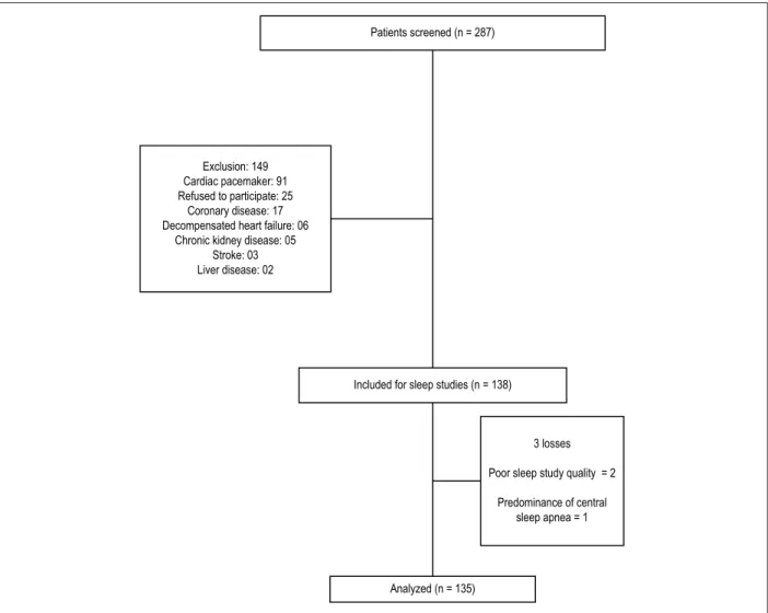
Figura 1 – Diagrama de fluxo do paciente.
Patients screened (n = 287)
Exclusion: 149 Cardiac pacemaker: 91 Refused to participate: 25
Coronary disease: 17 Decompensated heart failure: 06
Chronic kidney disease: 05 Stroke: 03 Liver disease: 02
Included for sleep studies (n = 138)
3 losses
Poor sleep study quality = 2
Predominance of central sleep apnea = 1
Analyzed (n = 135)
Results
We consecutively evaluated 287 patients with CD (41 [30%] with the indeterminate form of the disease and 94 [70%] with Chagas cardiomyopathy). Most of the exclusions were due to the presence of cardiac pacemakers (Figure 1). One patient had a predominance of central sleep apnea and was also excluded, resulting in a final sample of 135 patients (Figure 1).
The frequency of OSA in patients with CD was 58%. Mild OSA was diagnosed in 50 patients (37%) and
moderate‑to‑severe OSA (IAH ≥ 15 events/h) in 28 patients
(21%). None of the participants had a previous OSA diagnosis. Patients with moderate-to-severe OSA had larger neck and waist circumferences, a higher frequency of high blood pressure and a higher percentage of them were on diuretics, b-blockers and ACE inhibitors, as well as AT1 inhibitors compared to patients with mild and no OSA, respectively (Table 1).There was no difference in the degree of sleepiness (Epworth Sleepiness Scale) between the groups (Table 1). The demographics and clinical characteristics according with the absence or presence of OSA are described in Table 1.
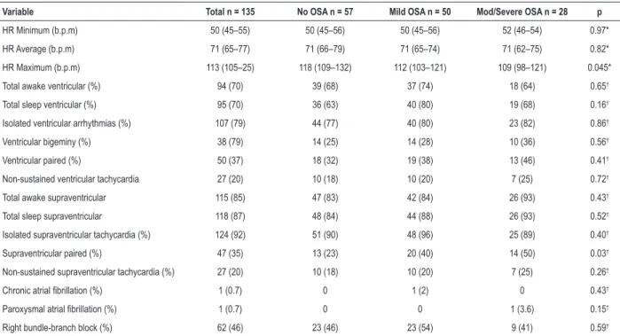
Overall, there were no clinically significant differences in supraventricular and ventricular arrhythmia frequencies across the three groups. However, there was a greater proportion of supraventricular paired in patients with moderate-to-severe OSA, compared with patients with mild and no OSA (50% vs 40% and 23%; p = 0.03), respectively. (Table 2)
Patients with moderate-to-severe OSA had increased nocturnal blood pressure (119 ± 17 vs. 113 ± 13 and 110 ± 11 mmHg; p = 0.01) compared to patients with mild and no OSA, respectively (Table 3). The aorta size, left ventricular systolic and diastolic diameters, septum and posterior wall thickness were similar between groups. Left atrial diameter [37 (33-42) vs. 35 (33-39) and 33 (30-36) mm, p < 0.01(Figure 2) and volume [66(54-95)vs. 46(39-65) and 42(35-56) mL/m2, p < 0.001] were larger in patients with moderate-to-severe OSA, compared with mild and no OSA groups, respectively(Table 3).
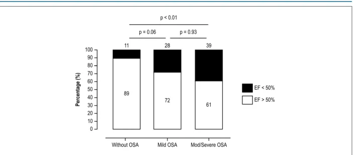
lower in patients with moderate-to-severe OSA compared with patients with mild and no OSA -16(-19/-13)% vs. -17(-20/-12)% and -19 (-21/-15)%, p = 0.04. The prevalence of ventricular dysfunction [LVEF < 50% (39% vs 28% and 11%), p < 0.01]
was also higher in the group with moderate-to-severe OSA than in participants with mild or no OSA (Figure 3). This higher prevalence was also observed in the subgroup of patients with Chagas cardiomyopathy.
Table 1 – Anthropometrics and clinical characteristics
Total n = 135 No OSA n = 57 Mild OSA n = 50 Mod/Severe OSA n = 28 p
Age, years 56 (45–62) 48 (42–58) 59 (55–63) 61 (51–63) < 0.001*
Male, n (%) 40 (30) 15 (26) 15 (30) 10 (36) 0.67†
BMI, kg/m2 26.3 ± 4.2 24.7 ± 3.9 27.4 ± 3.9 27.6 ± 4.5 0.001¥
Neck circumference, cm 35 (33–38) 34 (32–36) 36 (33–39) 38 (34–39) 0.02†
Waist, cm 92 (83–96) 87 (79-92) 96 (89–102) 96 (91–104) < 0.001*
Caucasians, n (%) 43 (32) 19 (33) 16 (32%) 8 (29%) 0.86†
Diabetes mellitus, n (%) 17 (13) 5 (9) 7 (14) 5 (18) 0.46†
Hypertension, n (%) 83 (62) 25 (44) 36 (72) 22 (79) 0.001†
Office Systolic BP, mmHg 131 ± 22 128 ± 20 135 ± 22 133 ± 26 0.27¥
Office Diastolic BP, mmHg 81 ± 11 83 ± 11 81 ± 12 78 ± 11 0.14¥
Use of antihypertensive drugs, n 1 (0–2) 1 (0–2) 2 (1–2) 2 (0–3) 0.001†
Diuretics, n (%) 54 (40) 13 (23%) 26 (52%) 15 (54%) < 0.001†
ACE inhibitors / AT1 inhibitors n (%) 76 (56) 23 (30%) 35 (46%) 14 (24%) < 0.001†
B-blockers, n (%) 57 (42) 17 (30%) 25 (50%) 13 (54%) 0.04†
Spironolactone, n (%) 14 (10) 5 (9%) 5 (10%) 4 (14%) 0.73†
Epworth sleepiness scale, Score, n (%) 9.8 ± 4.9 9.3 ± 5.2 10.2 ± 5.2 10.1 ± 3.8 0.57¥
Values are mean (±SD). Variables with skewed distribution presented as median (interquartile range). OSA: obstructive sleep apnea; BMI body mass index; BP: blood pressure. *: Test of Kruskal-Wallis; †: Chi-square test of Pearson; ¥: Anova test.
Table 2 – Holter evaluation – Distribution of atrial and ventricular arrhythmias
Variable Total n = 135 No OSA n = 57 Mild OSA n = 50 Mod/Severe OSA n = 28 p
HR Minimum (b.p.m) 50 (45–55) 50 (45–56) 50 (45–56) 52 (46–54) 0.97*
HR Average (b.p.m) 71 (65–77) 71 (66–79) 71 (65–74) 71 (62–75) 0.82*
HR Maximum (b.p.m) 113 (105–25) 118 (109–132) 112 (103–121) 109 (98–121) 0.045*
Total awake ventricular (%) 94 (70) 39 (68) 37 (74) 18 (64) 0.65†
Total sleep ventricular (%) 95 (70) 36 (63) 40 (80) 19 (68) 0.16†
Isolated ventricular arrhythmias (%) 107 (79) 44 (77) 40 (80) 23 (82) 0.86†
Ventricular bigeminy (%) 38 (79) 14 (25) 14 (28) 10 (36) 0.56†
Ventricular paired (%) 50 (37) 18 (32) 19 (38) 13 (46) 0.41†
Non-sustained ventricular tachycardia 27 (20) 10 (18) 10 (20) 7 (25) 0.72†
Total awake supraventricular 115 (85) 47 (83) 42 (84) 26 (93) 0.43†
Total sleep supraventricular 118 (87) 48 (84) 44 (88) 26 (93) 0.52†
Isolated supraventricular tachycardia (%) 124 (92) 51 (90) 48 (96) 25 (89) 0.40†
Supraventricular paired (%) 47 (35) 13 (23) 20 (40) 14 (50) 0.03†
Non-sustained supraventricular tachycardia (%) 27 (20) 10 (18) 10 (20) 7 (25) 0.26†
Chronic atrial fibrillation (%) 1 (0.7) 0 1 (2) 0 0.43†
Paroxysmal atrial fibrillation (%) 1 (0.7) 0 0 1 (3.6) 0.15†
Right bundle-branch block (%) 62 (46) 23 (46) 23 (54) 9 (41) 0.59†
Figure 2 – Left atrial diameter in patients with Chagas disease. OSA: obstructive sleep apnea. Kruskal-Wallis test, with Bonferroni post-hoc.
p < 0.01
p = 0.03 p = 0.54
55
45
35
25 50
40
30
Left
Atrial Diameter (mm)
Without OSA Mild OSA Mod/Severe OSA Table 3 – Echocardiography, Ambulatory Blood Pressure Monitoring and sleep study characteristics
Variable Total n = 135 No OSA n = 57 Mild OSA n = 50 Mod/Severe OSA n = 28 p
Echocardiography
LVFE (%) 60 (51–65) 61 (57–66) 59 (47–65) 57 (43–63) 0.09*
LA volume index (mL/m2)* 29 (23–38) 27 (21–33) 30 (23–38) 37 (51–55) < 0.001*
Aorta (mm) 30 (28–33) 30 (28–32) 30 (29–34) 30 (27–34) 0.21*
LV diastolic dimension (mm) 52 (49–58) 51 (48–55) 53 (49–58) 56 (50–61) 0.24*
LV systolic dimension (mm) 34 (30–41) 32 (30–37) 34 (30–40) 39 (30–48) 0.14*
Septum (mm) 8.0 (8.0–9.0) 8.0 (7.5–9.0) 8.0 (8.0–9.0) 8.0 (8.0–9.8) 0.10*
Posterior wall thickness (mm) 8.0 (7.0–9.0) 8.0 (7.0–9.0) 8.0 (8.0–9.0) 8.0 (8.0–9.0) 0.27* LV longitudinal strain (%)** –17 (–20/–13) –19 (–21/–15) –17 (–20/–12) –16 (–19/–13) 0.04*
ABPM
Systolic BP awake, mmHg 121 ± 12 118 ± 12 121 ± 12 123 ± 14 0.22¥
Diastolic BP awake, mmHg 74 ± 8 76 ± 8 73 ± 8 73 ± 9 0.34¥
Systolic BP Sleep, mmHg 113 ± 14 110 ± 11 113 ± 13 119 ± 17 0.01¥
Diastolic BP sleep, mmHg 67 ± 9 67 ± 8 66 ± 9 69 ± 9 0.41¥
Systolic non-dipping, % 78 75 79 89 0.29†
Diastolic non-dipping, % 48 40 41 75 < 0.01†
Sleep variables
AHI. Events/hour 5.8 (2.7–11.9) 2.4 (1.6–3.3) 7.2 (5.8–10.0) 20.3 (16.7–29.1) < 0.001¥
Mean SaO2, % 96 (95–97) 97 (94–97) 96 (94–97) 95 (94–97) < 0.001*
Lowest SaO2, % 88 (83–92) 92 (89–94) 86 (83–89) 83 (79–87) < 0.001*
Desaturation index. Number/hour 5.5 (2.2–12.8) 2.1 (0.9–3.4) 7.5 (4.9–10.4) 19.0 (5.9–26.5) < 0.001*
SaO2 < 90. % of night time 0 (0–3) 0 (0–0) 1 (0–4) 2 (1–5) < 0.001*
Values are mean (±SD). Variables with skewed distribution presented as median (interquartile range). OSA: obstructive sleep apnea; LVFE: left ventricular ejection fraction; LA: left atrial; LV: left ventricular; ABPM: ambulatory blood pressure measurement; BP: blood pressure; AHI: apnoea-hypopnoea index; SaO2: arterial
The only predictor of left atrial dimension in the multivariate analysis was Log10 (AHI) (β = 3.86, 95% CI: 1.91 to 5.81; p < 0.01) in the whole population (Table 4), as well as in the subgroup with Chagas cardiomyopathy.
The independent predictors of the presence of ventricular
dysfunction were AHI (≥ 15 events/h) (OR = 3.61, 95% C.I.:1.31
to 9.98; p = 0.01), systolic blood pressure (OR = 1.06, 95% C.I.: 1.02 – 1.10; p =< 0.01) and male gender (OR = 3.24, 95% C.I.: 1.31 – 8.01; p = 0.01) (Table 5).
Discussion
To the best of our knowledge, this is the first study that evaluated OSA in consecutive patients with CD. We found a high frequency of moderate to severe OSA in patients with CD (21%). None of the participants had a previous diagnosis of OSA, which suggests a low awareness of the disease in this population. Patients with moderate to severe OSA were more hypertensive, used more antihypertensive drugs and had increased nocturnal blood pressure. In addition, moderate Figure 3 – Frequency of Obstructive Sleep Apnea in patients with Chagas disease OSA: obstructive sleep apnea; Kruskal-Wallis test.
p < 0.01
p = 0.06 p = 0.93
11 28 39
89
72
61
EF < 50%
EF > 50% 100
90 80 70 60 50 40 30 20 10 0
Percentage (%)
Without OSA Mild OSA Mod/Severe OSA
Table 4 – Univariate and multiple linear regression predictors of left atrial dimension
Univariate Multivariate
Variable (β) p (β) CI p
Age (Years) 0.05 0.36
24h Systolic BP (mmHg) –0.03 0.46
BMI. kg/m2 0.32 0.01 0.16 –0.01 to 0.44 0.06
Log10AHI (events/hour) 3.86 < 0.001 3.86 1.91 to 5.81 < 0.01
BP: blood pressure; BMI: body mass index; AHI: Apnoea-hypopnoea; CI: Confidence interval
Table 5 – Logistic regression to assess predictors of ventricular dysfunction
Univariate Multivariate
Variable p OR CI p
Age (years) 0.49
BMI. kg/m2 0.09
Male % 0.01 3.24 1.31 – 8.01 0.01
24h Systolic BP (mmHg) 0.01 1.06 1.02 – 1.10 < 0.01
DM 0.95
AHI (≥ 15 events per hour) 0.02 3.61 1.31 – 9.98 0.01
Desaturation index. number/hour 0.25
SaO2 < 90%. % of night time 0.21
to severe OSA was independently associated with cardiac remodeling parameters, including larger left atrium dimension and a higher prevalence of ventricular dysfunction, especially in the subgroup of patients with Chagas cardiomyopathy. Taken together, our study emphasizes the concept that OSA may contribute to a poor prognosis in patients with CD.
OSA is a prevalent condition in patients with cardiovascular disease,20 and our study confirms its high frequency in patients with CD. Particular characteristics of studied OSA patients deserves some comments: First, the presence of higher BMI and neck circumference in patients with moderate to severe OSA as compared to patients with mild or no OSA denotes a typical phenotype of the disease (Table1). However, the lack of excessive daytime sleepiness, a characteristic of OSA patients from the overall population referred to sleep laboratories may explain the low awareness of OSA in patients with cardiovascular disease.21 This finding is in line with other populations with cardiovascular diseases, including heart failure22 and hypertrophic cardiomyopathy.23 The low prevalence of central sleep apnea in our sample is in contrast with a previous study that evaluated sleep disorders in patients with heart failure (LVEF < 45%), including CD. Silva et al.24 included 24 patients with CD, of which only 12% had OSA, but 44% of whom had Cheyne-Stokes respiration. The median LVEF of our sample was within the normal range (60%) and the majority of our sample did not have heart failure. Moreover, we excluded patients with decompensated heart failure from our study, which may explain the different frequencies.
OSA is recognized as being a cause of hypertension.25 In our sample, hypertension was more frequent in the group with moderate to severe OSA and this group used more antihypertensive drugs. However, only systolic BP during sleep was higher in patients with moderate-to-severe OSA. This finding may be related to the fact that the study took place in a reference center for cardiology, where most patients are being properly treated for hypertension. The relatively low frequency of atrial and ventricular arrhythmias in our study may be also explained by the exclusion of more severe cases of CD cardiomyopathy.
Our study demonstrated that in patients with CD, OSA is independently associated with left atrial enlargement. This finding is consistent with other OSA populations7,23,26 Rossi et al.27 conducted a meta-analysis of 1,157 patients who took part in 18 heart failure studies, and concluded that left atrial enlargement was associated with a worse prognosis, regardless of age, functional class, ejection fraction or diastolic function pattern,28 reinforcing the importance of our findings. In our study, moderate to severe OSA was also associated with a lower left ventricular longitudinal strain and a four-fold higher proportion of ventricular dysfunction than patients with no OSA. This finding is consonant with an earlier study that evaluated patients referred to a sleep laboratory and may impact negatively on mortality.6,29
There are several mechanisms that can contribute to cardiac remodeling in OSA patients30 One possible explanation for the heart remodeling in our study may be partially explained by the increased frequency of hypertension and higher nocturnal systolic blood pressure, as well as increased frequency of
group. Taken together, the increased hypertension burden may increase arterial stiffness and left ventricular afterload, contributing to these abnormalities.7 Moreover, inspiratory efforts during the apneas generate negative intrathoracic pressure, which leads to an increase in the left ventricular afterload and a decrease in the left ventricular preload, which in turn cause a reduction in the ejection volume and may induce left atrial enlargement, as demonstrated in our study. Intermittent hypoxia may also influence cardiac contractility, directly or indirectly, thereby reducing cardiac output.22 OSA induces hypoxia, hypercapnia and sleep arousals, thus promoting an increase in sympathetic activity and hence, in blood pressure.31 Long-term exposure to high sympathetic nerve activity can induce hypertrophy and apoptosis of the cardiac myocytes32 and thereby cause left ventricular dysfunction. These adverse hemodynamic effects may be more pronounced in individuals with heart failure,33 as shown in our subgroup with Chagas cardiomyopathy compared with patients with the indeterminate form.
The major strength of the present study is that it is the largest and one of the only cohorts evaluating the association between sleep apnea in patients with CD. The recruitment of consecutive patients with well-characterized CD not referred to a sleep laboratory may generalizes the findings of our study, as does the use of gold-standard techniques to assess blood pressure (ABPM) and respiratory effort (inductance plethysmography belts).11 The study has some potential limitations. A portable sleep monitor that does not measure sleep duration was used. Thus, measurements of the AHI were taken based on the total recording time and not on the total length of sleep, although this device has already been validated against full polysomnography.34 Furthermore, our findings are derived from a cross-sectional study and we cannot infer causality, but only an independent association between OSA and heart remodeling. The fact that we could not demonstrate the same atrial and ventricular remodeling findings in the subgroup of patients with the indeterminate form of CD could be due to the small number of patients in this category. Moreover, the absence of increased incidence of arrhythmias in OSA patients in this study should be analyzed with caution, as 24 h Holter monitoring could not detect intermittent arrhythmias. New studies with technologies that analyze long periods of time are warranted.
Conclusion
OSA is common and independently associated with atrial and ventricular remodeling in patients with CD. The improvement in OSA recognition and treatment may contribute to reducing the morbidity attributed to CD.
Author contributions
Potential Conflict of Interest
No potential conflict of interest relevant to this article was reported.
Sources of Funding
This study was funded by FACEPE and CNPq.
Study Association
This article is part of the thesis of master submitted by Carolina de Araújo Medeiros, from Programa de Pós Graduação em Ciências da Saúde da Universidade de Pernambuco (UPE).
Ethics approval and consent to participate
This study was approved by the Ethics Committee of the Complexo Hospitalar - Hospital Universitário Oswaldo Cruz - PROCAPE - Universidade de Pernambuco/UPE under approval the protocol ZAAE - 16127213.7.0000.5192, report number: 358162. All the procedures in this study were in accordance with the 1975 Helsinki Declaration, updated in 2013. Informed consent was obtained from all participants included in the study.
1. World Health Organization. (WHO). Chagas disease in Latin America: an epidemiological update based on 2010 estimates. Wkly Epidemiol Rec. 2015;(6):33-44.
2. World Health Organization. (WHO). Research priorities for Chagas Disease, human african trypanosomiasis and leishmaniasis: technical report of the TDR disease reference group on Chagas Disease, Human African Trypanosomiasis and Leishmaniasis. Genebra, Suiça; 2012;
3. Andrade JP, Marin-Neto JA, Paola AA, Vilas-Boas F, Oliveira GM, Bacal F, et al; Sociedade Brasileira de Cardiologia. [Latin American guidelines for the diagnosis and treatment of Chagas cardiomyopathy]. Arq Bras Cardiol. 2011;97(2 Suppl 3):1-48.
4. Albajar-Vinas P, Jannin J. Chagas disease in Europe. Eurosurveillance. 2011;22 Sep;16(38):special edition.
5. Morillo CA, Marin-Neto JA, Avezum A, Sosa-Estani S, Rassi A Jr, Rosas F, et al; BENEFIT Investigators. Randomized trial of benznidazole for chronic Chagas’ cardiomyopathy. N Engl J Med. 2015;373(14):1295-306.
6. Rosen D, Roux FJ, Shah N. Sleep and breathing in congestive heart failure. Clin Chest Med. 2014;35(3):521-34.
7. Drager LF, Bortolotto LA, Figueiredo AC, Silva BC, Krieger EM, Lorenzi-Filho G. Obstructive sleep apnea, hypertension, and their interaction on arterial stiffness and heart remodeling. Chest. 2007;131(5):1379-86.
8. Monahan K, Storfer-Isser A, Mehra R, Shahar E, Mittleman M, Rottman J, et al. Triggering of nocturnal arrhythmias by sleep-disordered breathing events. J Am Coll Cardiol. 2009;54(19):1797-804.
9. Bern C, Montgomery SP, Herwaldt BL, Marin-neto JA, Maguire JH, Acquatella H, et al. Evaluation and Treatment of Chagas Disease in the United States A Systematic Review. JAMA. 2007;298(18):2171–2181.
10. Gjevre JA, Taylor-Gjevre RM, Skomro R, Reid J, Fenton M, Cotton D. Comparison of polysomnographic and portable home monitoring assessments of obstructive sleep apnea in Saskatchewan women. Can Respir J. 2011;18(5):271-4.
11. Iber C, Chesson AL, Ancoli-Israel S, Quan SF. The AASM manual for the scoring of sleep and associated events:rules,terminology and technical specifications. Darien,IL:American Academy of Sleep Medicine(AASM);2007.
12. Collop NA, Goldberg R, Anderson WM, Boehlecke B, Gottlieb DJ, Hudgel D, et al; Portable Monitoring Task Force of the American Academy of Sleep Medicine. Clinical guidelines for the use of unattended portable monitors in the diagnosis of obstructive sleep apnea in adult patients. J Clin Sleep Med. 2007;3(7):737-47.
13. Johns M. A New Method for Measuring Daytime Sleepiness : The Epworth Sleepiness Scale. Sleep. 1991;14(6):540-5.
14. Bakris GL, Black HR, Cushman WC, Green LA, Izzo JL, Jones DW, et al; National Heart, Lung, and Blood Institute Joint National Committee on Prevention, Detection, Evaluation, and Treatment of High Blood Pressure; National High Blood Pressure Education Program Coordinating Committee. The Seventh Report of the Joint National Committee on Prevention, Detection, Evaluation, and Treatment of High Blood Pressure: the JNC 7 report. JAMA. 2003;289(19):2560-72. Erratum in: JAMA. 2003;290(2):197.
15. Pickering TG, Hall JE, Appel LJ, Falkner BE, Graves JW, Hill MN, et al; Council on High Blood Pressure Research Professional and Public Education Subcommittee, American Heart Association. Recommendations for blood pressure measurement in humans: an AHA scientific statement from the Council on high blood pressure research professional and public education subcommittee. J Clin Hypertens (Greenwich). 2005;7(2):102-9.
16. Crawford MH, Bernstein SJ, Deedwania PC, DiMarco JP, Ferrick KJ, Garson A Jr, et al. ACC/AHA guidelines for ambulatory electrocardiography. A report of the American College of Cardiology/American Heart Association Task Force on Practice Guidelines (Committee to Revise the Guidelines for Ambulatory Electrocardiography). Developed in collaboration with the North American Society for Pacing and Electrophysiology. J Am Coll Cardiol. 1999;34(3):912-48.
17. Lang RM, Bierig M, Devereux RB, Flachskampf FA, Foster E, Pellikka PA, et al; American Society of Echocardiography's Nomenclature and Standards Committee; Task Force on Chamber Quantification; American College of Cardiology Echocardiography Committee; American Heart Association; European Association of Echocardiography, European Society of Cardiology. Recommendations for chamber quantification. Eur J Echocardiogr. 2006;7(2):79-108.
18. Rudski LG, Lai WW, Afilalo J, Hua L, Handschumacher MD, Chandrasekaran K, et al. Guidelines for the echocardiographic assessment of the right heart in adults: a report from the American Society of Echocardiography endorsed by the European Association of Echocardiography, a registered branch of the European Society of Cardiology and the Canadian Society of Echocardiography. J Am Soc Echocardiogr. 2010;23(7):685-713.
19. Mancuso FJ, Almeida DR, Moisés VA, Oliveira WA, Mello ES, Poyares D, et al. Left atrial dysfunction in chagas cardiomyopathy is more severe than in idiopathic dilated cardiomyopathy: a study with real-time three-dimensional echocardiography. J Am Soc Echocardiogr. 2011;24(5):526–32.
20. Kasai T, Floras JS, Bradley TD. Sleep apnea and cardiovascular disease: a bidirectional relationship. Circulation. 2012;126(12):1495-510.
21. Costa LE, Uchôa CH, Harmon RR, Bortolotto LA, Lorenzi-Filho G, Drager LF. Potential underdiagnosis of obstructive sleep apnoea in the cardiology outpatient setting. Heart. 2015;101(16):1288-92.
22. Mentz RJ, Fiuzat M. Sleep-disordered breathing in patients with heart failure. Heart Fail Clin. 2014;10(2):243-50.
This is an open-access article distributed under the terms of the Creative Commons Attribution License
23. Pedrosa RP, Drager LF, Genta PR, Amaro AC, Antunes MO, Matsumoto AY, et al. Obstructive sleep apnea is common and independently associated with atrial fibrillation in patients with hypertrophic cardiomyopathy. Chest. 2010;137(5):1078-84.
24. Silva RS, Figueiredo AC, Mady C, Lorenzi-Filho G. Breathing disorders in congestive heart failure : gender, etiology and mortality. Braz J Med Biol Res. 2008;41(3):215-22.
25. Pedrosa RP, Drager LF, Gonzaga CC, Sousa MG, de Paula LK, Amaro AC, et al. Obstructive sleep apnea: the most common secondary cause of hypertension associated with resistant hypertension. Hypertension. 2011;58(5):811-7.
26. Chami HA, Devereux RB, Gottdiener JS, Mehra R, Roman MJ, Benjamin EJ, et al. Left ventricular morphology and systolic function in sleep-disordered breathing: the Sleep Heart Health Study. Circulation. 2008;117(20):2599-607.
27. Rossi A, Temporelli PL, Quintana M, Dini FL, Ghio S, Hillis GS, et al; MeRGE Heart Failure Collaborators. Independent relationship of left atrial size and mortality in patients with heart failure: an individual patient meta-analysis of longitudinal data (MERGE Heart Failure). Eur J Heart Fail. 2009;11(10):929-36.
28. Varol E, Akcay S, Ozaydin M, Ozturk O, Cerci SS, Sahin U. Influence of obstructive sleep apnea on left ventricular mass and global function: sleep apnea and myocardial performance index. Heart Vessels. 2010;25(5):400-4.
29. Alchanatis M, Tourkohoriti G, Kosmas EN, Panoutsopoulos G, Kakouros S, Papadima K, et al. Evidence for left ventricular dysfunction in patients with obstructive sleep apnoea syndrome. Eur Respir J. 2002;20(5):1239-45.
30. Floras JS. Sleep apnea and cardiovascular risk. J Cardiol. 2014;63(1):3-8.
31. Bradley TD, Floras JS. Sleep apnea and heart failure: Part II: central sleep apnea. Circulation. 2003;107(13):1822-6.
32. Bradley TD, Floras JS. Sleep apnea and heart failure: Part I: obstructive sleep apnea. Circulation. 2003;107(12):1671-8.
33. Bradley TD, Hall MJ, Ando S, Floras JS. Hemodynamic effects of simulated obstructive apneas in humans with and without heart failure. Chest. 2001;119(6):1827-35.