ISSN 1546-9239
© 2011 Science Publications
Corresponding Author: Qamar Uddin Ahmed, Department of Pharmaceutical Chemistry, Kulliyyah of Pharmacy, International Islamic University Malaysia, 25200-Kuantan, Pahang DM, Malaysia Tel: +609-5716400 (3096) Fax: +609-5716775
525
Bioassay Guided Isolation of Antibacterial
Compounds from Andrographis paniculata
1
Abubakar Sule,
2Qamar Uddin Ahmed,
1Othman Abd. Samah,
1Muhammad Nor Omar,
2Norazian M. Hassan,
2Laina Zarisa M. Kamal and
3M. Ambar Yarmo
1Department of Biomedical Sciences, Kulliyyah of Science,
2Department of Pharmaceutical Chemistry, Kulliyyah of Pharmacy,
International Islamic University Malaysia, Kuantan, Pahang DM,
3
School of Chemical Sciences and Food Technology,
Kulliyyah of Science and Technology, University Kebangsaan Malaysia,
43600 UKM Bangi, Selangor Malaysia
Abstract: Problem statement: Chronic disease-causing bacteria of medical importance have
developed resistance to antibiotics, hence, necessitating distinct and constant need for safe and efficient therapeutic agents. Plants are considered potent candidate for this aim. A way out of reducing antibiotic resistance and adverse effects on host is the employment of antibiotic resistance inhibitors of plant origin. Approach: About 5 kg pulverized Andrographis paniculata whole plant was macerated with MeOH at room temperature to get 305 g freeze dried MeOH extract. The bioautography of MeOH extract using Staphylococcus aureus and Proteus mirabilis as indicator organisms revealed the presence of two potent antibacterial compounds. MeOH extract was further fractionated and purified by silica gel column chromatography which led to the isolation of a diterpene lactone and an ent-labdane diterpene glycoside upon crystallization with absolute ethanol. Results: Two antibacterial compounds viz., 3-O-β-D-glucosyl-14-deoxyandrographolide and 14-deoxyandrographolide were successfully isolated and characterized. Their structures were exclusively elucidated through spectroscopic methods (UV, IR, 1H- and 13C NMR). Conclusion: A. paniculata possesses antibacterial activity and could be potential source of a new class of antibiotics that might be useful for infectious disease chemotherapy and control.
Key words: Andrographis paniculata, bioassay-guided isolation, antibacterial compounds
INTRODUCTION
Andrographis paniculata (Burm.f.) Wall. ex Nees.,
(Family-Acanthaceae) (English name-King of Bitters, Local malay name-Hempedu bumi) is an annual herbaceous plant and is extensively cultivated in Southern Asia, China and some parts of Europe. In traditional medicine, A. paniculata is widely used to get rid of body heat, dispel toxins from the body, prevent common cold, upper respiratory tract infections including sinusitis and fever (Gabrielian et al., 2002) and as an antidote against poisons of snakes and insects (Samy et al., 2008). A. paniculata has been reported to exhibit various mode of biological activities in vivo as well as in vitro viz., antibacterial (Singha et el., 2003; Mishra et al., 2009; Parvataneni and Koduru, 2010; Roy
et al., 2010; Abubakar et al., 2011), antiviral (Wiart et
al., 2000), anti-inflammatory (Wen et al., 2010),
antihuman immunodeficiency virus (HIV) (Calabrese et
al., 2000), immunomodulating/immunostimulatory (Iruretagoyena et al., 2005) and anticancer (Li et al., 2007; Geethangili et al., 2008). The characteristic secondary metabolites encountered in this plant have considerably enhanced its importance in the arena of medicinal plants. It is specifically rated high in therapeutic action in curing liver disorders, common cough and colds in human (Niranjan et al., 2010).
526 microbiologists and natural product chemists in tropical countries including Malaysia, with the richest flora and fauna placed right at their door step have a very central position. They are essential for building up international scientific cooperation, with the objective of expanding our understanding of biological and biochemical diversity and based on this bringing forward more biological solutions. The entire process is built on a principle of fairness and equity in sharing of the benefits and respecting the State's sovereign right to its own resources. After structure elucidation of secondary metabolites, it is considered crucial to know how useful these molecules might be in terms of medicinal properties. During the past 40 years, numerous novel compounds have been isolated from different plants and marine organisms and many of these have been reported to have core biological activities, some of which are of interest from the point of view of potential drug development (Gerald, 2001; Houghton, 2001). In this context, A. paniculata could be a potential source to develop new efficacious drugs. A. paniculata has already been reported for its significant antibacterial potential (Singha et al., 2003, Mishra et al., 2009; Parvataneni and Koduru, 2010; Roy et al., 2010; Abubakar et al., 2011). However, no attempt has ever been made to identify and isolate active principles responsible for unleashing its antibacterial activity. Identification and isolation of active principles from A.
paniculata might prove promising antibacterial agents
through foreseeable future endeavors. Hence, this study is a conscientious attempt to identify and isolate pure antibacterial compounds from the methanol extract of the whole plant of A. paniculata through bioassay guided isolation method.
MATERIALS AND METHODS
Collection of plant material: About 15 kg fresh whole
plant of A. paniculata was procured from the botanical garden of Forest Research Institute of Malaysia (FRIM), Kuala Lumpur, Malaysia, during the month of April, 2009. The plant was identified by Dr. Richard Chung Cheng Kong (Ph.D., Taxonomist, FRIM). The voucher specimen (NMPC-Q25) has been deposited in the Herbarium, Faculty of Pharmacy, IIUM, Kuantan, Pahang DM, Malaysia for future references.
Preparation of methanol (MEOH) extract: The fresh
whole plant (15 kg) of A. paniculata was cleaned and dried in a protech laboratory air dryer (LDD-720) at 40°C for 7 days and pulverized to powdered form (5.6 kg, 37.33%) using Fritsch Universal Cutting Mill-PULVERISETTE 19-Germany. It was then stored in a desiccator at 2°C until further use. The air dried powder
of whole plant (5 kg) of A. paniculata was extracted by macerating in double distilled methanol (20.0 L) at room temperature for 24 h, filtered and evaporated under reduced pressure. The whole process was repeated thrice to ensure maximum yield of methanol soluble compounds from the plant powder. Each time, filtrate was evaporated under reduced pressure (Buchi Rotary Evaporator, R-210) and combined. The dark blackish green residue so obtained was further freeze dried to yield 305 g (6.1%) MeOH extract and was stored at 2°C in a labeled sterile bottle until further antibacterial evaluation and isolation of antibacterial compounds.
Source of microorganism: Staphylococcus aureus
(IMR S-277), Streptococcus pyogenes (IMR S-526),
Micrococcus luteus (IMR B-7), Proteus mirabilis (IMR
P-74) and Pseudomonas aeruginosa (IMR P-84) were purchased directly from the Institute for Medical Research (IMR), Kuala Lumpur, Malaysia. The bacterial stock cultures were maintained on nutrient agar slants prior to use.
Preparation of standard bacterial suspensions: The
average number of viable, S. aureus, S. pyogenes, M.
luteus, P. mirabilis and P. aeruginosa organisms per
mL of the stock suspensions was determined by means of the surface viable counting technique (Hedges, 2002). About 107-108 CFU/mL was used. Each time, a fresh stock suspension was prepared; the experimental conditions were maintained constant so that suspensions with very close viable counts could be obtained successfully.
527 out for the extract against each of the test organism. After incubation the diameter of the results and growth inhibition zones were measured, averaged and the mean values were recorded.
Determination of Minimum Inhibitory
Concentration (MIC): Micro broth dilution method
was used for the determination of MIC values for each plant extract showing antibacterial activity against test pathogens (EUCAST, 2003; Jana et al., 2004). Serial dilutions of the extracts were carried out in 10% DMSO (which had no inhibitory activity against test microorganisms) to make 500 g mL−1 final concentration, this was then two fold serially diluted by adding to the broth media in a 96-wells microtiter plates to obtain 250, 125, 62.5, 31.3, 15.6 and 7.81 g mL−1. Thereafter, 100 L inoculum (108 CFU mL−1) was added to each well. Bacterial suspensions were used as negative control, while broth containing standard drug (vancomycin and gentamicin) were used separately as positive controls. The microtiter plates were incubated at 37°C for 24 h. Each extract was assayed in duplicate; one was kept for incubation while the other was kept at 4°C for comparing the turbidity in the wells of microtiter plate. The MIC values were taken as the lowest concentration of the extracts in the well of the microtiter plate that showed no turbidity after incubation. The turbidity of the wells in the microtiter plate was interpreted as visible growth of microorganisms.
Antibacterial Activity Index (AbI): Antibacterial
index (AbI) of MeOH whole plant extract of A.
paniculata was calculated separately as the average
value of zone of inhibition against the Gram-positive and Gram-negative bacteria, respectively (Mbwambo
et al., 2007).
Bioassay guided isolation: To sterilized 8×4 cm silica gel 60 F254 TLC plates (Merck, Germany), 10 L of MeOH extract was applied as small spots and the plates were developed in Hexane: Acetone (2:1) in duplicate (a TLC plate was used as the bioautogram while the other served as a chromatogram for reference in comparison with the bioautograph). The TLC plates were dried in an oven at 25°C for 7 h to activate the plates by absorbing the moisture content from the plates and removing all residual solvents (Veronica and Scott, 2005).
Bioautography technique: S. aureus and P. mirabilis
were used as the indicator microorganisms for the bioautography of antibacterial compounds from the MeOH extract of A. paniculata. 200 L each from broth cultures of S. aureus and P. mirabilis (adjusted to 108 CFU mL−1) were mixed with 35 mL molten
Mueller-Hinton agar (MHA) at 30°C separately. The suspensions of agar and bacteria were spread aseptically onto the already developed TLC plates in square Petri dishes (8×4 cm), allowed for 30 min to solidify and the plates were incubated at 37°C for 24 h. At the end of incubation time, 0.5%
p-iodonitrotetrazolium violet (INT) was uniformly sprayed over the TLC plates. The active antibacterial compounds in the plant extracts formed clear zones of inhibition on the TLC plates against a deep pink back ground of bacterial growth, allowing the chromatographic Retention factors (Rf) observation by viewing under UV light at 254 nm (Short wave) and 366 nm (Long wave) and comparing with the reference chromatogram (already sprayed with vanillin reagent and heated at 120°C) to note the antibacterial compounds. Vanillin reagent was prepared by dissolving 15 g of vanillin in ethanol (250 mL) and H2SO4 (2.5 mL). Vanillin reagent gives different colored spots with different compounds on TLC plate upon heating at 120°C (Rahalison et al., 2007).
Identification and isolation of antibacterial compounds (AB-1 and AB-2) from MeOH extract:
528 was made with the previous reported data of the same compounds.
3-O-β-D-glucosyl-14-deoxyandrographolides (AB-1): M.P. 242-244oC, UV λmax MeOH nm: 202. IR (cm
-1 ) ν: 3351, 1732, 165, 899. 1H NMR (600 MHz, in DMSO-d6), δ (ppm): 1.25 (o, 1H, C1-CH2), 1.71 (o, 1H, C1-CH2), 2.10 (m, 1H, C2-CH2), 1.98 (m, 1H, C2-CH2), 3.925 (o, 1H, C3-CH-), 1.3 (m, 1H, C5-CH-), 1.85 (m, 2H, C6-CH2), 2.4 (m, 2H, C7-CH2), 3.350 (d, J = 8.4 Hz, 1H, C9-CH-), 1.8 (m, 2H, C11-CH2), 2.6 (m, 1H, C12-CH2), 2.3 (m, 1H, C12-CH2), 7.105 (t, 1H, C14-CH-), 4.779 (brs, 2H, C15-CH2), 4.874 (brs, 1H, C17-CH2), 4.594 (brs, 1H, C17-CH2), 1.007 (brs, 3H, C18-CH3), 4.036 (d, J = 9.6 Hz, 1H, C19-CH2), 3.221 (d, J = 9.6, 1H, C19-CH2), 0.662 (brs, 3H, C20-CH3), 4.241 (d, J = 7.2 Hz, 1H, C1’-CH-), 3.379 (o, 1H, C2’-CH-), 3.394 (o, 1H, C3’-C2’-CH-), 3.570 (o, 1H, C4’-C2’-CH-), 3.595 (o, 1H, C5’-CH-), 3.842 (dd, J = 11.4, 4.8 Hz, 2H, C6’-CH2), “o” denotes overlapping signals; 13C NMR (125.76 MHz, in DMSOd6), δ (ppm): 38.19 (C1), 29.72 (C2), 75.04 (C3), 39.56 (C4), 56.44 (C5), 35.97 (C6), 38.44 (C7), 147.30 (C8), 56.18 (C9), 38.89 (C10), 21.72 (C11), 24.46 (C12), 136.02 (C13), 143.84 (C14), 70.11 (C15), 174.33 (C16), 107.08 (C17), 18.93 (C18), 62.61 (C19), 15.41 (C20), 103.10 (C1’), 71.72 (C2’), 72.73 (C3’), 74.03 (C4’), 76.23 (C5’), 70.72 (C6’). From this spectral data and their direct comparison with the previously published spectral data (Zhou et al., 2008) of the same compound, AB-1 was unambiguously identified as 3-O-β-D-glucosyl-14-deoxyandrographolide (Fig. 1).
14-deoxyandrographolide (AB-2): M.P. 172-174oC, UV λmax MeOH nm: 223. IR (cm−1) ν: 3367, 1736, 1646, 896. 1H NMR (600 MHz, in DMSO-d6), δ (ppm): 1.327 (m, 2H, C1-CH2), 2.045 (brs, 1H, C2-CH2), 1.999 (o, 1H, C2-CH2), 3.353 (o, 1H, C3-CH-), 1.463 (o, 1H, C5-CH-), 2.182 (brs, 1H, C6-CH2), 1.987 (brd, J = 6 Hz, 1H, C6-CH2), 2.451 (t, 2H, C7-CH2), 3.506 (o, 1H, C9-CH-), 2.433 (dd, J = 4.2, 2.4 Hz, 2H, C11-CH2), 2.557 (dd, J = 12, 7.2 Hz, 2H, C12-CH2), 7.10 (brs, 1H, C14-CH-), , 4.91 (brs, 2H, C15-CH2), 4.595 (brs, 1H, C17-CH2), 4.455 (brs, 1H, C17-CH2), 1.599 (o, 3H, C18-CH3), 4.207 (brs, 1H, C19-CH2), 4.189 (brs, 1H, C19-CH2), 0.71 (s, 3H, C20-CH3), “o” denotes overlapping signals; 13C NMR (125.76 MHz, in DMSOd6), δ (ppm): 38.93 (C1), 37.74 (C2), 80.50 (C3), 64.13 (C4), 55.21 (C5), 28.22 (C6), 37.06 (C7), 148.94 (C8), 55.96 (C9), 42.96 (C10), 24.88 (C11), 23.76 (C12), 146.64 (C13), 127.83 (C14), 66.31 (C15), 172.17 (C16), 108.86 (C17), 22.67 (C18), 74.61 (C19), 15.16 (C20). From this spectral data and their direct comparison with the previously published data (Poonam et al., 2010) of the same compound, AB-2 was unambiguously identified as 14-deoxyandrographolide (Fig. 2).
Fig. 1: Structure of AB-1 (3-O-β-D-glucosyl-14-deoxyandrographolide) based on 1H- and 13C NMR spectra
Fig. 2: Structure of AB-2 (14-deoxyandrographolide) based on 1H- and 13C NMR spectra
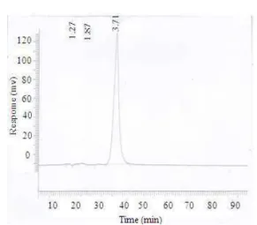
529 Fig. 4: HPLC analysis of compound AB-2 from the
MeOH extract of Andrographis paniculata. Mobile phase: Water: Acetonitrile: Methanol (11:6:3) at Wave length: 214 nm, Flow rate: 1mL/min and Injection volume: 1µL
Minimum Inhibitory Concentration (MIC) of Isolated Compounds: The minimum inhibitory concentrations of the isolated compound 1 and AB-2 were determined using the agar dilution method following the standard protocol of the European Committee for Antimicrobial Susceptibility Testing (EUCAST, 2003). The compounds were dissolved in 10% DMSO and 2-fold diluted in MHA to obtain 250, 125, 62.5, 31.3, 15.6 and 7.81 g mL−1. The mixture of the media and compounds were thoroughly mixed and poured onto pre-labeled sterile Petri dishes on a level surface. Additional Petri dishes containing only the growth media were prepared in the same way so as to serve for comparison of growth of the respective bacteria. The plates were then set at room temperature and dried. The suspensions of the respective bacteria (corresponding to 108 CFU mL−1) were inoculated onto the series of agar plates. The plates were then incubated at 37°C for 24 h. The experiments were performed in duplicate and MIC values expressed as the lowest concentration of the plant extracts that produced complete suppression of colony of respective bacteria.
Statistical analyses: The experimental results were
expressed as mean ± Standard Deviation (STD) of triplicate experiments. Statistical differences between the antibiotics and inhibition zones formed by the plant extracts were detected by Analysis Of Variance (ANOVA) using SPSS 19.0 statistical software (SPSS, Chicago, Illinois, USA) followed by the Tukey test for multiple comparisons between means. P values lower than 0.05 (p<0.05) were considered significantly
different whereas P values lower than 0.01 (p<0.01) were considered highly significant.
RESULTS
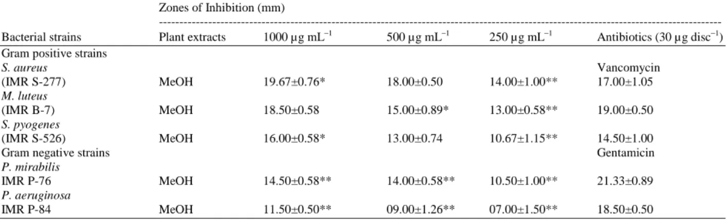
The results of the cup-plate agar diffusion method showed that MeOH extract of the whole plant of A.
paniculata do possess antibacterial activity against all 5
bacteria taken into account in vitro (Table 1). Maximum antibacterial activity was observed against S.
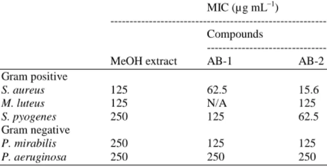
aureus (19.67 ± 0.76 mm) at 1000 gmL−1 and the lowest activity was detected against P. aeruginosa (7.00 ± 1.50 mm) at 250 gmL−1. MIC values for MeOH extract and isolated compounds are shown in Table 2. MIC of MeOH extract ranged from 125-250 g mL−1 with the highest MIC value exerted by the extract against S. pyogenes, P. mirabilis and P.
aeruginosa (250 g mL−1) and the lowest against S.
aureus and M. luteus (125 g mL−1). The bioassay-guided isolation of antibacterial compounds from MeOH extract led to the identification and subsequent isolation of an ent-labdane diterpene glycoside (AB-1) and a diterpene lactone (AB-2) as the main active principles. Both compounds were active against S.
aureus (Fig. 5-6) and P. mirabilis which were used as
indicator organisms by the bioautography technique on TLC plates forming clear zones against pink background of the living microorganisms when compared to the reference chromatogram. MIC values for both isolated compounds ranged from 15.6-250 g mL−1. Highest MIC value was exerted by compound AB-1 against P. aeruginosa (250 g mL−1) while the lowest was exerted by compound AB-2 against S.
aureus (15.6 g mL−1), however, no activity was exerted by compound AB-1 against M. luteus (Table 2). The MeOH extract’s antibacterial index (AbI) was found to be the best against Gram-positive strains tested as compared to the Gram-negative strains with mean inhibition zones of 13.9 mm and 10.4 mm, respectively (Table 3).
530
(a) (b)
Fig. 5: Bioautography of AB-1 (3-O-β-D-glucosyl-14-deoxyandrographolide) against S. aureus. (a) Referenced chromatogram sprayed with vanillin/H2SO4 spray reagent. (b) Bioautogram against S. aureus
(a) (b)
Fig. 6: Bioautography of AB-2 (14-deoxyandrographolide) against S. aureus. (a) Bioautogram against S. aureus. (b) Referenced chromatogram viewed under UV light
Table 1: Antibacterial activity of MeOH extract of A. paniculata whole plant. Numbers indicate the mean diameters of inhibition of triplicate experiments ± standard deviation (SD)
Zones of Inhibition (mm)
---
Bacterial strains Plant extracts 1000 µg mL−1 500 µg mL−1 250 µg mL−1 Antibiotics (30 µg disc−1)
Gram positive strains
S. aureus Vancomycin
(IMR S-277) MeOH 19.67±0.76* 18.00±0.50 14.00±1.00** 17.00±1.05
M. luteus
(IMR B-7) MeOH 18.50±0.58 15.00±0.89* 13.00±0.58** 19.00±0.50
S. pyogenes
(IMR S-526) MeOH 16.00±0.58* 13.00±0.74 10.67±1.15** 14.50±1.00
Gram negative strains Gentamicin
P. mirabilis
IMR P-76 MeOH 14.50±0.58** 14.00±0.58** 10.50±1.00** 21.33±0.89
P. aeruginosa
IMR P-84 MeOH 11.50±0.50** 09.00±1.26** 07.00±1.50** 18.50±0.50
531
Table 2: Minimum inhibitory concentrations (MIC) of the MeOH extract of A. paniculata whole plant and isolated compounds against bacterial strains
MIC (µg mL−1)
--- Compounds
---
MeOH extract AB-1 AB-2
Gram positive
S. aureus 125 62.5 15.6
M. luteus 125 N/A 125
S. pyogenes 250 125 62.5 Gram negative
P. mirabilis 250 125 125
P. aeruginosa 250 250 250 *NA: No Activity
Table 3: Antibacterial activity indexes (AbI) of MeOH extract of A.
paniculata whole plant.
Activity index (mm)
Bacterial strains MeOH extract
Gram positive
S. aureus, M. luteus, S. pyogenes 13.9 Gram negative
P. mirabilis, P. aeruginosa 10.4
The characteristic NMR spectral data indicated that compound AB-2 was a labdane-type diterpene with α, β-unsaturated γ-lactone. In the 1H-NMR spectrum of AB-2, two methyl singlets were observed at δ 0.71 and 1.599, respectively. The characteristic exocyclic methylene protons for AB-2 diterpenoids were observed at δ 4.595 (brs, 1H) and 4.455 (brs, 1H) in 1HNMR as well as at
δ 108.86 in 13C, respectively. The 1H- and 13C-NMR (in CDCl3) spectra of AB-2 suggested a diterpenoid compound with a structure similar to that of 14-deoxyandrographolide (Poonam
et al., 2010) (Fig. 2).
DISCUSSION
Antibiotics provide the main basis for the therapy of chronic bacterial infections. However, the high genetic variability of bacteria enables them to rapidly evade the action of antibiotics by developing antibiotic resistance. As resistance becomes more common, there becomes a greater need for alternative treatments. However despite a push for new antibiotic therapies there has been a continued decline in the number of newly approved drugs (Bachi, 2002; Nagi et al., 2010). According to the World Health Report on infectious diseases 2000, overcoming antibiotic resistance is the major issue of the WHO for the next millennium. Hence, the last decade witnessed an increase in the investigations on plants as a source of human disease management (Paul et al., 2006). A. paniculata is
common throughout Southeast Asia and India and is extensively used by traditional healers for the treatment of a wide variety of ailments (Coon and Ernst, 2004). The antibacterial activity of A. paniculata extracts are well known (Singha et al., 2003; Mishra et al., 2009; Parvataneni and Koduru, 2010; Roy et al., 2010; Abubakar et al., 2011). Whilst many studies have isolated and characterized A. paniculata compounds, no study has ever determined the antimicrobial activity of isolated compounds so far. In the present experiment, the MeOH extract of the whole plant of A. paniculata showed broad spectrum antibacterial activity. 3-O-β-D-glucosyl-14-deoxyandrographolide and 14-deoxyandrographolide were isolated as active principles, which may serve as lead for the development of new pharmaceuticals that might address the unmet therapeutic needs. The obvious fields where the natural product chemist can harvest benefits from a cooperation with the microbiologists are development of bioassay for efficient monitoring of isolation and purification of new compounds; bioassay fingerprinting to help early de-selection of known compounds (hereby supplementing the chemical data and giving additional avenues for tapping into the computerized data bases); activity spectrum to help de-selecting the very toxic compounds; obtaining a sharper focus in the natural product chemistry work on biologically active compounds. Novel and potentially useful may be of more interest than to go exclusively for just novelty (Houghton, 2001). Bio-autography provides more information about plant compounds requires a smaller weight of sample and can be used for the bioassay-guided isolation of biological active compounds, simplifying the process of the identification and isolation of the active compounds (Rahalison et al., 2007).
The antibacterial activity measured by the cup-plate agar diffusion method was more pronounced on the Gram-positive bacteria (S. aureus, M. luteus and S.
pyogenes) than the Gram-negative bacteria (P. mirabilis
and P. aeruginosa). Gram-positive bacteria were more susceptible to growth inhibition by MeOH extract of A.
paniculata whole plant. The greater susceptibility of
532 The Gram-positive bacteria tested were more susceptible to the plant extracts because it is well known that all Gram-positive bacteria have an outer peptidoglycan layer which is not an effective permeability barrier. The cell walls of Gram-negative organisms are more complex in lay out than the Gram-positive ones acting as a diffusion barrier and making them less susceptible to the antimicrobial agents than are Gram-positive bacteria (Nikaido, 2003). In the present study, after the first chromatography of the MeOH extract of the whole plant of A. paniculata on a silica gel column, the antibacterial activity of the collected fractions were tested against S. aureus and P.
mirabilis using bio-autography on a TLC plate. This
revealed that all the fractions except nine were active against S. aureus and P. mirabilis. Isolation of these compounds in pure form was achieved by repeated washing of the crystalline matter off the green coloring material with toluene and repeated recrystallization with absolute ethanol and final washing of the crystals with cold methanol. The purity of the sample at every stage of recrystallization was monitored through TLC and HPLC.
CONCLUSION
The TLC bioautography-guided strategy was used to separate the antibacterial compounds from the MeOH plant extract. Two antibacterial compounds were successfully isolated from MeOH extract of the whole plant of A. paniculata for the first time. The isolated 3-O-β-D-glucosyl-14-deoxyandrographolide and 14-deoxyandrographolide demonstrated significant antibacterial activities against the selected microbial strains. Quantitative HPLC and TLC analysis confirmed that these isolated compounds are predominant components in whole plant MeOH extract, indicating their significant contribution to the overall antibacterial activity. Further investigation of the activities of these compounds and their potential use in the treatment of bacterial diseases are still warranted. This is the first report on the isolation of antibacterial components through bioassay-guided isolation from A. paniculata.
Conflict of interest: None declared.
ACKNOWLEDGEMENT
All researchers wish to thank the Research and Innovation Centre (RIC), International Islamic University Malaysia (IIUM), for financial support (EDW B 0904-267) and the School of Chemical Sciences and Food Technology, Faculty of Science and Technology, University Kebangsaan Malaysia (UKM),
Malaysia for the usage of its NMR facility to obtain required spectra to accomplish this research.
REFERENCES
Abubakar, S., Q.U. Ahmed, O.A. Samah and M.N. Omar. 2011. Bacteriostatic and bactericidal activity of the polar and non-polar extracts of Andrographis
paniculata against skin disease causing pathogenic
bacteria. J. Medicinal Plants Res., 5: 7-14. http://www.academicjournals.org/jmpr/PDF/pdf20 11/4Jan/Sule%20et%20al.pdf
Bachi, B.B., 2002. Resistance mechanisms of Gram-positive bacteria. J. Med. Microb., 292: 27-35. DOI: 10.1078/1438-4221-00185
Calabrese, C., S.H. Berman, J.G. Babish, M. Xinfang and L. Shinto, et al., 2000. A phase I trial of andrographolide in HIV positive patients and normal volunteers. Phytother. Res., 14: 333-338. DOI: 10.1002/1099-1573(200008) 14:5<333: AID-PTR584>3.0.CO;2-D
Coon, J.T. and E. Ernst, 2004. Andrographis paniculata in the treatment of upper respiratory tract infections: A systematic review of safety and efficacy. Planta Med., 70: 293-298. DOI:
10.1055/s-2004-818938http://www.ncbi.nlm.nih.gov/pubmed/1509 5142
EUCAST, 2003. Discussion document, determination of Minimum Inhibitory Concentrations (MICs) of antibacterial agents by broth dilution. Clin. Microb.
Infect., 9: 1-7.
http://www.eucast.org/fileadmin/src/media/PDFs/2 News_Discussions/3Discussion_Documents/E_Def _5_1_03_2003.pdf
Gabrielian, E.S., A.K. Shukarian, G.I. Goukasova, G.L. Chandanian and A.G. Panossian et al., 2002. A double blind, placebo-controlled study of
Andrographis paniculata fixed combination Kan
Jang in the treatment of acute upper respiratory tract infections including sinusitis. Phytomed., 9: 589-597. DOI: 10.1078/094471102321616391 Geethangili, M., Y.K. Rao, S.H. Fang and Y.M. Tzeng,
2008. Cytotoxic constituents from Andrographis
paniculata induce cell cycle arrest in jurkat cells.
Phytother. Res., 22: 1336-1341. DOI: 10.1002/ptr.2493
Gerald, B., 2001. Biologically active compounds from marine organisms. Phytother. Res., 15: 89-94. DOI: 10.1002/ptr.982/pdf
533 Houghton, P.J., 2001. Old yet new-pharmaceuticals
from plants. J. Chem. Educ., 78: 175. DOI: 10.1021/ed078p175
Iruretagoyena, M., J.A. Tobar and P.A. Gonzalez, 2005. Andrographolide interferes with T-cell activation and reduces experimental autoimmune encephalomyelitis in the mouse. J. Pharmacol. Exp.
Ther., 312: 366-372. DOI:
10.1124/jpet.104.072512
Jana, M.S., G.E. Killgore and F.C. Tenover, 2004. Antimicrobial susceptibility testing of
Acinetobacter spp. by NCCLS broth microdilution
and disk diffusion methods. J Clin. Microbiol., 42: 5102-5108. DOI: 10.1128/JCM.42.11.5102-5108.2004
Kokoska, L., Z. Polesny, V. Rada, A. Nepovim and T. Vanek, 2002. Screening of some Siberian medicinal plants for antimicrobial activity. J. Ethnopharmacol., 82: 51-53. DOI: 10.1016/S0378-8741(02)00143-5
Kudi, A.C., J.U. Umoh, L.O. Eduvie and J. Gefu, 1999. Screening of some Nigerian medicinal plants for antibacterial activity. J. Ethnopharmacol., 67: 225-228. DOI: 10.1016/S0378-8741(98)00214-1 Li, W., X. Xu, H. Zhang, C. Ma, H. Fong, R.V.
Breemen and J. Fitzloff, 2007. Secondary metabolites from Andrographis paniculata. Chem. Pharm. Bull., 55: 455-458. DOI: 10.1248/cpb.55.455
Mbwambo, Z.H., M.J. Moshi, P.J. Masimba, M.C. Kapingu and R.S. Nondo, 2007. Antimicrobial activity and brine shrimp toxicity of extracts of
Terminalia brownii roots and stem. BMC Comp.
Alter. Med., 7: 1-9. DOI: 10.1186/1472-6882-7-9 Mishra, U.S., A. Mishra, R. Kumari, P.N. Murthy and
B.S. Naik, 2009. Antibacterial activity of ethanol extract of Andrographis paniculata. Ind. J. Pharm. Sci., 71: 436-438. DOI: 10.4103/0250-474X.57294 Nagi, A.A.H., N.I. Mashan, M.N. Shamsudin, H. Mohamad and C.S. Vairappan et al., 2010. Antibacterial activity of marine source extracts against multidrug resistance organisms. Am. J. Pharm. Toxicol., 5: 95-102. DOI: 10.3844/ajptsp.2010.95.102
Nikaido, H., 2003. Molecular basis of bacterial outer membrane permeability revisited. Microb. Mol.
Bio. Rev., 67: 593-656. DOI:
10.1128/MMBR.67.4.593-656.2003
Niranjan, A., S.K. Tewari and A. Lehri, 2010. Biological activities of Kalmegh (Andrographis
paniculata Nees) and its active principles-A
review. Ind. J. Nat. Prod. Resour., 1: 125-135. http://nopr.niscair.res.in/bitstream/123456789/9819 /1/IJNPR%201(2)%20125-135.pdf
Palombo, E.A. and S.J. Semple, 2001. Antibacterial activity of traditional Australian medicinal plants. J. Ethnopharmacol., 77: 151-157. DOI: 10.1016/S0378-8741(01)00290-2
Parvataneni, R. and R.L. Koduru, 2010. Antimicrobial activity of the chloroform extracts of the root and the stem of Andrographis paniculata Nees. Int.
Res. J. Microb., 1: 037-039.
http://interesjournals.org/IRJM/Pdf/2010/May/Rad hika%20and%20Lakshmi.pdf
Paul, C.J., V. Arnold, V.B. Dirk and M. Louis, 2006. Anti-infective potential of natural products: How to develop a stronger in vitro ‘proof-of-concept’. J. Ethnopharmacol., 106: 290-302. DOI: 10.1016/j.jep.2006.04.003
Paz, E.A., M.P. Cerdeiras, J. Fernandez, F. Ferreira and P. Moyna et al., 1995. Screening of Uruguayan medicinal plants for antimicrobial activity. J.
Ethnopharmacol., 45: 67-70.
www.ncbi.nlm.nih.gov/pubmed/7739229
Poonam, K., U.K. Tiwari, A. Shukla and A.K. Gaur, 2010. Chemical constituents isolated from
Andrographis paniculata. Ind. J. Chem., 49:
356-359. DOI: 10.1002/chin.201029197
Rahalison, L., M. Hamburger, K. Hostettmann, M. Monod and E. Frenk, 2007. A Bioautographic agar overlay method for the detection of antifungal compounds from higher plants. Phytochem. Anal., 2: 199-203. DOI: 10.1002/pca.2800020503 Roy, S., K. Rao, C. Bhuvaneswari, A. Giri and L.N.
Mangamoori, 2010. Phytochemical analysis of
Andrographis paniculata extract and its antimicrobial activity. World J. Microb. Biotech., 26: 85-91. DOI: 10.1007/s11274-009-0146-8 Samy, R.P., M.M. Thwin, P. Gopalakrishnakone and S.
Ignacimuthu, 2008. Ethnobotanical survey of folk plants for the treatment of snake bites in southern part of Tamilnadu, India. J. Ethnopharmacol., 115: 302-312. DOI: 10.1016/j.jep.2007.10.006.
Singha, P.K., S. Roy and S. Dey, 2003. Antimicrobial activity of Andrographis paniculata. Fitoterapia 74: 692-694. DOI: 10.1016/S0367-326X(03)00159-X
Veronica, G.C. and A.R. Scott, 2005. Bioautography and chemical characterization of antimicrobial compound(s) in ommercial water-soluble Annatto extracts. J. Agri. Food Chem., 53: 2524-2529. DOI: 10.1021/jf048056q
Wen, W.C., K.H. Yueh and L.B. Fong, 2010. Anti-inflammatory activity of new compounds from
534 Wiart, C., K. Kumar, M.Y. Yusof, H. Hamimah, Z.M.
Fauzi and M. Sulaiman, 2000. Antiviral properties of ent-labdene diterpenes of Andrographis paniculata Nees. Phytother. Res., 19: 1069-1070.
DOI: 10.1002/ptr.1765
Zhou, K.L., L.X. Chen, Y.L. Zhuang, N.L. Wang and X.S. Yao et al., 2008. Two new ent-labdane diterpenoid glycosides from the aerial parts of
Andrographis paniculata. J. Asian. Nat. Prod. Res.,
10: 939-943.