Article
J. Braz. Chem. Soc., Vol. 26, No. 1, 3-8, 2015. Printed in Brazil - ©2015 Sociedade Brasileira de Química 0103 - 5053 $6.00+0.00
A
*e-mail: [email protected]
Alkaloids from
Acorus gramineus
Rhizomes and their Biological Activity
Ki H. Kim,a Eunjung Moon,b Ki S. Kang,c Sun Y. Kim,b Sang U. Choid and Kang R. Lee*,a
aNatural Products Laboratory, School of Pharmacy, Sungkyunkwan University, 440-746 Suwon, Republic of Korea
bCollege of Pharmacy, Gachon University, 406-799 Incheon, Republic of Korea
cCollege of Korean Medicine, Gachon University, 461-701 Seongnam, Republic of Korea
dKorea Research Institute of Chemical Technology, 305-600 Deajeon, Republic of Korea
Como parte de nossa pesquisa em andamento por constituintes bioativos de fontes medicinais coreanas, um fracionamento biomonitorado e uma investigação química de extrato metanólico de rizomas de Acorus gramineus resultaram no isolamento e identificação de dois alcaloides, incluindo um novo alcaloide do tipo aporfina, chamado gramichunosina e um alcaloide pirrol conhecido. Suas estruturas foram determinadas por uma combinação de análises espectroscópicas de ressonância magnética nuclear (NMR) 1D e 2D e espectrometria de massas de alta resolução (HRMS). Este é o primeiro relato de alcaloides de A. gramineus. Os compostos isolados apresentaram atividade antiproliferativa contra as linhagens de células A549, SK-OV-3, SK-MEL-2, e HCT-15 com EC50 na faixa de 7,46-45,23 µM. Ademais, as atividades antineuroinflamatórias desses compostos foram determinadas através da medida dos níveis de óxido nítrico (NO) meio utilizando células de microglia murina BV-2. O composto gramichunosina inibiu a produção de NO em células BV-2 estimuladas com lipopolisacarídeo com valores de IC50 de 7,83 µM.
As part of our ongoing search for bioactive constituents from natural Korean medicinal resources, a bioassay-guided fractionation and a chemical investigation of the methanolic extract from the rhizomes of Acorus gramineus resulted in the isolation and identification of two alkaloids, including a new aporphine-type alkaloid, named gramichunosin, and a known pyrrole alkaloid. Their structures were determined by a combination of 1D and 2D nuclear magnetic resonance (NMR) spectroscopic analysis and high resolution mass spectrometry (HRMS). This is the first report of alkaloids from
A. gramineus. The compoundsisolated showed antiproliferative activities against A549, SK-OV-3, SK-MEL-2 and HCT-15 cell lines with EC50 values in the range of 7.46-45.23 µM. Moreover, the anti-neuroinflammatory activities of these compounds were determined by measuring the nitric oxide (NO) levels in the medium using murine microglia BV-2 cells. Compound namedgramichunosin inhibited NO production in lipopolysaccharide-stimulated BV-2 with IC50 values of 7.83 µM.
Keywords:Acorus gramineus, alkaloid, antiproliferative activity, anti-neuroinflammatory activity
Introduction
Alkaloids are a chemically heterogenous group of natural substance and compose more than 6000 basic nitrogen containing organic compounds that occur in about 15% of all vascular terresterial plants and in more than 150 different plant families. The alkaloids exhibit diverse structures as well as an extraordinary spectrum of pharmacological activities.1-3 Many researchers have been
attracted by these characteristics of alkaloids for their chemical, biological and taxonomical studies.
Acorus gramineus Soland (Araceae) is an aquatic perennial herbaceous plant with semi-evergreen grasslike foliage. It is distributed in Korea, Japan and China, and its rhizomes have been used as a traditional medicine in China to treat various disorders including cognitive disorders, sedation, stomach ache and edema.4-6 Previous
phytochemical studies of this plant reported some pharmacologically active phenolics, such as β-asarone,
which were associated with antibacterial, antifungal, anthelminthic, pesticidal and cytotoxic activities.7-12 As
part of our ongoing search for bioactive constituents from natural Korean medicinal resources, we found that the methanolic (MeOH) extract from the rhizomes of A. gramineus had excellent cytotoxic activity against A549, SK-OV-3 and SK-MEL-2 cells and showed inhibitory effects on nitric oxide (NO) production using an activated murine microglial cell line in our preliminary studies.
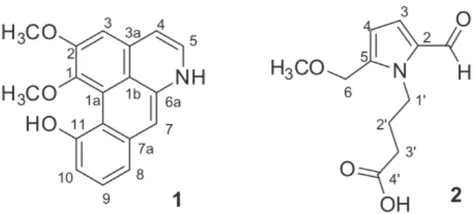
Our continuing interest in the research on bioactive constituents from this plant led us to investigate bioactive metabolites of A. gramineus rhizomes. A bioassay-guided fractionation and chemical investigation of its MeOH extract resulted in the isolation and identification of two alkaloids, including a new aporphine-type alkaloid (1), named gramichunosin, and a known pyrrole alkaloid (2). This report will discuss the isolation and structural elucidation of the two alkaloids from A. gramineus rhizomes (Figure 1), as well as their antiproliferative activities against A549, SK-OV-3, SK-MEL-2 and HCT-15 cell lines and their inhibitory effects on NO production in a lipopolysaccharide (LPS)-activated BV2 cell line.
Experimental
General experimental procedures
Melting point was determined on a Gallenkamp melting point apparatus and is uncorrected. Infrared (IR) spectra were recorded on a Bruker IFS-66/S FT-IR spectrometer (Bruker, Karlsruhe, Germany). UV spectra were recorded with a Shimadzu UV-1601 UV-Vis spectrophotometer (Shimadzu, Tokyo, Japan). Electrospray ionization mass spectrometry (ESIMS) and high resolution electrospray ionization mass spectrometry (ESI-MS) (HR-ESIMS) spectra were recorded on a Micromass QTOF2-MS (MicroMass, Waters, Milford, MA, USA). Nuclear magnetic resonance (NMR) spectra, including 1H-1H COSY, HMQC, and HMBC experiments,
were recorded on a Varian UNITY INOVA 500 NMR spectrometer (Varian, Palo Alto, CA, USA) operating at 500 MHz (1H) and 125 MHz (13C), with chemical shifts
given in ppm (d). Semi-preparative high performance liquid chromatography (HPLC) was conducted using a Gilson 306 pump (Gilson, Middleton, WI) with Shodex refractive index detector (Shodex, New York, NY). Silica gel 60 and RP-C18
silica gel (230-400 mesh, Merck, Darmstadt, Germany) were used for column chromatography. The packing material for molecular sieve column chromatography was Sephadex LH-20 (Pharmacia, Uppsala, Sweden). Merck precoated silica gel F254 plates and RP-18 F254s plates
(Merck, Darmstadt, Germany) were used for thin layer chromatography (TLC). Spots were detected on TLC under UV light or by heating after spraying with 10% H2SO4 in
C2H5OH (v/v).
Plant material
The rhizomes of A. gramineus were collected from Jeju Island, Korea, in March 2009, and the plant was identified by one of the authors (K. R. L.). A voucher specimen (SKKU-NPL-0910) has been deposited in the herbarium of the School of Pharmacy, Sungkyunkwan University, Suwon, Korea.
Extraction and isolation
The rhizomes of A. gramineus (15 kg) were extracted with 80% aqueous MeOH at room temperature and filtered. The filtrate was evaporated under vacuum to obtain a MeOH extract (825 g), which was suspended in distilled H2O
(2 L) and successively solvent-partitioned with n-hexane, CHCl3, EtOAc and n-BuOH, yielding 166, 14, 5 and 47 g
of residues, respectively. Each fraction was evaluated for cytotoxicity against A549, SK-OV-3 and SK-MEL-2 cells using a sulforhodamine B (SRB) assay. The n -hexane-soluble and EtOAc--hexane-soluble fractions showed significant cytotoxic activity against tested cancer cell lines. The active fractions also inhibited NO production in LPS-stimulated BV-2 cells. The n-hexane-soluble fraction (62 g) was separated over a silica gel column chromatography with n-hexane-EtOAc (11:1) to yield eight fractions (H1-H8). Fraction H8 (2 g) was applied to RP-C18 silica gel column
chromatography using a solvent system of MeOH-H2O
(1:1) to give six subfractions (H8a-H8f). Fraction H8d (156 mg) was subjected to a Sephadex LH-20 column using a solvent system of CH2Cl2-MeOH (1:1), and
further purified by semi-preparative reversed-phase HPLC using a 250 mm × 10 mm i.d., 10 µm, EconosilRP-18 column (Alltech, Nicholasville, KY, USA) with a solvent system of 100% MeOH (flow rate; 2 mL min−1) to obtain
silica gel column chromatography using a solvent system of MeOH-H2O (1:1) to provide eight fractions (E1-E8).
Fraction E3 (870 mg) was applied to a silica gel column chromatography with CHCl3-MeOH (25:1) to obtain
five subfractions (E3a-E3e). Fraction E3d (83 mg) was subjected to a Sephadex LH-20 column using a solvent system of MeOH-H2O (4:1), and further purified by
semi-preparative reversed-phase HPLC with a solvent system of MeOH-H2O (2:3, flow rate of 2 mL min−1) to furnish
compound 2 (13 mg, tR = 18.0 min).
Gramichunosin (1)
Yellowish powder; m.p. 308-309 °C; UV (MeOH)
λmax/nm (log ε) 392 (1.8), 272 (3.5), 232 (3.1); IR (KBr)
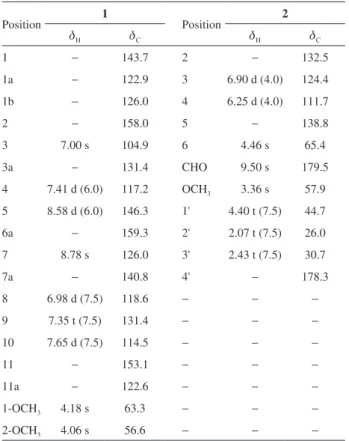
νmax/cm–1 3357, 2945, 2832, 1451, 1116, 1032, 674; 1H (500 MHz) and 13C (125 MHz) NMR data, see Table 1;
ESIMS (positive-ion mode) m/z 294 [M + H]+; HR-ESIMS
(positive-ion mode) m/z 316.0951 [M + Na]+ (calcd. for
C18H15NO3Na, 316.0950).
4-(2-Formyl-5-(methoxymethyl)-1H-pyrrol-1-yl)butanoic acid (2)
Brown gum; UV (MeOH) λmax/nm (log ε) 290 (3.8);
IR (KBr) νmax/cm
–1 3435, 2929, 1733, 1660, 1450, 1352,
1031, 670; 1H (500 MHz) and 13C (125 MHz) NMR data,
see Table 2; ESIMS (positive-ion mode) m/z 226 [M + H]+.
Cell cultures
All tumor cell cultures were maintained using RPMI1640 cell growth medium (Gibco, Carlsbad, CA), supplemented with 5% fetal bovine serum (FBS) (Gibco), 100 units mL−1
penicillin and 100 µg mL−1 streptomycin. Human tumor cell
lines such as A549 (non-small cell lung carcinoma), SK-OV-3 (ovary malignant ascites), SK-MEL-2 (skin melanoma) and HCT-15 (colon adenocarcinoma) were provided by the National Cancer Institute (NCI). Murine microglial BV2 was generously provided by Dr E. Choi from Korea University (Seoul, Korea). It was maintained in Dulbecco’s modified Eagle (DMEM) medium supplemented with 5% FBS, 100 units mL−1 penicillin and 100 µg mL−1 streptomycin.
All cells were incubated at 37 °C in a humidified incubator with 5% CO2.
Cytotoxicity assessment
The cytotoxicity of the compounds against cultured human tumor cell lines was evaluated by the sulforhodamine B (SRB) method.13 Each tumor cell line was inoculated
over standard 96-well flat-bottom microplates and then incubated for 24 h at 37 °C in a humidified atmosphere
of 5% CO2. The attached cells were then incubated with
the serially diluted samples. Control cultures received the carrier solvent (0.1% dimethyl sulfoxide). After continuous exposure to the compounds for 48 h, the culture medium was removed from each well and the cells were fixed with 10% cold trichloroacetic acid at 4 °C for 1 h. After washing with tap water, the cells were stained with 0.4% SRB dye and incubated for 30 min at room temperature. The cells were washed again and then solubilized with 10 mM unbuffered Tris base solution (pH 10.5). The absorbance was measured spectrophotometrically at 520 nm with a microtiter plate reader. Doxorubicin (purity ≥ 98%, Sigma) was used as a positive control. Tested compounds were demonstrated to be pure as evidenced by NMR and HPLC analysis (purity ≥ 95%).
Measurement of NO production
BV-2 cells were plated into a 96-well plate (3 × 10⁴ cells well−1). After 24 h, cells were pretreated
with the compounds for 30 min, and stimulated with 100 ng mL−1 of LPS for another 24 h. Control cultures
received the carrier solvent (0.1% dimethyl sulfoxide). Nitrite, a soluble oxidation product of NO, was measured in the culture media using the Griess reaction. The supernatant (50 µL) was harvested and mixed with an equal volume of Griess reagent (1% sulfanilamide, 0.1% N-1-napthylethylenediamine dihydrochloride in 5% phosphoric acid). After 10 min, the absorbance at 570 nm was measured using a microplate reader. Sodium nitrite was used as a standard to calculate the NO2− concentration.
NG-monomethyl-L-arginine (NMMA, Sigma, St. Louis,
MO, USA), a well-known NO synthase inhibitor, was tested as a positive control.14
Results and Discussion
The MeOH extract of A. gramineus rhizomes was subjected to liquid-liquid solvent-partitioning to yield n-hexane, CHCl3, EtOAc and n-BuOH soluble
factions. Among them, the active n-hexane-soluble and EtOAc-soluble fractions were further separated by silica gel or Sephadex LH-20 column chromatography, and subsequent HPLC to give two alkaloids (1 and 2). To the best of our knowledge, the presence of alkaloids is reported from A. gramineus for the first time in this study.
Compound 1, obtained as yellowish powder, gave a positive test with Dragendorff’s reagent. The molecular formula was determined to be C18H15NO3 from the
[M + Na]+ peak at m/z 316.0951 (calcd. for C
18H15NO3Na,
indicated that 1 possessed hydroxyl (3357 cm−1) and
aromatic (1451 cm−1) groups. The UV spectrum exhibited
absorption bands at λmax 232, 272 and 392 nm, suggesting
the presence of the conjugated double-bond system. The
1H NMR spectral data (Table 1)showedthe presence of
seven protons consisting of two vicinal coupled aromatic protons at dH 8.58 (d, J 6.0 Hz) and 7.41 (d, J 6.0 Hz), three
vicinal coupled aromatic protons at dH 7.65 (d, J 7.5 Hz),
7.35 (t, J 7.5 Hz) and 6.98 (d, J 7.5 Hz) and two singlets of uncoupled aromatic protons at dH 8.78 (s) and 7.00 (s). In
addition, two sharp singlets at dH 4.18 (3H, s) and 4.06 (3H,
s), representing aromatic methoxy groups, were observed in the 1H NMR spectrum. The 13C NMR spectrum (Table 1) of 1 showed 18 carbon signals including two methoxy signals (dC 63.3 and 56.6) and five oxygenated or nitrogenated
carbon signals (dC 159.3, 158.0, 153.1, 146.3 and 143.7),
all of which were classified as olefinic or aromatic carbon signals except for two methoxy signals. Overall, the proton and carbon signals in the 1H and 13C NMR data of 1 implied
that compound 1 may be a trioxygenated aporphine-type alkaloid by the aforementioned evidence and reported literatures.15-18
The structure of the aporphine-type skeleton was ultimately elucidated by the interpretation of the 1H-1H
correlated spectroscopy (COSY), heteronuclear multiple quantum correlation (HMQC), and heteronuclear multiple bond correlation (HMBC) data. The HMQC spectrum permitted the correlation of all protonated carbons. The HMBC data was crucial to confirming the structural assignments and correctly positing the methoxy and hydroxy groups and all non-protonated carbons. The COSY spectrum showed two different spin systems of H-4/H-5 and H-8/H-9/H-10 by their COSY correlations (Figure 2). The down-field shifted proton at H-5 (dH 8.58)
suggested that the proton is adjacent to nitrogen atom, while the HMBC cross-peaks of 3a and H-5/C-6a as well as H-3/C-1, H-3/C-2, H-3/C-1b, H-4/C-3 and H-4/C-1b allowed us to establish the partial structure from C-1 to C-6a (Figure 2). Particularly, the positions of two methoxy groups were confirmed to be at C-1 and C-2 by the HMBC correlations between 1-OCH3 (dH 4.18)
and C-1 (dC 143.7) and between 2-OCH3 (dH 4.06)
and C-2 (dC 158.0), respectively. In addition, the other
remaining structure was elucidated by the H-8/H-9/H-10 COSY system and the HMBC correlations of H-7/C-1b, H-7C-8, H-7/C-11a, H-8/C-11a, H-9/C-7a, H-9/C-11, H-10/C-8 and H-10/H-11a (Figure 2). Finally, even though HMBC correlation between C-1a and any protons was not observed, the connectivity of C-1-C-1a-C-11a was clearly deduced based on the molecular formula after connecting other partial structures. Thus, the gross
structure of 1 was established as shown in Figure 1 and named gramichunosin. To our knowledge, an aporphine alkaloid with similar structural feature was isolated from the stem bark of Enantia chlorantha, and identified as 6a,7-dehydro-1,2-dimethoxy-7-hydroxyaporphine.19 It
shares 6a,7-dehydro-1,2-dimethoxy-aporphine skeleton, whereas it has hydroxyl group at C-7 and saturation at C-4/C-5.
Compound 2 was obtained as a brown gum. The molecular formula was determined as C11H15NO4 from
the molecular ion peak [M + H]+ at m/z 226 [M + H]+ in
the ESIMS. The IR spectrum exhibited absorptions of
Table 1.1H and 13C NMR data of compounds 1 and 2 inCDCl 3a
Position 1 Position 2
dH dC dH dC
1 − 143.7 2 − 132.5
1a − 122.9 3 6.90 d (4.0) 124.4
1b − 126.0 4 6.25 d (4.0) 111.7
2 − 158.0 5 − 138.8
3 7.00 s 104.9 6 4.46 s 65.4
3a − 131.4 CHO 9.50 s 179.5
4 7.41 d (6.0) 117.2 OCH3 3.36 s 57.9
5 8.58 d (6.0) 146.3 1' 4.40 t (7.5) 44.7
6a − 159.3 2' 2.07 t (7.5) 26.0
7 8.78 s 126.0 3' 2.43 t (7.5) 30.7
7a − 140.8 4' − 178.3
8 6.98 d (7.5) 118.6 − − −
9 7.35 t (7.5) 131.4 − − −
10 7.65 d (7.5) 114.5 − − −
11 − 153.1 − − −
11a − 122.6 − − −
1-OCH3 4.18 s 63.3 − − −
2-OCH3 4.06 s 56.6 − − −
a1H and 13C NMR data were recorded at 500 and 125 MHz, respectively. Coupling constants (in Hz) are given in parentheses.
carboxylic acid (1733 cm−1) and aldehyde (1660 cm−1)
groups. Compound 2 had a UV spectrum characteristic of pyrrole-2-aldehyde with an absorption maximum at 290 nm.20 Inspection of the 1H chemical shifts and
coupling constants of signals at dH 6.90 (d, J 4.0 Hz) and
6.25 (d, J 4.0 Hz) implied the presence of a heterocyclic ring containing a nitrogen atom and the coupling constant of 4.0 Hz indicated 2,5-disubstituted pyrrole ring.21 The
characteristic signal at dH 9.50 in the 1H NMR spectrum
and at dC 179.5 in the
13C NMR spectrum (Table 1) were
indicative of an aldehyde, and a singlet signal at dH 4.46
(dC 65.4) was assigned to an oxygenated methylene group.
The partial structure of 2,5-disubstituted pyrrole ring was established by the analysis of the 1H-1H COSY and HMBC
spectroscopic data (Figure 3), and the location of a methoxy group was confirmed to be C-6 by the HMBC correlation between the methoxy proton (dH 3.36) and C-6 (dC 65.4).
A butanoic acid unit was also assigned as a side chain by the COSY correlations from H-1' to H-3' and HMBC cross peaks of H-2'/C-4 and H-3'/C-4 (Figure 3). Finally, the connectivity of the side chain to the pyrrole ring was suggested by the observation of HMBC correlations from H-1' to C-2 and C-5. According to these pieces of evidence, the structure of 2 was elucidated to be 4-(2-formyl-5-(methoxymethyl)-1H-pyrrol-1-yl)butanoic acid. As a result of the literature survey, compound 2 was already reported from Lycium chinense fruits.21 However, the reported 13C NMR data did not match completely with our 13C NMR
assignments, even though the NMR data were obtained in the same solvent (CDCl3). In particular, most of
13C NMR
data of 2 were not much different from the reported data in the difference-range of 0.1-0.4 ppm, whereas the 13C
chemical shift of C-5 (dC 138.8) was obviously different
(DdC-5 −5.1 ppm) and that of C-4' was also slightly different
(DdC-4' +2.3 ppm). Thus, we suggest that the assignments
of 13C NMR for 2 should be corrected as the data we are
publishing in this study.
The antiproliferative activities of the alkaloids 1-2
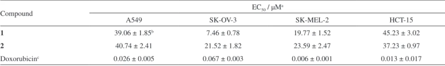
were evaluated by determining their inhibitory effects on four human tumor cell lines, namely A549 (non-small cell lung carcinoma), SK-OV-3 (ovary malignant ascites), SK-MEL-2 (skin melanoma) and HCT-15 (colon adenocarcinoma) using the SRB bioassay.13 The
results (Table 2) showed that compounds 1 and 2 showed moderate antiproliferative activities against SK-OV-3 and SK-MEL-2 cell lines with EC50 values ranging from 7.46
to 23.59 µM. Moreover, they showed weak inhibitory activity against the proliferation of the A549 and HCT-15 cell lines with EC50 values in the range of 37.23-45.23 µM
(Table 2).
Next, the anti-neuroinflammatory activities of the compounds 1 and 2 were evaluated by measuring the produced NO levels in LPS-activated microglia BV-2 cells. Microglia cells, the unique population of the central nervous system resident immune cells, have been implicated in the pathogenesis of a variety of neurodegenerative diseases including Parkinson’s disease and Alzheimer’s disease.22,23 Excessive production of NO
and proinflammatory cytokines from activated microglia play an important role in neurodegenerative disorders. Therefore, regulation of the microglial activation will be an important strategy to prevent the progressive damage of the neurodegenerative diseases. In this test, compound
1 significantly inhibited LPS-stimulated NO production with IC50 values of 7.83 µM, which displayed more
activity than a positive control, NG-nonomethyl-L-arginine
(L-NMMA; a well-known NOS inhibitor, IC50 value of
14.27 µM). However, compound 2 had no influence on NO levels in the medium (IC50 > 200 µM). The isolates 1-2 had no influence on cell viability (data not shown) at concentrations up to 20 µM.
Table 2. Antiproliferative activities of compounds 1 and 2 isolated from A. gramineus
Compound EC50 / µM
a
A549 SK-OV-3 SK-MEL-2 HCT-15
1 39.06 ± 1.85b 7.46 ± 0.78 19.77 ± 1.52 45.23 ± 3.02
2 40.74 ± 2.41 21.52 ± 1.82 23.59 ± 2.47 37.23 ± 0.97
Doxorubicinc 0.026 ± 0.005 0.067 ± 0.003 0.006 ± 0.001 0.013 ± 0.017
aEC
50 value of compounds against each cancer cell line. EC50 value was defined as concentration (µM) causing 50% inhibition of cell growth in vitro; bdata are expressed as mean ± SEM of three independent experiments; cdoxorubicin as a positive control.
Conclusions
The bioassay-guided fractionation and chemical investigation of A. gramineus rhizomes resulted in the isolation and identification of two alkaloids, including a new aporphine-type alkaloid, gramichunosin (1). The corrected assignments in 13C NMR of compound 2 were
also suggested in this study. To our knowledge, this is the first report to investigate alkaloidal compounds from A. gramineus. The isolated alkaloids displayed antiproliferative activities by suppressing the survival of human cancer cells (A549, SK-OV-3, SK-MEL-2 and HCT-15). Particularly, compound 1 exhibited significant anti-neuroinflammatory activity by inhibiting the NO production in LPS-stimulated microglial BV-2. These results suggest that compound 1 can be applied as the key compound in the drug development for various cancers and neuroinflammatory diseases. In addition, its anti-neuroinflammatory activity supports the folk usage of A. gramineus in the treatment of cognitive disorders, and it is possible that compound 1 may be one of the active ingredients responsible for the cognitive improvement.
Supplementary Information
Supplementary data are available free of charge at http://jbcs.sbq.org.br as a PDF file.
Acknowledgments
This work was supported by the KIST Institutional Program (Project No. 2Z04210-14-124).We are thankful to the Korea Basic Science Institute (KBSI) for the measurements of NMR and MS spectra.
References
1. Fattorusso, E.; Taglialatela-Scafati, O.; Modern Alkaloids: Structure, Isolation, Synthesis, and Biology; Wiley-VCH: Weinheim, 2008.
2. Souto, A. L.; Tavares, J. F.; Silva, M. S.; Diniz, M. F. F. M.; Athayde-Filho, P. F.; Barbosa-Filho, J. M.; Molecules 2011, 16, 8515.
3. Lu, J. J.; Bao, J. L.; Chen, X. P.; Huang, M.; Wang, Y. T.; Evid. Based Complement. Alternat. Med. 2012, 2012, 485042.
4. Liao, J. F.; Huang, S. Y.; Jan, Y. M.; Yu, L. L.; Chen, C. F.; J. Ethnopharmacol. 1998, 61, 185.
5. Tang, W.; Eisenbrand, G.; Chinese Drugs of Plant Origin; Springer: New York, 1992.
6. Wang, H. Z.; Cheng, Y. G.; Fan, C. S.; Acta Bot. Yunnanica
1998, 5, 96.
7. Greca, M. D.; Monaco, P.; Previtera, L.; Aliotta, G.; Pinto, G.; Pollio, A.; Phytochemistry 1989, 28, 2319.
8. Lee, J. Y.; Lee, J. Y.; Yun, B. S.; Hwang, B. K.; J. Agric. Food Chem. 2004, 52, 776.
9. Perrett, S.; Whitfield, P. J.; Phytother. Res. 1995, 9, 405. 10. McGaw, L. J.; Jager, A. K.; van Staden, J.; S. Afr. J. Bot. 2002,
68, 31.
11. Kim, K. H.; Kim, H. K.; Choi, S. U.; Moon, E.; Kim, S. Y.; Lee, K. R.; J. Nat. Prod. 2011, 74, 2187.
12. Kim, K. H.; Moon, E.; Kim, H. K.; Oh, J. Y.; Kim, S. Y.; Choi, S. U.; Lee, K. R.; Bioorg. Med. Chem. Lett. 2012, 22, 6155. 13. Skehan, P.; Storeng, R.; Scudiero, D.; Monks, A.; MaMahon, J.;
Vistica, D.; Warren, J. T.; Bokesch, H.; Kenney, S.; Boyd, M. R.; J. Natl. Cancer Inst. 1990, 82, 1107.
14. Reif, D. W.; McCreedy, S. A.; Arch. Biochem. Biophys. 1995, 1, 170.
15. Priestap, H. A.; Magn. Reson. Chem. 1989, 27, 460. 16. Priestap, H. A.; Phytochemistry 1985, 24, 849.
17. Kim, S. K.; Ryu, S. Y.; No, J.; Choi, S. U.; Kim, Y. S.; Arch. Pharm. Sci. Res. 2001, 24, 518.
18. Kim, K. H.; Piao, C. J.; Choi, S. U.; Son, M. W.; Lee, K. R.; Planta Med. 2010, 76, 1732.
19. Wafo, P.; Nyasse, B.; Fontaine, C.; Sondengam, B. L.; Fitoterapia 1999, 70, 157.
20. Shigagetsu, H.; Shibata, S.; Kurata, T.; Kato, H.; Fujimaki, M.; Agric. Biol. Chem. 1977, 41, 2377.
21. Chin, Y. W.; Lim, S. W.; Kim, S. H.; Shin, D. Y.; Suh, Y. G.; Kim, Y. B.; Kim, Y. C.; Kim, J.; Bioorg. Med. Chem. Lett. 2003, 13, 79.
22. Wilms, H.; Zecca, L.; Rosenstiel, P.; Sievers, J.; Deuschl, G.; Lucius, R.; Curr. Pharm. Des. 2007, 13, 1925.
23. McGeer, P. L.; McGeer, E. G.; Brain Res. Rev. 1995, 21, 195.
Submitted: July 31, 2014