DOI: 10.5562/cca2739
Structural and Magnetic Properties of Iron Oxide
Nanoparticles in Shells of Hollow Microcapsules
Designed for Biomedical Applications
I. S. Lyubutin,1 S. S. Starchikov,1,* Chun-Rong Lin,2,# N. E. Gervits,1 N. Yu. Korotkov,1 T. V. Bukreeva1,3
1 Shubnikov Institute of Crystallography, Russian Academy of Sciences, Moscow 119333, Russia 2 Department of Applied Physics, National Pingtung University, Pingtung County 90003, Taiwan 3 National Research Center „Kurchatov Institute“, pl. Akademika Kurchatova 1, Moscow 123182, Russia
* Corresponding author’s e-mail address: [email protected]
# Corresponding author’s e-mail address: [email protected]
RECEIVED: September 12, 2015 ACCEPTED: October 13, 2015
THIS PAPER IS DEDICATED TO DR.SVETOZAR MUSIĆ ON THE OCCASION OF HIS 70TH BIRTHDAY
Abstract: The functional hollow biodegradable microcapsules modified with the maghemite γ-Fe2O3 nanoparticles and the hollow spherical CoFe2O4/SiO2 nanocomposites were synthesized. Mössbauer spectroscopy data reveal that the main part maghemite nanoparticles have evi-dent superparamagnetic behavior which is retained up to room temperature. This allows directing the microcapsules by an external magnetic field, which is very important for the problem of target drug delivery. On the other hand, the hollow spherical CoFe2O4/SiO2 nanocomposites with the small size particles do not show superparamagnetic behavior, but transit from magnetic to paramagnetic state by jump-like magnetic transition of the first order. This effect is a specific property of the magnetic nanoparticles isolated by an inert material. The method of synthesis can be modified with various bioligands on the silane surface, and such materials can have great prospects for use in diagnostics and bio-separation.
Keywords: nanocomposites, Mössbauer spectroscopy, core-shell spheres, maghemite, cobalt-ferrite, size-dependent magnetic properties, first-order magnetic transition.
1. INTRODUCTION
AGNETIC nanoparticles (MNP) of iron oxides hav-ing unique properties are very promishav-ing in many applications, in particular, their use in biology and medi-cine is of great interest. Targeted drug delivery[1] and theranostics,[2] hyperthermia treatment,[3] contrast me-dia in magnetic resonance imaging,[4] immunoassays,[5] separation and manipulation of biochemical products[6] are the main fields of investigations. The possibility to tune the magnetic properties of iron oxides nanoparticles (Fe3O4, α-Fe2O3, γ-Fe2O3, CoFe2O4) by the specific meth-ods of synthesis is very important, and this is the key fac-tor of practical applications of these materials.
For applications in the field of targeted drug delivery, iron oxides MNPs should be non-toxic, chemical stable,
biocompatible with cells and tissues of body. MNPs are often used to control and deliver the biodegradable pol-ymeric microcapsules with drug to the cancer tumor. In order to manipulate the microcapsule, MNPs must be able to be controlled by magnetic field and not aggre-gate after releasing the drug. This is possible if MNPs show the superparamagnetic behavior. The superpara-magnetic MNPs reveal zero value of coercitivity and can have high value of saturation magnetization. This allows MNPs to reply on the external magnetic field and do not aggregate after field is removed. The properties of na-noparticles depend on their morphology, agglomera-tion, surface structure, interparticle interactions and interaction with environment media. These factors can significantly influence the functional properties of the material.
Croat. Chem. Acta2015, 88(4), 397–403 DOI: 10.5562/cca2739 In this paper two types of microcapsules modified by
γ-Fe2O3 and CoFe2O4 magnetic nanoparticles were synthe-sized and investigated. Along with magnetic measure-ments, XRD, SEM, TEM and Mössbauer spectroscopy were used to characterize the structural and magnetic properties of the nanoparticles.
2. PREPARATION OF THE SAMPLES
In the first series, hollow microcapsules with the shell com-posed of biodegradable polyelectrolytes modified with the Fe3O4/Fe2O3 nanoparticles were fabricated by in situ syn-thesis. Firstly, the spherical CaCO3 particles with diameter of about 5 μm were prepared as the core templates for mi-crocapsules. Polyelectrolyte capsules were fabricated by layer-by-layer deposition technique.[7] To obtain hollow capsules, the CaCO3 core was decomposed by EDTA (triso-dium salt of ethylenediamine tetraacetic acid). Then, the in situ synthesis of magnetic nanoparticles in a suspension of polyelectrolyte capsules was performed by chemical con-densation of Fe3+ and Fe2+ ions (FeCl3 and FeCl2) on capsule shells by adding a base (the Elmore method).[8] The details of this process could be find in.[9]
In the second series, the methyl methacrylate (MMA) and methacrylic acid (MAA) microspheres were used as a core template to prepare the magnetic core-shell nano-composites of CoFe2O4/SiO2. The CoFe2O4/poly(MMA-co-MAA) core-shell particles were prepared by adding FeCl3 and CoCl2 into the poly(MMA-co-MAA) latex and stirred for 4 h. Tetraethyl orthosilicate (TEOS) was used to cover these particles and obtain the SiO2/CoFe2O4/poly(MMA-co-MAA) composite spheres. The hollow spheres with shell consisting of SiO2/CoFe2O4 nanocomposites were obtained after annealing of SiO2/CoFe2O4/poly(MMA-co-MAA) spheres in air at temperatures TAbetween 300 and 900 °C
for 4 h. During the calcination, the core MMA-co-MAA microspheres were dissolved.
3. EXPERIMENTAL TECHNIQUES
X-ray diffraction studies of Fe3O4/Fe2O3 nanoparticles were performed on a “Belok” station installed on the synchro-tron source from a bending magnet of the storage ring in the National Research Center “Kurchatov Institute” (NRC-KI), Moscow, Russia.[10] The wavelength was λ = 0.9823 Å. The crystal structure and phase purity of the CoFe2O4 sam-ples were examined by powder X-ray diffraction (XRD) us-ing Mutiflex MF2100, Rigaku Co. Ltd.
The morphology and microstructure of the particles were characterized by scanning (SEM) and high resolution transmission electron microscopy (HRTEM). In case of Fe3O4/Fe2O3 nanoparticles a Tecnai G230ST (FEI, US)
trans-mission/scanning electron microscope (TEM/STEM) oper-ating at an acceleroper-ating voltage of 300 kV was used.[9] Also a Tecnai G2 F20, FEGTEM, Philips Co. Ltd. was used to ana-lyze SiO2/CoFe2O4 nanocomposites.
The Mössbauer absorption spectra from 57Fe nuclei were recorded at temperatures between 10 and 295 K using a standard MS1100Em spectrometer operating in the constant acceleration mode. The gamma-ray source 57Co(Rh) was at room temperature. The closed-cycled he-lium cryostat was used for the low temperature Mössbauer measurements.[11] Isomer shifts were measured relative to the reference α-Fe sample (18-μm-thick iron foil annealed in hydrogen) at room temperature. Computer processing of spectra was carried out using Univem MS software.
4. EXPERIMENTAL RESULTS
4.1. Sample Characterization by XRD and
Electron Microscopy.
X-ray diffraction patterns of microcapsules modified by Fe3O4/Fe2O3 nanoparticles and CoFe2O4/SiO2 composite recorded after annealed the samples at different tempera-tures TA are shown in Figure 1a and Figure 1b.
The Rietveld method was used for the crystal struc-ture refinement of iron oxide Fe3O4/Fe2O3 nanoparticles. For the peak interpolation, the symmetric pseudo-Voigt function was applied. The Bragg R-factor RBr = 2.1 % was ob-tained. All reflections in Figure 1a correspond to the cubic spinel-type structure (sp. gr. Fd m3 ) and the calculated unit cell parameter is a = 8.3545(3) Å. Assuming the spherical shape of the particles we estimate the average crystallite size of D = 12(1) nm by Scherrer’s formula of the peak broadening.[12] It is difficult to separate magnetite Fe3O4 and maghemite γ-Fe2O3 phases in XRD pattern because both have a spinel-type crystal structure. In order to verify reliably the iron oxide phase, we carried out Mössbauer spectroscopy measurements which very sensitive for iron valence state (Fe3+, Fe2+).
All peaks in the XRD patterns of CoFe2O4/SiO2 nano-composites (Figure 1b) can be indexed to the Fd m3 space group in the cubic symmetry related to the spinel structure of cobalt ferrite.[13] The peak of SiO2 should be located at about 2Ɵ = 24° but it is not visible due to amorphous state of silica. The calculated unit cell parameter is a = 8.380(1) Å which is closed to nanoscale cobalt ferrites.[13] We calcu-lated an average crystallite size of the coated CoFe2O4 na-noparticles from the width of (311) peak using Scherrer method.[12] The particle size d is about 2.2 and 10.2 nm in the samples annealed at TA = 300 and 900 °C, respectively.
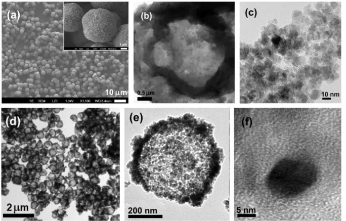
DOI: 10.5562/cca2739 Croat. Chem. Acta2015, 88(4), 397–403 Figure 2a,b,c. The microcapsules have uniform spherical
shape with diameter of about 6.7 μm while the average thickness of the capsule shell is about 0.9 μm (Figure 2b). It was established from the HRTEM data that the average size of the Fe3O4/Fe2O3 nanoparticles in shell of the microcapsules varies from 6 to 9 nm for more than 94 % nanoparticles with an average value of about 7.5 nm (Figure 2c).
The SEM and TEM images of microcapsules consisting
of the CoFe2O4/SiO2 nanocomposites are shown in Figure 2d,e,f. The capsules have spherical shape and uniform size of about 450 nm in diameter (Figure 2d,e). The thickness of the shell consisting of CoFe2O4/SiO2 is about 40–50 nm (Figure 2e). As seen in the HRTEM image of Figure 2f, the size of CoFe2O4 nanoparticles annelid at 900 oC is about 10 nm, which correlates with estimation of the particle size from the XRD data by the Sherrer method.
Figure 1. X-ray diffraction patterns of the Fe3O4/Fe2O3 (a) and CoFe2O4/SiO2 (b) composites in shells of microcapsules. The
reflection indexes correspond to the cubic spinel-type structure (sp. gr. Fd m3 ).
Figure 2. SEM (a) and TEM (b,c) images of the hollow polyelectrolyte microcapsules modified by iron oxides Fe3O4/Fe2O3, and
Croat. Chem. Acta2015, 88(4), 397–403 DOI: 10.5562/cca2739
4.2. Mössbauer Spectroscopy
Measure-ments
4.2.1. IRON OXIDES NANOPARTICLES IN SHELLS OF HOLLOW BIODEGRADABLE POLYELECTROLYTES
MICROCAPSULES
The temperature evolution of Fe57-Mossbauer spectra of Fe3O4/Fe2O3 nanoparticles between 10 and 295 K is showed in Figure3a. Low-temperature spectra indicate that all iron ions are in the magnetically ordered state. These spectra can be fit to two dominating and one small-inten-sive magnetic components A, B, and C, respectively (Figure 3b). The hyperfine parameters for A and B components at 10 K are: isomer shifts δ(A) = 0.41 mm/s, δ(B) = 0.46 mm/s, quadrupole shifts ε(A)= –0.03, ε(B) = 0.01 mm/s, and mag-netic hyperfine fields Hhf(A) = 50.5 T and Hhf(B) = 52.6 T. The
values of isomer shifts are typical of ferric Fe3+ ions, and no ferrous Fe2+ ions were found in the sample. This indicates that magnetite Fe3O4 phase is not present in these nano-particles. Taken into account the XRD data (indicating the spinel structure of these NPs), we have to conclude that the particles have the crystal structure of maghemite γ-Fe2O3. Maghemite and magnetite are iron oxides with the same cubic spinel structure, but their electronic and magnetic properties are different. In the spinel structure, iron ions occupy tetrahedral (A) and octahedral [B] sites, and in the chemical formula of magnetite ferrous ions are located in the octahedral sites (Fe3+)[Fe3+Fe2+]O4. The chemical for-mula of maghemite γ-Fe2O3 can be represented as non-stoichiometric magnetite containing vacancies □ in the octahedral sites (Fe)[Fe5/6□1/6]2O4. All iron ions in ma-ghemite are ferric.
Two main magnetic components in the Mössbauer spectrum of our NPs correspond to the Fe3+ iron ions in tet-rahedral (A) and octahedral [B] sites of maghemite γ-Fe2O3.[14] The intensity ratio of the (A) and [B] Mössbauer components in the vacant γ-Fe2O3 is expected to be near 1 : 1.67. In our case, the observed A/B ratio in nanoparticles is about 1.65 that is very close to the expected value for maghemite. This is an additional support for the ma-ghemite phase present in the shells of the microcapsules obtained.
The third magnetic component (C) in Figure 3b has the isomer shift δ(C) = 0.44 mm/s, ε(C) = –0.06 mm/s, and <Hhf(C)> = 47.0 T. These parameters are also should be
at-tributed to the ferric ions. The lines of this component are much broadened, and its relative area is about 15 %. Obvi-ously the third component (C) corresponds to iron ions on the surface of the nanoparticles.[9]
As temperature increases, at T > 70 K the paramag-netic doublet component appears in the central part of the spectra (Figure 3a). This indicates that some part of the
na-noparticles undergoes the transition from magnetically or-dered to paramagnetic state. The isomer shift of the doublet is δ = 0.43(1) mm/s, the quadrupole splitting Δ = 0.79 (1) mm/s, and the half-line width Г = 0.81(2) mm/s. These values are the typical characteristics of the high spin Fe3+ ions in maghemite.[14–16] The area of the doublet in-creases with increasing temperature. It should be noted that we did not observed remarkable transformations of the spectra shape at temperatures where the Verwey tran-sition occurs in magnetite Fe3O4 (around 120 K). This addi-tionally confirms an absence of magnetite in the sample.
The line broadening increases as temperature rises, and at room temperature the spectrum consists of the overlapping broad magnetic component and paramagnetic doublet (Figure 3b). The parameters of the doublet δ = 0.34(1) mm/s, Δ = 0.70(1) mm/s and Г = 0.61(1) mm/s are closed to nano-maghemite.[14,15] Such behavior of the spec-tra is the signature of superparamagnetic relaxation of the iron magnetic moments. Thus, at room temperature the γ-Fe2O3 nanoparticles reveal superparamagnetism.
4.2.2. HOLLOW MICROCAPSULES MADE OF CoFe2O4 / SiO2 NANOCOMPOSITES
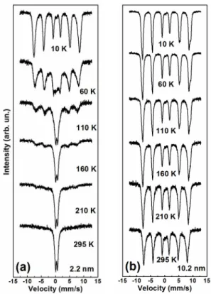
The Fe57-Mossbauer spectra of CoFe2O4/SiO2 nanocompo-sites at temperatures between 10 and 295 K are shown in Figure 4. Well resolved six-line magnetic pattern is clearly seen at low temperatures. However, the evolution of the spectra is different for the samples with different size. The
Figure 3. The Fe57-Mössbauer spectra of the iron oxide
γ-Fe2O3 nanoparticles in shells of microcapsules recorded at
DOI: 10.5562/cca2739 Croat. Chem. Acta2015, 88(4), 397–403 transition from the magnetically ordered state to a
para-magnetic state occurs at about T > 60 K in the samples with the particle size of (2.2–3.4) nm (Figure 4a), whereas a mag-netic order persists for all iron ions in the 10.2 nm sample even above room temperature (Figure 4b).
At 10 K, the spectra of 10.2 nm sample can be fit to several magnetic components corresponding to nonequiv-alent iron ion states in cubic spinel structure of CoFe2O4. The isomer shift values δ in all components are about 0.37– 0.49 mm/s, which are typical for Fe3+ ions. The ferrous ions Fe2+ were not found. Thus, the cobalt ions should be biva-lent Co2+, and the general formula of cobalt ferrite can be given as: (Co2+xFe3+1–x)[Co2+1–xFe3+1+x]O4.
Several nonequivalent iron sites in CoFe2O4 appear due to different number of Fe and Co neighbors in the near-est sublattice. The isomer shift δ and magnetic hyperfine field Hhf values for iron ions in A-site are less than in B-site
due to stronger covalence of the Fe-O bonds in A-site. Thus, we can assign the Mössbauer components to Fe3+ ions in (A)- and [B] sites, and the fraction of Fe3+ ions in tetrahedral and octahedral sublattices can be determined from the ar-eas of the corresponding components. Thus, we found the cation distribution in the 10.2 nm sample in the form: (Co2+0.33Fe3+0.67)[Co2+0.67Fe3+1.33]O4. The inversion degree λ
which is defined as the fraction of (A) sites occupied by tri-valent cations, is of λ = 0.67(1), and this is close to the val-ues obtained in the bulk cobalt ferrite.[18,19]
5. DISCUSSION
Mössbauer spectroscopy is an accurate method to study the superparamagnetic relaxation phenomenon. The time-scale of measurements in Mössbauer spectroscopy is an or-der of nanoseconds, which is much more less than in DC magnetization measurements and applying of the external magnetic field is not necessary. This allows investigating fast spin relaxation processes in nanoparticles caused by the thermal energy. The time of thermal fluctuation τ of magnetic moments among the various easy directions of magnetization is given by well-known formula:[20]
τ = τ0exp(V K/kBT) (1)
where τ0 is the material constant (~10–9 s),[21]K is the
ani-sotropy constant, kB is the Boltzmann constant, V is the
vol-ume of nanoparticles, and T is the temperature. The magnetization is stable below the spin-blocking tempera-ture Tb, and the thermal equilibrium state of the assembly
of uniaxial single-domain nanoparticles can be given in the form:[21]
VK = 2.3 kBTb , (2)
From the temperature dependence of the area of paramagnetic component we estimated the Tb values as ≈
90 and ≈ 250 K for the γ-Fe2O3 nanoparticles with size of d
≤ 5 and 6 nm, respectively. Using the values of V and Tb
ob-tained in our experiments we estimated the anisotropy constants K = 4 × 10⁵ erg/сm3 and 7 × 10⁵ erg/сm3 for the particles with the sizes of 5 and 6 nm, respectively. These data show that the surface anisotropy (which relative con-tribution to the total anisotropy in 5 nm particles is higher than in 6 nm particles) is less than the core particle anisot-ropy. This can be attributed to coating of the nanoparticles by nonmagnetic polyelectrolyte media.
Contrary to the γ-Fe2O3 nanoparticles in shells of bi-odegradable polyelectrolytes microcapsules, the CoFe2O4 particles covered by amorphous silica SiO2 do not reveal su-perparamagnetic behavior even in very small samples of (2.2–3.4) nm. At temperature increasing from 10 K, the area of magnetic six-linear component starts to decrease at about T > 60 K due to the appearance of the paramagnetic doublet (Figure 4a), meanwhile, the line width remains un-changed, and the shape of the spectra does not show relax-ation broadening typical of superparamagnetism. The average values of the magnetic hyperfine field Hhf also did not change significantly. The magnetic sextet directly
Figure 4. Temperature evolution of the Fe57-Mössbauer
spectra between 10 and 295 K in the CoFe2O4 / SiO2 samples
Croat. Chem. Acta2015, 88(4), 397–403 DOI: 10.5562/cca2739 transits into a paramagnetic doublet without relaxation
phenomena typical of superparamagnetic behavior. This indicates that in small particles (2.2, 2.4 and 3.4 nm), a cer-tain part of magnetic Fe ions transits to the paramagnetic state by the jump-like first order magnetic transition (JMT) without an intermediate relaxation state.
Such a transition was discovered previously in ferri-hydrite (Fe5HO8 ∙ 4H2O) nanoparticles with the size of 2 nm; located in the copolymer pores,[22] and in nanocomposites of α-Fe2O3/SiO2, in which the 2–4 nm size nanoparticles of hematite α-Fe2O3 were located in the amorphous silica channels.[23] In both cases, the magnetic nanoparticles were isolated by inert material, which is similar to our CoFe2O4/SiO2 samples.
Thermodynamical analysis indicated[22] that such a transition can be initiated by internal pressure created in the surface layer of the particle. The temperature of the transition depends on many parameters such as the parti-cle size, degree of isolation of the partiparti-cles from each other, the particle aggregations in nanoclusters, and intercluster interaction. The interaction between the surface Fe3+ ions and the wall of the coating material can influences the sur-face tension and pressure, thus changing the value of the magnetic transition temperature. Spread of values of the transition temperature can also be explained by the inter-cluster interactions.[23] The covered material can induce pressure on the nanoparticles and this effect can shift the transition temperature. Considering the size distribution of CoFe2O4 nanoparticles one can expect a gradual transition from magnetically ordered to paramagnetic state.
6. CONCLUSION
The functional hollow biodegradable microcapsules modi-fied with the iron oxide nanoparticles in shells and the hol-low spherical CoFe2O4/SiO2 nanocomposites were synthesized.
Mössbauer spectroscopy data revealed that the iron oxide nanoparticles in shells of the capsules have the crys-tal structure of maghemite γ-Fe2O3. The most important data reveals that approximately 80 % of all maghemite na-noparticles with the size of 7–9 nm have evident superpar-amagnetic behavior which is retained up to room temperature due to slow spin relaxation. This allows direct-ing the microcapsules to a place determined by an external magnetic field, which is very important for the problem of target drug delivery. Mild conditions in the synthesis of magnetic nanoparticles incorporated in the microcapsules may enable encapsulating bioactive substances without loss of its biological activity.
On the other hand, the hollow spherical CoFe2O4/SiO2 nanocomposites with the small size of the
CoFe2O4 particles (2.2, 2.4 and 3.4 nm) do not show super-paramagnetic behavior, but transit from magnetic to para-magnetic state by jump-like para-magnetic transition of the first order. This effect is a specific property of the magnetic na-noparticles isolated by an inert material. Such a transition is related to the surface effects, and in particular, can be initiated by internal pressure creating in a surface layer of the particle. The method of synthesis can be modified with various bio-ligands on the silane surface in order to cova-lently attach the specific bioligands to the surfaces of the hollow magnetic spheres. Such materials can have great prospects for use in diagnostics and bio-separation.
Acknowledgements. Support by the Russian Scientific Founda-tion (Project #14-12-00848) is acknowledged.
REFERENCES
[1] H.-S. Cho, Z. Dong, G. M. Pauletti, J. Zhang, H. Xu, H. Gu, L. Wang, R. C. Ewing, C. Huth, F. Wang, D. Shi,
ACS Nano 2010, 4(9), 5398.
[2] Z. Li, J. C. Barnes, A. Bosoy, J. F. Stoddart, J. I. Zink,
Chem. Soc. Rev.2012, 41(7), 2590.
[3] S. Mornet, S. Vasseur, F. Grasset, E. Duguet, J Mater Chem 2004, 14(14), 2161.
[4] F. Hu, K. W. MacRenaris, E. A. Waters, E. A. Schultz-Sikma, A. L. Eckermann, T. J. Meade, Chem. Commun.
2010, 46(1), 73.
[5] H. Nakayama, A. Arakaki, K. Maruyama, H. Takeyama, T. Matsunaga, Biotechnol. Bioeng.2003, 84(1), 96. [6] J. Ugelstad, A. Berge, T. Ellingsen, R. Schmid, T.-N.
Nilsen, P. C. Mørk, P. Stenstad, E. Hornes, Ø. Olsvik,
Prog. Polym. Sci.1992, 17(1), 87.
[7] G. B. Sukhorukov, E. Donath, H. Lichtenfeld, E. Knippel, M. Knippel, A. Budde, H. Möhwald, Colloids Surf. Physicochem. Eng. Asp.1998, 137(1–3), 253. [8] W. C. Elmore, Phys. Rev.1938, 54(4), 309.
[9] I. S. Lyubutin, S. S. Starchikov, T. V. Bukreeva, I. A. Lysenko, S. N. Sulyanov, N. Y. Korotkov, S. S. Rumyantseva, I. V. Marchenko, K. O. Funtov, A. L. Vasiliev, Mater. Sci. Eng. C 2014, 45, 225.
[10] D. M. Kheiker, M. V. Kovalchuk, Y. N. Shilin, V. A. Shishkov, S. N. Sulyanov, P. V. Dorovatovskiĭ, A. A. Rusakov, Crystallogr. Rep.2007, 52(2), 358. [11] P. G. Naumov, I. S. Lyubutin, K. V. Frolov, E. I.
Demikhov, Instrum. Exp. Tech.2010, 53(5), 770. [12] J. I. Langford, A. J. C. Wilson, J. Appl. Crystallogr.
1978, 11(2), 102.
[13] L. Kumar, P. Kumar, A. Narayan, M. Kar, Int. Nano Lett.2013, 3(1), 1.
[14] A. G. Roca, J. F. Marco, M. del P. Morales, C. J. Serna,
DOI: 10.5562/cca2739 Croat. Chem. Acta2015, 88(4), 397–403 [15] K. Závěta, A. Lančok, M. Maryško, E. Pollert, D.
Horák, Czechoslov. J. Phys.2006, 56(3), E83. [16] G. M. Da Costa, E. De Grave, L. H. Bowen, R. E.
Vandenberghe, P. M. A. De Bakker, Clays Clay Miner.
1994, 42(5), 628.
[17] G. A. Sawatzky, F. Van Der Woude, A. H. Morrish,
Phys. Rev.1969, 187(2), 747.
[18] T. A. S. Ferreira, J. C. Waerenborgh, M. H. R. M. Mendonça, M. R. Nunes, F. M. Costa, Solid State Sci.
2003, 5(2), 383.
[19] G. A. Sawatzky, F. V. D. Woude, A. H. Morrish, J. Appl. Phys.1968, 39(2), 1204.
[20] L. Neel, Ann. Geophys.1949, 5, 99.
[21] B. D. Cullity, Introduction to magnetic materials. Addison-Wesley Pub. Co., 1972.
[22] I. P. Suzdalev, V. N. Buravtsev, V. K. Imshennik, Y. V. Maksimov, V. V. Matveev, S. V. Novichikhin, A. X. Trautwein, H. Winkler, Z. Für Phys. At. Mol. Clust.
2014, 37(1), 55.