DEPARTAMENTO DE QUÍMICA ANALÍTICA E FISICO-QUÍMICA PROGRAMA DE PÓS-GRADUAÇÃO EM QUÍMICA
DAVINO MACHADO ANDRADE NETO
MAGNETIC RESSONANCE IMAGING CONTRAST AGENTS OBTAINED BY FAST SONOCHEMISTRY APPROACH
MAGNETIC RESSONANCE IMAGING CONTRAST AGENTS OBTAINED BY FAST
SONOCHEMISTRY APPROACH
Dissertação apresentada ao Programa de Pós-Graduação em Química da Universidade Federal do Ceará, como requisito parcial à obtenção do título de mestre em química. Área de concentração: química.
Orientador: Prof. Dr. Pierre Basílio Almeida Fechine.
A566m Andrade Neto, Davino Machado.
Magnetic ressonance imaging contrast agents obtained by fast sonochemistry approach / Davino Machado Andrade Neto. – 2016.
84 f. : il. color.
Dissertação (mestrado) – Universidade Federal do Ceará, Centro de Ciências, Programa de Pós-Graduação em Química, Fortaleza, 2016.
Orientação: Prof. Dr. Pierre Basílio Almeida Fechine.
1. Sonochemistry. 2. Magnetic nanoparticles. 3. Functionalization of nanomaterials. 4. Magnetic resonance imaging. I. Título.
À Universidade Federal do Ceará pela infraestrutura oferecida para que o trabalho fosse realizado.
Aos membros participantes da Banca examinadora, Profs. Amauri Jardim e Igor Frota.
A todos os professores que contribuíram para a minha formação profissional desde a graduação. Destaque à professora Izaura Cirino, Ronaldo Nascimento, Francisco Belmino, Nágila Ricardo, Nádja Ricardo e Otília Deusdênio.
À professora Selma Mazzeto por todo o apoio que foi dado, principalmente nos momentos de maior dificuldade e necessidade.
A meu amigo Wanderson Moreira, pelas análises termogravimetria.
Ao Laboratório de Raios-X da UFC pelas análises de difração de raio-X. Agradecimento especial à aluna Ana Cláudia Abreu pelas análises feitas em momentos de maior necessidade.
Ao professor Giuseppe Mele, pelas análises de microscopia eletrônica de transmissão.
Ao professor Igor Frota, pelas análises Mössbauer feitas de forma bastante rápidas e em momentos de grande necessidade.
Ao Danilo Queiroz e Carol Moura pelas análises de relaxatividade.
Ao Manuel Banobre, pela tremenda ajuda nas análises magnéticas e de relaxatividade e pela contribuição científica ao trabalho.
Ao Laboratório Internacional Ibérico de Nanotecnologia, pelas análises de relaxatividade e medidas magnéticas.
Ao Laboratório de Espectroscopia Vibracional e Microscopia, pelas análises de espectroscopia na região do infravermelho. Agradecimento especial aos colegas, Juliene Toméh e Antonio César Honorato, pela ajuda na realização dos experimentos.
Ao professor Odair Pastor, pela disponibilização do equipamento para realizar as medidas de espalhamento dinâmico de luz. Especialmente à colega Laís Helena, pelo treinamento de utilização no referido equipamento.
A todos os amigos, familiares e padrasto, Luciano Fernandes, que sempre me ajudaram e entenderam os momentos de ausência devido ao desenvolvimento desta pesquisa.
A minha namorada, Dayany Barros, pelo apoio e compreensão.
aplicadas como agente de contraste para imagem por ressonância magnética nuclear e sua metodologia de síntese e funcionalização afetam fortemente sua performance in vivo. A
metodologia mais utilizada para a produção dessas NPs funcionalizadas é a decomposição térmica, a qual tem provado ser financeiramente desfavorável, laboriosa além de requisitar longos tempos de execução. Portanto, este trabalho tem como objetivo descrever uma metodologia fácil e rápida, através do método sonoquímico, para a síntese e funcionalização de NPs de Fe3O4 com excelentes propriedades físico-químicas com objetivo de serem
aplicadas como agente de contraste para imagem por ressonância magnética nuclear. Neste trabalho, o método sonoquímico foi usado para produzir, em 12 min, NPs de Fe3O4
funcionalizadas com policrilato de sódio, citrato de sódio, polietilenamina ramificada e oleato de sódio. Difração de raios-X e microscopia eletrônica de transmissão demonstraram que as NPs produzidas são compostas de uma única fase de espinélio inverso de 9-11 nm de diâmetro e uma distribuição de tamanho estreita. Foi confirmado por meio da espectroscopia Mössbauer e medidas magnéticas que as NPs sintetizadas estão em transição para o regime superparamagnético e que possuem excelente valor de magnetização de saturação (59-77 emu/g) para aplicações biomédicas. Espectroscopia de infravermelho por transformada de Fourier provou que a radiação sonoquímica forneceu condições adequadas para que acontecesse uma interação forte entre o núcleo magnéticos e os agentes fucionalizantes. Além disso, experimentos de espalhamento dinâmico de luz confirmaras que as amostras recobertas com moléculas orgânicas hidrofílicas possuem estabilidade coloidal em solventes aquosos. Destaque deve ser dado a nanopartícula magnética recoberta com poliacrilato de sódio, que demonstrou excelente estabilidade coloidal em condições fisiológicas simuladas. Finalmente, as NPs obtidas se mostraram serem promissores candidatos a agentes de contraste. Uma vez que seus valores de relaxatividade transversal foram maiores que os agentes de contraste comerciais e daqueles preparados por outras metodologias sintéticas. Portanto, este trabalho trás um grande avanço no que se refere a preparação de NPs de Fe3O4 funcionalizadas para
aplicações biológicas, uma vez que materiais de alta qualidade foram preparados de forma rápida e fácil.
Palavras-chave: Sonoquímica; Nanopatículas magnéticas, funcionalização de nanomateriais;
Functionalized Fe3O4 nanoparticles (NPs) have emerged as a promising contrast agent for
magnetic resonance imaging (MRI). Their synthesis and functionalization methodology strongly affects their performance in vivo. Although thermal decomposition is the most
commonly used growth methodology reported in the literature, it has proven to be time-consuming, expensive, and laborious. Therefore, this work describes a rapid and facile sonochemical methodology to synthesize and functionalize Fe3O4 NPs with excellent
physico-chemical properties for MRI. In this work, a sonochemistry approach was used to produce, in 12 min, Fe3O4 NPs functionalized with polysodium acrylate (PAANa), trisodium citrate
(CIT), branched polyethylenimine (BPEI), and sodium oleate. X-ray diffraction and transmission electron microscopy demonstrated that the NPs were composed of a single inverse spinel phase with an average diameter of 9–11 nm and a narrow size distribution. It was confirmed by Mössbauer spectroscopy and magnetic measurements that the obtained NPs were transitioning to the superparamagnetic regime and possessed excellent magnetization saturation values (59–77 emu/g). Fourier transform infrared spectroscopy proved that the sonochemistry approach provided conditions that induced a strong interaction between Fe3O4
and the capping agents. Furthermore, dynamic light scattering experiments evidenced that samples coated with PAANa, CIT, and BPEI possess colloidal stability in aqueous solvents. Emphasis must be placed on PAANa-coated NPs, which also presented remarkable colloidal stability under simulated physiological conditions. Finally, the obtained NPs exhibited great potential to be applied as an MRI contrast agent. The transverse relaxativity values of the NPs synthesized in this work (277–439 mM-1s-1) were greater than those of commercial NPs and those prepared using other methodologies. Therefore, this work represents significant progress in the preparation of Fe3O4 NPs, providing a method to prepare high-quality
materials in a rapid, cost-effective, and facile manner.
Keywords: Sonochemistry; Magnetic Nanoparticles; Functionalization of nanomaterials;
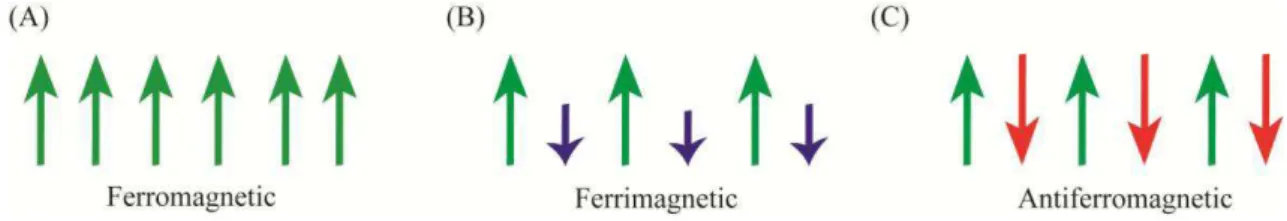
Figure 2 - Magnetic behavior in (A) Ferromagnetic, (B) Ferrimagnetic and (C) Antiferromagnetic ... 18
Figure 3 - Schematic illustration of the particle size effect on: (A) on the domain state and (B) in Hysteresis loops of spinel ferrite materials. Blue and red lines are the first and the second runs, respectively ... 20
Figure 4 - (A) Schematic representation of magnetic drug delivery system acting at cancer infected tissue. (B) Illustration of the synthesis mechanism for a pH-sensitive drug delivery system. (C) TEM images showing the structure before and after the insertion of a CaP shell. (D) Drug profiles realized in solutions with different pH values. ... 22
Figure 5 - (A) TEM image of extremely small Fe3O4 NPs. (B) T1-weighted MR image of cell
pellets after 24 h of incubation with ultra-small MNPs. (C) High-resolution blood pool MR image after intravenous injection of ESIONs (JV = jugular vein, AV = axillary vein, SVC = superior vena cava, IVC = inferior vena cava, CA = carotid artery, SV = subclavian vein, and AA = aortic arch). ... 24
Figure 6 - (A) External magnetic field varied in region of interest. (B) Using an infrared camera, it is possible to see that the heat is concentrated in the extracranial tumor. (C) Animal trial data on hyperthermia treatments in rabbits, showing preferential heating of a tumor using intra-vascularly infused ferromagnetic microspheresμ ( ■) tumor edge, (♦) tumor centre, (▲)
normal liver 1–2 cm from tumor, (×) alternative lobe, and (◊) core body temperature. ... 25
Figure 7 - Schematic showing in situ co-precipitation synthesis process for MNPs in polymer
chains. ... 27
Figure 8 - Schematic illustration of shape evolution of hematite nanostructures at different reaction times and different ferric concentrations. ... 29
Figure 9 - Schematic illustration of LaMer model. ... 30
Figure 12– (A) Timeline of synthesis and functionalization of MNPs, sequence for addition of reactants, and proposed mechanism of sonochemical synthesis and functionalization of MNPs. (B) Magneto-fluid response of the samples herein synthesized. ... 46
Figure 13 – XRD patterns of obtained NPs. The black dots and red line show the experimental and calculated data, respectively. The green line shows the difference between these two parameters. ... 47
Figure 14 – TEM micrographs and size-distribution curves of (A) Fe3O4@PAANa, (B)
Fe3O4@BPEI, (C) Fe3O4@CIT, and (D) Fe3O4@OLNa samples. ... 49
Figure 15 – (A) MS spectra of synthesized samples. The black dots and red lines represent the experimental and calculated data, respectively. Furthermore, for the Fe3O4@OLNa sample,
the green and blue lines are the site distributions for the populations of NPs under ferri- and superparamagnetic regimes, respectively. (B) The image shows the hyperfine field distribution curves of each sample. The table at the top of the image summarizes the data extracted from the site distribution. Duple: doublet; sex: sextet; IS: isomer shift; B: magnetic hyperfine field; wid: line width. ... 51
Figure 16 – ZFC and FC magnetization curves for functionalized NPs prepared in this work.52
Figure 17 – VSM curves of (A) Fe3O4@PAANa, (B) Fe3O4@BPEI, (C) Fe3O4@CIT, and (D)
Fe3O4@OLNa samples; insets: expanded views of low-field regions. ... 53
Figure 18 – FT-IR spectra of (A) Fe3O4@PAANa, (B) Fe3O4@CIT, (C) Fe3O4@OLNa, and
(D) Fe3O4@BPEI samples. For each sample, the spectra of the functionalized NPs and free
CA molecules are shown. ... 55
Figure 19 – Model of interactions between CA molecules and Fe ions of IONPs in (A) Fe3O4@PAANa, (B) Fe3O4@CIT, (C) Fe3O4@OLNa, and (D) Fe3O4@BPEI samples. ... 57
Figure 20 – TGA and DTG data for (A) Fe3O4@PAANa, (B) Fe3O4@CIT, (C) Fe3O4@OLNa,
Table 2 – Parameters extracted from the Rietveld refinement and average size of the NPs calculated through TEM. ... 48
Table 3 – Results from DLS experiments. ... 60
Table 4 - Summary of the already reported MRI contrast agents based on Fe3O4 NPs prepared
1.1. Magnetite (Fe3O4): structure and magnetic properties ... 16
1.1.1 Structure ... 16
1.1.2 Magnetic Properties ... 18
1.2. In Vivo Applications of Fe3O4 ... 21
1.2.1 Drug delivery ... 21
1.2.2 Magnetic resonance imaging (MRI)... 22
1.2.3 Magnetic hyperthermia... 24
1.3. Main Methodologies for Synthesis and Functionalization of MNPs ... 26
1.3.1 Co-precipitation ... 26
1.3.2 Hydrothermal or solvothermal method ... 28
1.3.3 Thermal decomposition ... 29
1.4. Ultrasound: Theory and Application in Synthesis of Fe3O4 ... 31
2. JUSTIFICATION FOR RESEARCH ... 36
3. OBJECTIVES ... 39
3.1 General Objective ... 39
3.2 Specific Objectives ... 39
4. MATERIALS AND METHODS ... 40
4.1 Chemicals ... 40
4.2Synthesis and Functionalization of Fe3O4 with PAANa, BPEI, and CIT ... 40
4.3Functionalization of Fe3O4 NPs with OLNa ... 41
4.4 Characterizations of NPs ... 42
5. RESULTS AND DISCUSSION ... 45
5.2.1 XRD... 47
5.2.2 TEM... 49
5.2.3 Mössbauer spectroscopy... 50
5.2.4 Magnetic measurements ... 52
5.3 Coating ... 55
5.3.1 FTIR ... 55
5.3.2 TGA ... 58
5.4 Colloidal Properties ... 60
1. BIBLIOGRAPHIC REVIEW
1.1 Magnetite (Fe3O4): structure and magnetic properties 1.1.1Structure
Fe3O4 belongs to a huge class of ceramics materials called spinel ferrites. All
oxides of this class posses the same structure of the natural spinel MgAl2O4. According to the
literature(1), over 140 oxides and 80 sulphides were already synthesized and their physicochemical properties studied. This large variety of spinels is due to their capacity to incorporate cations with different charges into the structure. However, the total positive charge should not be higher than 8 to balance to the charge of the anions. Another requirement is about the cation radii. The values must be in the range of 0.4-0.9 Å. Magnetic spinels usually have the general formula of M2+Fe2O4 (or MO.Fe2O3), where the divalent
cation can be Mn, Ni, Fe, Co, Zn, Mg, etc. Fe3O4 is the most important and abundant among
other oxides of this class (2).
The crystalline structure of spinel ferrites was firstly determined by Bragg (3) in 1915. In the spinel structure, the metallic ions are coordinated to oxygen with two different ways, which generate two coordination sites. The first one is called A site and the cation is coordinated in tetrahedral symmetry. The second one, namely B sites, is coordinated in octahedral symmetry as it can be seen in the Figure 1. Spinel space group is Fd3m and the unit cell contains eight units AB2O4 counting 56 ions per unit cell (16 cations in A sites, 32 cations
in B sites and 32 oxygen directly bonded to the cations).
According to distribution of trivalent and divalent cations, spinels can be classified as:
a) Normal Spinel – Divalent cations are present in A sites, while the trivalent
cations are in B sites. For instance, zinc ferrite (ZnFe2O4) presents Zn2+ in
tetrahedral sites (A) and Fe3+in octahedral sites (B). The general formula for normal spinels is (M12+)[Fe3+]2O4, where the square brackets indicate B sites
occupancy and parentheses denote the cation in A sites;
b) Inverse Spinel – Divalent cations are in B sites, while the trivalent cations are in
A and B sites. Fe3O4 is an example of inverse spinel. Half of its Fe3+ is located in
c) Mixed Spinel –In some cases, the divalent cation has no defined preference
between A or B site. Then, it distributes in both sites leading to a formation of
mixed spinel. Its general formula is (M12+1- δFe3+δ)[ M12+δFe3+2- δ]O4, where δ is the degree of inversion. The degree of inversion depends mainly on the preparation technique and synthetic conditions (4). For instance, Galvão et al. (5) evidenced the migration of Fe3+cations among tetrahedral and octahedral sites increasing the microwave heating time.
Figure 1 -Representative structure of Fe3O4
Source: Super-paramagnetic Nanoparticles with Spinel Structure: A Review of Synthesis and Biomedical Applications (6)
1.1.2 Magnetic Properties
Ferrimagnetic materials exhibit a net magnetization similar to the ferromagnetism. The difference between them is the alignment of the spins. For ferromagnetic materials, all spin moments are aligned parallel to each other. Unlike, ferrimagnetic materials display spin moments aligned anti-parallel. However, they are not completely canceled keeping a net magnetic moment. Figure 2. A and B illustrate the alignment of the spins as well as the interactions achieved for ferromagnetism and ferrimagnetism, respectively (1). The remaining magnetism of the ferromagnetic materials can be explained by exchange interactions, which emerge from the presence of oxygen between the cations. In spinel ferrites, the magnetic interactions can occur between the cations occupying A and B sites (A-B), B sites (B-B) and A sites (A-A). It is worth to mention that when these interactions occur the spins for each site become anti-parallel.
Figure 2 - Magnetic behavior in (A) Ferromagnetic, (B) Ferrimagnetic and (C) Antiferromagnetic
Source: Super-paramagnetic Nanoparticles with Spinel Structure: A Review of Synthesis and Biomedical Applications (6)
Usually, net magnetic moment happens with inverse or mixed spinel ferrites where the A-B sites interactions predominate. To illustrate this situation, we can consider inverse spinel Fe3O4. All Fe2+ ions are in B sites and Fe3+ are divided among A and B sites.
Thus, the magnetic moment of Fe3+ ions situated in A and B sites are anti-parallel cancelling
each other. The magnetization found for this ferrite is due to the five unpaired electrons of Fe2+ ions. On the other hand, normal spinels are quite different. The cations in A sites do not present net magnetization and the absence of A-B interactions is observed. As a consequence, B-B interactions dominate. Therefore, the spinel will not present net magnetic moment owing to the moments in B sites. Since an anti-parallel alignment is achieved, therefore, no magnetism is observed. For instance, ZnFe2O4 is a normal spinel and the non-magnetic Zn is
(4). Besides the organization of di- and trivalent cation, the magnetic behavior of the spinel ferrites is also size- and temperature-dependent. These parameters play a very important role. For instance, when size of particles of some inverse-spinel reach a critical value, its magnetic properties at room temperature exhibit the superparamagnetism (SPM). Which brought new possible applications for magnetic nanoparticles (MNPs), especially for the low-toxic Fe3O4.
The difference between of SPM for ferri- and ferromagnetism is that, SPM materials do not display remnant magnetization (Mr). This means that Materials with this
remarkable property exhibit no magnetization upon removal of the external magnetic field (6). Thus, no aggregation due to the remained magnetic field can be found, which enables applications in the biological environment.
The SPM is strongly related to the NPs size. Therefore, the understanding about this phenomenon is necessary to know what magnetically happens for bigger and smaller NPs. There will be few regions of uniform magnetization separated by boundaries (domain walls, Figure 3 (A)) when the NPs with a certain size have multi-domain state. However, more energy is required to create a domain wall when NPs size starts to decrease. Therefore, if a material is reduced below the so-called critical size to single-domain (DSD), the energy
required to hold a domain-wall is greater than support the magnetostatic energy. In this case, it is more energetically favourable for the material to become single-domain (SD) (9). This means that the magnetic moment of each particle will point to the same direction, that is determined by magnetocrystalline anisotropy (Figure 3 (A)) (10). In other words, for a particle to reach the SD state, the size should be greater than the thickness of the domain wall and SD materials differ from each other in relation to the coercive field (Hc), which must be
applied in the negative direction to bring the magnetization of the sample back to zero (9). In SD particles, the magnetization/demagnetization processes are dominated by magnetization rotation which requires relatively high external field. Unlike, in multi-domain state, lower external fields are necessary, once magnetization/demagnetization process is dominated by domain-wall motion (11,12). As a consequence of this fact, materials with single-domain display a larger hysteresis. Therefore, it is possible to observe bigger Mr and Hc values
(Figure 3 (B)). However, continuously decreasing the NPs size below the DSD, there is a
decreasing of the Hc and thermal energy starts to play an important role over relaxation
processes of the spins. A further decrease of the NPs size leads to the formation of a SPM material. This kind of MNPs exhibit Hc and Mr value equal to zero. Thus, no hysteresis is
barrier (anisotropy forces) and the relaxation of the spins happens even in the absence of a magnetic field.
In summary, the SPM behaviour displayed by Fe3O4 NPs brought the possibility
to solve problems in the field of biomedicine, mainly in the treatment of cancer. Besides the SPM, magnetic properties of NPs can be used in vivo in three main ways. The first is
incorporate some anti-cancer drug on the NPs and use an external magnetic field to orientate directly to the infected tissue. Second, it is as magnetic contrast agent in magnetic resonance imaging. Last, as hyperthermia or thermoblation agents, where MNPs, through varying of the magnetic field, heat and kill selectively cancer cells. Thus, scientists around the world start to intensively study which structural and magnetic features of the NPs affects the their in vivo
performance (13).
Figure 3 - Schematic illustration of the particle size effect on: (A) on the domain state and (B) in Hysteresis loops of spinel ferrite materials. Blue and red lines are the first and the second runs, respectively
1.2 In Vivo Applications of Fe3O4 1.2.1 Drug delivery
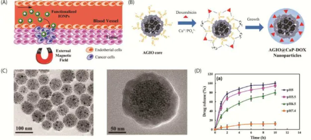
In traditional drug delivery methods such as oral ingestion and intravascular injection, the drug is distributed throughout the body by the systemic bloodstream. Thus, the drug loading must be higher than the required dosage to reach the affected tissue at a sufficient concentration to treat the disease (13). However, using Fe3O4 NPs as a carrier for
some neoplastic agents, along with an external magnetic field, would make it possible to concentrate the medication in the tissue of interest. This would decrease the concentration of the medication in healthy tissues, which would decrease the side effects (Figure 4 (A)). Therefore, magnetic drug delivery systems are promising candidates for the treatment of cancer. The use of this method will prevent the side effects caused by conventional chemotherapy by reducing the systemic distribution of drugs and the doses of cytotoxic compounds (14). Generally, magnetic drug delivery systems are composed of a magnetic core covered by a biocompatible component that acts as a shell. The drugs are usually bound or encapsulated within this shell. In a drug carrier system, the sizes, surface properties, and colloidal stability are crucial features that determine the usefulness of the carrier (15). Therefore, the synthetic methodology for the synthesis of the core, composition of the shell, and appropriate strategy for incorporating the drug play key roles in obtaining a good performance from a magnetic drug carrier.
When the carrier reaches the target site, the drug is released to the cells via an enzymatic activity or a change in a physiological condition such as the pH, osmolality, or temperature. This has allowed the development of smart control release systems that respond to changes in physiological conditions and deliver drugs with a higher specificity. For instance, Li et al. developed a pH-responsive amphiphilic gelatin–iron oxide core/calcium
molecules was clearly observed at acidic pH levels. For example, at pH 5.0, 100% of the DOX was eluted when immersed for 10 h (Figure 4 (D)).
Figure 4 - (A) Schematic representation of magnetic drug delivery system acting at cancer infected tissue. (B) Illustration of the synthesis mechanism for a pH-sensitive drug delivery system. (C) TEM images showing the structure before and after the insertion of a CaP shell. (D) Drug profiles realized in solutions with different pH values.
Source: Recent progress on magnetic iron oxide nanoparticles: synthesis, surface functional strategies, and biomedical applications (13), and In situ doxorubicin–CaP shell formation on amphiphilic gelatin–iron oxide core as a multifunctional drug delivery system with improved cytocompatibility, pH-responsive drug release, and MR imaging (18)
1.2.2 Magnetic resonance imaging (MRI)
MRI is a non-invasive imaging technique used to produce high-quality images of the human body. This technique has been used to make diagnoses for clinical purposes. It is based on the principle of nuclear magnetic resonance. Briefly, 1H, 11B, 13C, 19F, or 31P can be used as the signal source for MRI. However, the water proton (1H) is the most commonly used source for clinical purposes because of its sensitivity and abundance in biological tissues (19). The image formation process begins with spin excitation caused by the application of radiofrequency pulses. When the pulse is removed, the spin returns to its ground state, emitting energy. This process is called relaxation and the MRI time of the process. There are two signals recorded: longitudinal (T1) and transverse (T2). The measured discrepancy
tumor can be easily detected, allowing the patient to be treated. However, in many cases, the contrast difference between the target and normal tissues is negligible, making it harder to obtain the correct diagnosis. To solve this problem, contrast agents were developed to increase the signal difference by improving the contrast between pathological lesions and normal tissues (21). However, the lack of specificity is still a problem for MRI contrast agents. Their poor sensitivity makes their use unfeasible for accurately detecting small targets such as tumors in an early stage of cancer (22). Therefore, MNPs appear to be an excellent alternative because they can be easily functionalized with molecules that selectively bind with cancer cells and can be delivered to the targeted site using an external magnetic field (13). Additionally, their unique superparamagnetic properties generate significant susceptibility effects, which result in strong T2 and T1 values at very low concentrations for MRI (23–25).
Therefore, MRI is one of the most promising applications of magnetic MNPs.
Because the relaxation process is an environment-dependent parameter, the accumulation of MNPs in the target tissue can change the local physiochemical properties and enable the detection of a pathological lesion. Furthermore, the MRI properties of MNPs are dependent on their shape and size. For instance, Young-wook et al. (26) synthesized
water-soluble MNPs with diameters raging from 4 nm to 12 nm (Figure 5(A)). They found that 4, 6, 9, and 12 nm NPs had values of 25, 43, 80, and 120 emu g-1 Fe, respectively. Size-dependent
magnetization leads to a significant effect under T2 contrast, which enhances the MRI signal.
However, sometimes an extreme enhancement of the T2 signal produces intrinsic dark signals
that can be confused with other hypointense areas such as those containing bleeding, calcification, or metal deposition, which makes diagnosis difficult (19). This problem was solved by preparing extremely small Fe3O4 NPs less than 4 nm in size, with a value of 35 emu
g-1 at 3 T. Because the ultra-small MNPs exhibited a very weak T2 contrast effect, cell pellets
labeled with the NPs showed significant signal enhancement in a T1-weighted MR image,
Figure 5 - (A) TEM image of extremely small Fe3O4 NPs. (B) T1-weighted MR image of cell pellets
after 24 h of incubation with ultra-small MNPs. (C) High-resolution blood pool MR image after intravenous injection of ESIONs (JV = jugular vein, AV = axillary vein, SVC = superior vena cava, IVC = inferior vena cava, CA = carotid artery, SV = subclavian vein, and AA = aortic arch).
Source: Large-scale synthesis of uniform and extremely small-sized iron oxide nanoparticles for high-resolution t1 magnetic resonance imaging contrast agents (27)
1.2.3 Magnetic hyperthermia
The most active companies involved in hyperthermia treatment involving MNPs are MagForce Nanotechnologies AG (Berlin, Germany; www.magforce.de) and Dr. Sennewald Medizintechnik GmbH (Munich, Germany; www.sennewald.de) (30). Both companies have developed very useful software platforms for planning the thermal therapy (see each company’s website). For example, NanoPlan® (the software developed by MagForce Nanotechnologies AG) makes it possible to define some parameters such as the estimation time, along with an assessment of the risks during the temperature elevation stage (6).
Recently, Riccardo et al. made great progress in understanding how the cellular
environment affects the magnetic properties of NPs and, as consequence, the hyperthermia efficiency (31). They synthesized a wide range of different types of MNPs, including
nanocubes, nanoflowers, nanoparticles in liposomes, Au@ -Fe2O3 dimmers, and CoFe2O4 (in
or close to the ferromagnetic regime). They measured their hysteresis loops and specific power losses in water, on cell membranes, and inside the cells. The authors found that the heating power began to fall as soon as the NPs attached to the cell membranes. However, the amplitude of this fall was dependent on the magnetic behavior of the material. For instance, superparamagnetic materials were minimally affected by the cellular environment, in contrast to materials in the ferromagnetic regime. This demonstrated the essential importance of the superparamagnetic behavior.
Figure 6 - (A) External magnetic field varied in region of interest. (B) Using an infrared camera, it is possible to see that the heat is concentrated in the extracranial tumor. (C) Animal trial data on hyperthermia treatments in rabbits, showing preferential heating of a tumor using intra-vascularly infused ferromagnetic microspheres: ( ■) tumor edge, (♦) tumor centre, (▲) normal liver 1–2 cm from
tumor, (×) alternative lobe, and (◊) core body temperature.
1.3. Main Methodologies for Synthesis and Functionalization of MNPs
As mentioned in the previous sections, the size and composition play important roles in the magnetic properties of NPs, which consequently affect their performance in in
vivo applications. All of these properties are a result of the type of synthetic methodology
selected to prepare them. Therefore, the selection of the synthetic methodology is of fundamental importance. However, it is not enough to simply synthesize uncoated MNPs for biomedical applications because if they are not functionalized they tend to agglomerate in order to reduce their surface energy, which can lead to serious problems for patients. Therefore, colloidal stability is an absolute requirement for NPs in a physiological medium (33). To solve this problem, it is necessary to coat NPs with a suitable biocompatible compound to suppress the aggregation of iron oxide NPs by suppressing their magnetic interactions and, additionally, provide functional groups for the attachment of a bioactive compound (e.g., drug) (34). In order to achieve efficient functionalization, it is preferable to
perform the synthesis and coating of MNPs in a single step, because an irreversible agglomeration occurs after Fe3O4 NPs are completely dried, making it impossible to achieve
good dispersibility. Therefore, this section focuses on the most important established methodologies to synthesize and functionalize MNPs.
1.3.1 Co-precipitation
Co-precipitation is one of the oldest synthetic methodology to obtain spinel ferrites (35). This process uses a mixture of metallic salts containing (cation I)2+ and (cation
II)3+ ions. Under vigorous stirring, the metallic mixture is heated, and the precipitant agent
(usually an alkaline chemical like sodium hydroxide (NaOH)) is added. Then, the formation of the precipitate should be observed. Basically, this process consists of condensation reactions where the species in solution bond together through oxygenated bridges. Initially, the concentration of the metal hydroxide species (M-OH) quickly increases with the addition of the base. Subsequently, the M-OH is converted into metal oxides (M-O) through the replacement of the hydroxide bridges by oxygen bonds (36). For some spinel ferrites such as Fe3O4, this is the process that is needed to obtain a crystalline phase, as well as magnetic
Fe3+(aq) + 3OH–(aq)
→
Fe(OH)3(s)Fe(OH)3(s)
→
FeOOH (s) + H2O(l)Fe2+(aq) + 2OH–(aq)
→
Fe(OH)2(s)2FeOOH (s) + Fe(OH)2(s)
→
Fe3O4(s)+ 2H2O(l)Fe2+
(aq)+ 2Fe3+(aq)+ 8OH–(aq)→ Fe3O4(s)+ 4H2O(l) (General equation)
The size, shape, and composition of the MNPs depend on the experimental parameters, including the types of iron salts (chlorides, perchlorates, sulfates, nitrates, etc), concentrations of these salts, Fe(II)/Fe(III) ratio, final pH value, and ionic strength of the medium (13). Recently, Hans et al. conducted a study that focused on the impact of the
synthesis parameters on the co-precipitation process for superparamagnetic iron oxide NPs (37). The authors performed an experimental design study and found that the magnetization saturation (Ms) of the particles could be enhanced by employing high iron salt concentrations
and a molar Fe3+/Fe2+ ratio of less than 2:1. Furthermore, the particle size could be increased using a higher iron salt concentration and hyperstoichiometric normal ratio of hydroxide ions to iron ions of 1.4:1.
After the first reported co-precipitation synthesis performed by Massart (38), this methodology was widely studied for the preparation of Fe3O4 NPs because of its
extraordinary advantages, which include gram-scale production capacity (13). Additionally, Suh et al. reported the in situ synthesis of nonspherical MNPs in a carboxyl functionalized
polymer matrix, in which iron ions were diffused into polymer particles. They then chelated with the deprotonated carboxyl groups, nucleated, and finally grew into the MNPs in the polymer particles (39) (Figure 7).
Figure 7 - Schematic showing in situ co-precipitation synthesis process for MNPs in polymer chains.
Although co-precipitation is a successful and classic technique for synthesizing MNPs, it still has the great drawback of poor size distribution control, which results in the generation of polydispersed NPs (40).
1.3.2 Hydrothermal or solvothermal method
In the mid-19th century, geologists and analytical chemists needed to dissolve substances with poor solubility under natural conditions. They accomplished this using the hydrothermal/solvothermal method (41), where a substrate was placed in a closed reactor and heated to a temperature higher than the boiling point of the solvent. As a result, the analyte was dissolved and analyzed. This approach later became one of the most important methods for fabricating MNPs with improved properties. The essential difference between the hydrothermal and solvothermal processes is the solvent. In the hydrothermal method, the solvent used is water, whereas the solvothermal method utilizes other chemicals such as methanol, ethanol, ethylene glycol, and glycerol. Freire et al. (42) prepared mixed ferrite
Mn0.5Zn0.5Fe2O4 NPs using different solvent compositions of water and/or ethylene glycol.
Under hydro/solvothermal conditions, the results showed the strong correspondence between the MNPs properties and the composition of the reaction environment.
One great advantage of the hydro/solvothermal method is the ability to control the shape of the NPs. For instance, Wu et al. presented a facile approach for producing magnetic
Figure 8 - Schematic illustration of shape evolution of hematite nanostructures at different reaction times and different ferric concentrations.
Source: Large-scale and controlled synthesis of iron oxide magnetic short nanotubes: shape evolution, growth mechanism, and magnetic properties (43).
1.3.3 Thermal decomposition
Figure 9 - Schematic illustration of LaMer model.
Source: Synthesis and characterization of monodisperse nanocrystals and close-packed nanocrystal assemblies
(45)
In addition to monodispersed NPs, thermal decomposition provides highly crystalline and isolated NPs (40). Generally, thermal decomposition is performed at a high temperature (>200 °C) using metal-organic precursors in nonpolar solvents (47). The most commonly used ferric organometalic compounds are Fe(CO)5 (48), Fe(acac)3 (acac =
acetylacetonate) (49), iron oleate (50), Fe(Cup)3 (Cup = N-nitrosophenylhydroxylamine)
(51,52), Prussian blue (Fe4[Fe(CN)6·14H2O] (53,54), Fe–urea complex
([Fe(CON2H4)6](NO3)3) (55), ferrocene (Fe(C5H5)2) (56), and Fe3(CO)12 (57).
As previously mentioned, a stabilizing agent must be used to prevent the agglomeration and formation of polydispersed NPs. In some cases, this stabilizing agent can act as a organometalic precursor, like an iron oleate complex. However, it is also possible to obtain NPs covered with a hydrophilic coating, like poly-acrylic acid (PAA), as reported by Li et al. (58). The mechanism for the formation of MNPs using this method is presented
Fe(C5H7O2)3
→
FeOOH (step 1)C4H7NO (2-pyrrolidone)
→
C3H7N (azetidine) + CO (step 2)FeOOH + CO + PAA
→
Fe3O4@PAA + CO2 + H2O (step 3)In step 1, iron(III) acetyl acetonate decomposes at 210 °C to the intermediate FeOOH. As expected, the FeOOH particles obtained in this step are nonmagnetic. In step 2, after increasing the reaction temperature to 245 °C, 2-pyrrolidone decomposes to form azetidine and carbon monoxide (CO). Simultaneously, the FeOOH will be partially reduced by the CO, leading to the formation of Fe3O4 (containing Fe2+ and Fe3+) nanoparticles (step
3). These Fe3O4 nanoparticles then diffuse through the reaction medium and adsorb on the
PAA polymer chains, through the coordination of the COO‒ groups of PAA with Fe2+ cations on the Fe3O4 nanoparticle surface (40).
It is worth mentioning that this methodology also has great disadvantages, including a large synthesis time (>1 h), higher temperature (>200 °C), operational difficulty, high cost, and the use of toxic organic precursors (22).
1.4 Ultrasound: Theory and Application in Synthesis of Fe3O4
Some form of energy (e.g., heat, light, radiation, electric potential, etc.) is required for the occurrence of a chemical reaction. Each type of energy has its own specific reaction condition features, which are determined by its inherent reaction parameters, as demonstrated in Figure 10 (A) (59). For example, ultrasonic irradiation provides high temperatures and pressures in a short period of time (60), which cannot be provided by other methods. This makes ultrasound radiation a very interesting approach to synthesize a wide range of chemical compounds.
Surprisingly, such extraordinary conditions are not derived directly from the ultrasound itself once the acoustic wavelengths are much larger than the molecular dimensions. Therefore, there is no direct interaction between the ultrasound and the chemical species (61). When a liquid is subjected to ultrasonic irradiation, the acoustic waves create
∆ 210
∆ 245°C
bubbles and make these bubbles oscillate (see Figure 10 (B)). The energy of the ultrasound method comes from this oscillation, with the ultrasonic energy accumulating as the bubbles grow to a certain size (typically tens of micrometers). When the bubbles grow too large and subsequently collapse, this concentrated energy is released within a very short time. This cavitation is very localized and transient, with a temperature of ~5000 K and pressure of ~1000 bar (62). For these reasons, ultrasound radiation has been applied to synthesize a wide range of different materials from different sources (59), including Fe3O4 NPs.
Figure 10 - (A) Individual methods used in chemistry as function of time, pressure, and energy, and (B) schematic representation of transient acoustic cavitation.
Source: Applications of ultrasound to the synthesis of nanostructures materials (59)
Table 1 lists all of the studies (to the best of our knowledge) that used an ultrasound methodology to synthesize non-functionalized and functionalized Fe3O4 NPs. It is
easy to note that several studies employed a large synthesis time (60–180 min; see Table 1 entries 1, 2, 3, 8, 10, 12, and 13), which was not expected because sonochemistry makes it possible to reach a high energy level within a very short reaction time.
Table 1 - Use of ultrasound method to synthesize Fe3O4 NPs
Entry Ref. Brief description Time required for
synthesis (min) Features of the NPs
1 (64) Fe3O4 NPs were obtained by the sonochemical oxidation of an
aqueous solution of iron(II) acetate. 180 The NPs were 10 nm in size, as calculated by XRD.
2 (65)
Functionalized Fe3O4 NPs were obtained through the sonolysis of an
aqueous solution of Fe(CO)5 in the presence of sodium dodecyl
sulfate.
180 Highly dispersed 9 nm NPs were obtained. The zeta potential of the NPs was -49.2 mV. An FTIR analysis suggested a chemical bond between the surfactant and Fe3O4 NPs.
3 (66) Fe3O4 NPs were coated with SiO2 using the alkaline hydrolysis of
tetraethyl orthosilicate in ethanol-water under an ultrasonic field. 60–180
A hydrodynamic size of 49–53 nm and PDI of 0.01–0.012 were obtained for Fe3O4@SiO2
NPs. The Ms value of the NPs was 48 emu g-1.
4 (67) Magnetic iron oxide NPs were prepared by reverse precipitation
under ultrasound radiation. 30
NPs with a diameter of 10 nm and Ms value of 32 emu g-1 were obtained. The mechanisms of
sonochemical oxidation were also discussed.
5 (68)
The binding of a semi-essential amino acid, L-arginine, onto the surface of nano-magnetite was reported, creating a stable aqueous suspension in an in situ one-step method using sonochemical
synthesis.
10–60
The initial amino acid concentration was found to play an important role in controlling the particle size and binding motif. Dynamic light scattering and zeta potential measurements indicated that the synthesized NPs were monodispersed and colloidally stable.
6 (69)
Two-dimensional plate-like Fe3O4 nanocrystals were synthesized by
ultrasonic irradiation in an aqueous solution at a low temperature without protection from oxygen.
30 Nanoplates with thicknesses in the range of 10–20 nm and lateral sizes of 50–90 nm were prepared. These NPs had an Ms value of 54 emu g-1.
7 (70)
Magnetic Fe3O4 nano-powder was synthesized by ultrasonic-assisted
chemical coprecipitation utilizing high purity iron separated from iron ore tailings by an acidic leaching method.
30 The prepared NPs had a diameter of 15 nm and an Ms value of 74.8 emu g-1.
8 (71) A sonochemical approach was proposed for the large-scale synthesis
of iron oxide NPs. 105 The synthesized NPs had a diameter of 11 nm and an Ms value of 80 emu g-1.
9 (72)
MNPs were prepared through the ultrasonic irradiation of Fe(OH)2 in
di- and tri-ethylene glycol/water solutions with the volume ratio varying between 7:3 and 3:7.
Table 1 – Continuation
10 (73)
Ionic liquid (IL)-stabilized iron oxide (Fe2O3) nanoparticles were
synthesized by the ultrasonic decomposition of iron carbonyl precursors in [EMIm][BF4] without any stabilizing or capping agents.
90
It was found that the size distribution of the maghemite nanoparticles was 2–6 nm and the Ms
value was 14.5 emu g-1 .The physicochemical properties of ILs containing magnetic Fe 2O3
nanoparticles (denoted as Fe2O3@[EMIm][BF4]), including the surface properties, density,
viscosity, and stability, were investigated in detail.
11 (74)
Fe3O4 NPs were synthesized in a single reaction sonochemical
method using inexpensive and nontoxic metal salt (FeSO4.7H2O).
Subsequently, the crystallinity of the magnetite nanocubes was enhanced by annealing treatment at a temperature of up to 600 °C.
75 Monodisperse magnetite nanocubes with a uniform particle size of about 80 nm were synthesized. The Ms value of the calcined NPs was 90.2 emu g-1.
12 (75)
The incorporation of iron cations in graphene oxide was achieved using ultrasonic radiation. Fe3O4 NPs were synthesized using
calcination at 450 °C for 3 h. Further, the synthesized NPs were used for an H2O2 sensor.
180 The Fe3O4 crystals at the graphene oxide had a diameter of 25 nm, and the whole composite
displayed an Ms value of 30 emu g-1
13
(76)
Fe3O4 NPs were synthesized by ultrasonic waves from the chemical
reaction and precipitation of ferrous and ferric iron chloride (FeCl3·
6H2O y FeCl2· 4H2O) in a basic medium.
105 The prepared NPs had a diameter of 11 nm and an Ms value of 80.0 emu g-1.
14 (77) In this work, the sonosynthesis of Fe3O4 NPs coated with foleate and
cisplatin was performed. 15
The NPs had a size range of 21–31 nm, and an Ms range of 60–93 emu g-1. The role of the
frequency in the absorption of the molecules was evaluated.
15 (78)
Fe3O4 NPs coated with chitosan were prepared using an ultrasound
bath. The Gd complex was attached to the coated NPs via electrostatic interaction and covalent bonding for MRI applications.
40 The authors obtained NPs with excellent magnetic properties (Ms = 75 emu g-1) and relaxativity properties (r2= 361.4 mM-1 s-1).
16 (63)
Chitosan-coated NPs were produced in situ in a very short time. The
NPs produced in this work were shown to be promising candidates for applications in electroanalytical chemistry.
2
The spheroidal NPs had a diameter range of 10–24 nm and an Ms range of 32–57 emu g-1. An
exceptional electrochemical signal was obtained using the NPs as a modifier for a glassy carbon electrode.
2. JUSTIFICATION FOR RESEARCH
MNPs have brought new possibilities in the treatment and diagnosis of cancer diseases, overcoming the drawbacks of conventional treatment (79). These MNPs can be used in drug/gene delivery (80), magnetic separation (81), magnetic hyperthermia (82), and stem cell tracking (83). Another application is as a contrast agent for MRI (84), which is a powerful platform for the real-time visualization of cancer-related, cardiovascular, liver, and neurodegenerative diseases (85). Furthermore, MRI is also used to detect diseases associated with the central nervous system, like Alzheimer’s (86). MNPs have been extensively applied as a contrast agent for MRI, as summarized in several reviews (19,84,85,87–90). This is mainly attributed to their low toxicity and strong T2 effects (84), which make MNPs
promising candidates for use in the early detection and diagnosis of atherosclerosis, cancer, and many other human diseases (91–93). When using as a T2 contrast agent, which is the
common use for MNPs, the efficiency of the magnetic material is measured in terms of the transverse relaxativity (r2). This parameter is determined by calculating the slope of the plot
of 1/T2 versus the concentration of the contrast agent (89), with a higher r2 value associated
with a more effective MRI contrast agent (22).
However, the successful achievement of a medical diagnosis using an MRI contrast agent is directly related to the structural properties of the contrast agent, including the size, surface coating, and shape (79). As consequence, the saturation magnetization, colloidal stability, interaction with water molecules, and inhomogeneous induced magnetic field also affect the performance of the contrast agent(19,94,95). The effects of all these variables on r2
were evaluated in terms of the quantum-mechanical outer-sphere theory(96,97).
When the size of the MNPs is in the motional average regime, which is the case with superparaMNPs, an increase in their dimensions leads to higher r2 values, as a result of
the enhancement of the Ms value (22,89). The surface coating is another crucial factor that
influences r2. First, the surface coating provides the NPs with colloidal stability, which is a
circumstantial requirement for in vivo applications. Second, the CA attached to the surface of
the NPs can hinder water diffusion, or immobilize nearby water molecules by hydrogen bonds (95). This causes the water molecules to be more significantly influenced by the induced magnetic field, which affects the nuclear proton relaxation and consequently increasing
Therefore, the synthesis and functionalization steps play a key role in the performance of MRI contrast agents, allowing their physical-chemical properties to be tailored by selecting a favorable methodology. Moreover, the desired implementation of MNPs in clinical usage will require the development of a simplified and fast process that produces NPs with significant physical and chemical properties(98). The most commonly used approach to synthesize and functionalize MNPs is thermal decomposition(85). This method produces crystalline and size monodispersed MNPs. Nevertheless, the process is laborious and time-consuming, with two days sometimes needed because of the preparation and purification requirements(98). For instance, the Fe-oleate complex is prepared on the first day of the procedure, and the second day is used to thermally decompose the Fe-oleate complex and obtain hydrophobic coated MNPs. A third day may even be necessary to perform ligand exchange in order to obtain MNPs with colloidal stability in aqueous solvents(98). Therefore, it is desirable to develop a methodology to overcome these drawbacks of thermal decomposition while simultaneously generating NPs with excellent physical and chemical properties.
Sonochemistry has received a tremendous amount of attention in relation to the synthesis of materials because of its ability to achieve unique hot spots with temperatures greater than 5000 K and a pressure of 1000 atm(59,60,99). Such conditions make this technique distinctive from other conventional methods, and thus make this approach a satisfactory option to the laborious thermal decomposition method. Sonochemistry has already been used to synthesize Fe3O4 NPs(64–77). However, in most of the published papers,
the process time was still large (60–180 min)(63–66,71,73,75,76,100). Furthermore, although sonochemistry has already been used to functionalize MNPs, there is still a lack of information about its effects in the functionalization of Fe3O4 NPs. Moreover, no paper has
reported an easy and fast functionalization protocol that can be used with a wide range of CAs. To date, this approach has mainly been used to coat Fe3O4 NPs with chitosan, as
reported by Szpak et al. (100) and, recently, by our group (63).
In the work reported here, we proposed a new, straightforward, and fast methodology to produce functionalized Fe3O4 NPs with outstanding physico-chemical
properties and the potential to be applied as an MRI contrast agent. It is important to emphasize that the proposed method provides, in just 12 min, Fe3O4 NPs exhibiting
significant magnetic properties, colloidal stability, and high r2 values. The CAs used in this
and branched-polyethylenimine (BPEI). Moreover, several characterization techniques were employed to evaluate the structure and magnetic properties of the core, interaction between the CA and Fe3O4, and colloidal stability of the synthesized NPs. Finally, we demonstrated
3. OBJECTIVES
3.1 General Objective
To prove that ultrasound irradiation is a facile, rapid, and efficient approach to synthesize and functionalize Fe3O4 NPs with potential biomedical applications.
3.2 Specific Objectives
To synthesize and functionalize Fe3O4 NPs with PAANa, BPEI, OLNa, and CIT;
To characterize the obtained NPs using X-ray diffraction (XRD), Mössbauer spectroscopy (MS), transmission electron microscopy (TEM), a vibrating sample magnetometer (VSM), field-cooled (FC) and zero-field-cooled (ZFC) magnetization curves, thermo-gravimetric analysis (TG), Fourier transform infrared spectroscopy (FTIR), and dynamic light scattering (DLS) techniques.
4. MATERIALS AND METHODS
4.1 Chemicals
Iron chloride (III) hexahydrate (FeCl3.6H2O) and iron sulfate heptahydrate
(FeSO4.7H2O) were purchased from Vetec Química. PAANa (Mw = 5100), BPEI (Mw =
25000), CIT, and OLNa (82%) were purchased from Sigma-Aldrich. Ammonium hydroxide (29%) was purchased from Dinâmica Química.
4.2Synthesis and Functionalization of Fe3O4 with PAANa, BPEI, and CIT
The synthesis and functionalization of Fe3O4 NPs were performed in a two-step
synthesis, using an ultrasound probe (Ultrasonique Desruptor) with a frequency of 20 KHz and 750 W of power. The experimental apparatus is shown in Figure 11 (A). The structures of the CAs used in this work are presented in Figure 11 (B).
Figure 11 – (A) Experimental apparatus used in synthesis and functionalization of MNPs. (B) Structures of CAs used in this work.
Initially, two solutions were prepared, an iron salt solution (solution A) and a CA solution (solution B). Solution (A) contained 1.16 g (4 mmol) of FeSO4.7H2O and 1.85 g (7
mmol) of FeCl3.6H2O dissolved in 15 mL of deionized water. Solution B contained 1.0 g of
CA in 4.0 mL of deionized water.
First, solution A was sonicated for 4 min until it reached a temperature of 60 °C. Then, 7.0 mL of concentrated NH4OH was added under sonication using a burette. Thereafter,
the color of solution A changed from orange to black, evidencing the formation of Fe3O4 NPs,
and the reaction medium were sonicated for more 4 min. Finally, solution B was added to the reaction medium, which was kept under sonication for another 4 min. Thus, the ultrasound-assisted synthesis and functionalization of Fe3O4 NPs occurred in just 12 min.
To remove the excess NH4OH and unbounded CA, the resultant NPs were washed
several times with distilled water and precipitated with acetone. Finally, the NPs were dispersed in water and centrifuged for 10 min at 3000 rpm to remove the less stable NPs. The remaining functionalized NPs showed colloidal stability in water. Thus, they were stored in deionized water and de-aerated with argon to remove the dissolved oxygen. This procedure was used to generate three samples labeled as Fe3O4@PAANa, Fe3O4@BPEI, and
Fe3O4@CIT.
4.3Functionalization of Fe3O4 NPs with OLNa
The difference between this procedure and the former one is that the CA was dissolved with the iron salt solution. The reactant amounts remained the same. In this procedure, 1.0 g of OLNa was dissolved in 10.0 mL of deionized water under 60 °C. This solution was added to the iron salt solution (solution A) and sonicated for 4 min. At this point, the reaction medium reached a temperature of 60 °C. Then, 7.0 mL of concentrated NH4OH
was added under sonication, and the reaction medium was sonicated for an additional 8 min.
The NPs were washed with distilled water to remove the excess NH4OH.
4.4 Characterizations of NPs
The core structure of the obtained NPs was evaluated by XRD using an X’Pert
MPD X-ray powder diffractometer (PANalytical, Westborough, USA) with 40 kV and a 30 mA in a scanning range of βθ = β0–80°. A CuKα tube was used for the Fe3O4@PAANa,
Fe3O4@BPEI, and Fe3O4@OLNa samples, while a CoKα tube was employed in the analysis
of the Fe3O4@CIT sample. The diffraction patterns were obtained using a Bragg–Brentano
geometry in the continuous mode with a speed of 0.5°/min and step size of 0.0β° (βθ). The
Rietveld structure refinement was used to interpret and analyze the diffraction data using the program DBWstools 2.4 (101). The full-width at half maximum (FWHM) of the instrument was calculated with the standard hexaboride lanthanum. The crystallite size of each sample was calculated using Scherrer's equation.
A TEM analysis of the synthesized nanoparticles was performed using a JEOL JEM 1011 (JEOL, Tokyo, Japan) operating at 100 kV and equipped with a CCD camera (Gatan Orius 831). A drop of each aqueous NP suspension was deposited onto a carbon coated Cu grid. Subsequently, the specimens were dried at 60 °C overnight before being analyzed. The size distribution curves were obtained by manually measuring the sizes of 200 particles (102), using the software Image J (US National Institutes of Health, Bethesda, Maryland, USA). The polydispersity index for the TEM analysis (PDITEM) was calculated as
reported in the literature (103).
The Mössbauer spectroscopy (MS) data were recorded at room temperature (300 K) with a FAST (ConTec) Mössbauer system spectrometer using the transmission geometry. A 57Co radioactive source was used. The data analysis was performed using the NORMOS program written by R. A. Brand (distributed by Wissenschaftliche Elektronik GmbH, Germany). The isomer shifts (d) relative to α-Fe were found at room temperature.
The presence of the CAs on the surface of the NPs was confirmed by FTIR. The samples were ground in an agate mortar and pressed into discs of KBr at a ratio of 1:10 (sample:KBr). The spectra were recorded in vacuum to avoid interference from water and carbon dioxide using a Vertex 70v. The range used was 4000–400 cm-1, with a resolution of 2 cm-1 and 128 scans.
The number of CAs and how they were organized on the surface of the NPs were evaluated using a thermogravimetric analysis (TGA). The measurements were performed using a Mettler Toledo TGA/simultaneous differential thermal analysis (TGA/SDTA) 851e machine. A nitrogen atmosphere was used (50 cm3/min), with a heating rate of 10 °C/min, sample mass of 10 mg, and temperature programs in the range of 25–800 °C. The method previously reported in the literature was used for the quantitative analysis of the CA molecules (104).
The hydrodynamic size of the NPs in solution was measured in DLS experiments using a Malvern zetasizer NS 3601 at 25 °C. The DLS measurements were performed on a diluted solution of the NPs (0.066 mg/mL), with a single scattering angle of 173°. The hydrodynamic size of the synthesized samples was further analyzed using a phosphate buffer with a pH of 7.4 (PB 7.4) and that with a high salt concentration and a pH of 7.4 (PBS 7.4), which were prepared as previously reported (105). The DLS sizes of the samples were expressed as Z-average values, and the polydispersity index (PDIDLS) values were calculated
using the cumulate method. Five measurements were performed for each sample. The surface zeta potential (ζ) of each sample was measured using the same machine at 25 °C. The ζvalue was also measured using the PB 7.4 as a solvent.
4.5Relaxativity Measurements and MRI Weighted Images
Four aqueous dilutions with different nanoparticle concentrations (between 0 and 0.25 mM Fe) were prepared for each sample. To conduct measurements in the Minispec (1.41
T), β00 L of each water-dispersed sample was prepared in a relaxometer tube. The Fe concentrations of the solutions were calculated using an inductively coupled plasma-atomic emission spectroscopy (ICP-AES) analysis.
5. RESULTS AND DISCUSSION
5.1 Synthesis and Magneto-fluid Response
Sonochemistry has been proven to be a powerful tool for preparing a wide range of materials, as presented in two reviews (59,60). The interesting feature of this methodology is that under ultrasound irradiation, the alternating acoustic waves create bubbles in a oscillating manner. These waves make the bubbles grow to a certain size and then collapse, releasing a concentrated burst of energy within a short time. The energy is so localized that the collapsing bubbles can quickly increase the temperature up to 5000 K and reach pressures as large as 1000 bar (13,59,106). These conditions are capable of speeding up chemical reactions (68). We believe that these events occurred during the synthesis and functionalization steps of the NPs reported here. Figure 12 (A) summarizes the timeline of all the events that occur and the possible physical mechanisms underlying the experimental stages of the NP synthesis and functionalization. The mechanism presented in Figure 12 (A) has been adapted from a previously reported route describing the sonochemical synthesis of hydroxyapatite NPs (107–109).
During the first 4 min of the synthesis, a mixture of Fe2+ and Fe3+ was sonicated to reach a temperature of ~60 °C, after which concentrated NH4OH was added to the reaction
medium. Then, ferrous, ferric, and hydroxide ions were adsorbed on the cavitation bubbles, followed by their implosive collapse. This event induced a collision between the ions at high temperatures and pressures, which subsequently led to the formation of Fe3O4 NPs (Figure 12
(A)). The reaction medium was kept under sonication for more than 4 min to allow the particles to grow to the desired size and crystallinity. At this time, the selected CA was added, and the reaction was left under sonication for an additional 4 min, which produced CA-functionalized Fe3O4 NPs (see Figure 12 (A)).
We believe that the sonochemical component was essential for the successful CA-Fe3O4 interaction and, consequently, for the achievement of the particle functionalization, in
addition to the synthetic efficacy.
The presence of a CA during the nucleation of the Fe3O4 NPs hindered the NP
growth, which led to an amorphous MNPs and low Ms (110). Therefore, we decided to add
the hydrophilic CAs (PAANa, CIT, and BPEI) after the NH4OH solution. However, when the
hydrophilic CAs were added, the Fe3O4 NPs still had enough surface energy to be
magnetic properties and colloidal stability in aqueous solvents, which are two key factors for biological applications.
Figure 12– (A) Timeline of synthesis and functionalization of MNPs, sequence for addition of reactants, and proposed mechanism of sonochemical synthesis and functionalization of MNPs. (B) Magneto-fluid response of the samples herein synthesized.
Source: Present author
5.2 Structural and Magnetic Characterizations
5.2.1 XRD
The lattice structure and composition of the NPs were evaluated using XRD combined with Rietveld structural refinement. The diffraction patterns of the prepared samples are shown in Figure 13. A Rietveld analysis was performed to acquire additional information about the structures of the synthesized samples. The results are summarized in Table 2. To express the quality of the refinement, the percentage of obtained errors (RWP) and goodness of
fit (S) are reported (Table 2). The data were found to be in the range of a refinement of good quality (112).
Figure 13– XRD patterns of obtained NPs. The black dots and red line show the experimental and calculated data, respectively. The green line shows the difference between these two parameters.
Table 2 – Parameters extracted from Rietveld refinement and average size of NPs calculated using TEM. Sample XRD TEM Lattice parameters (a) (Å) Rwp
(%) S Average crystallite size (nm)
Average particle size (nm)
PDITEM
Fe3O4@PAANa 8.372 15.41 0.87 12.7 ± 0.22 11 ± 3 0.293
Fe3O4@BPEI 8.374 15.18 0.89 10.2 ± 0.16 10 ± 3 0.280
Fe3O4@CIT 8.360 13.75 1.01 12.9 ± 0.2 11 ± 3 0,256
Fe3O4@OLNa 8.362 15.48 0.89 7.0 ± 0.09 9 ± 3 0.284
Source: Present author
All of the diffraction peaks indicated that the core structures of the NPs were composed of a cubic inverse spinel structure Fd3m (ICSD code: 84611)(113), which is characteristic of Fe3O4. Moreover, the crystalline and nanosized components of the obtained
samples are evidenced by the background and broad peaks of the patterns presented in Figure 13. Such features are attenuated in the pattern of the Fe3O4@OLNa sample (Figure 13), in
which OLNa was added before the formation of Fe3O4 crystals. Therefore, it is reasonable to
assume that the carboxylate groups of OLNa molecules tended to strongly chelate iron ions, thus slowing or hindering the growth of the particles, and leading to smaller and even amorphous NPs (110).
The cubic cell parameter (a) values for all the samples are in agreement with that
of nanosized magnetite (27,114,115) (Table 2). However, a value changes were found for the
NPs functionalized with different CAs. This indicates that the CA slightly affected the stoichiometry of the Fe3O4 NPs. Lu et al. reported similar results when evaluating the effects
of the surfactants on the structure of Fe3O4 NPs (115). Furthermore, the crystallite sizes of the
NPs were calculated using Scherrer's equation, and the values are shown in Table 2. These values range from 7.0 to 12.7 nm for the Fe3O4@OLNa to Fe3O4@CIT samples, respectively.
The smallest crystallite size obtained for the Fe3O4@OLNa NPs was confirmed by its XRD
5.2.2 TEM
The morphology and structure of the NPs were evaluated using TEM. The micrographs and size-distribution curves of the Fe3O4@PAANa, Fe3O4@BPEI, Fe3O4@CIT,
and Fe3O4@OLNa samples are shown in Figure 14. Additionally, the average diameters of the
NPs are listed in Table 2. The TEM micrographs show that the synthesized NPs possessed a sphere-like morphology, which is expected when the precipitation of Fe3O4 NPs is performed
using iron salts and ammonium hydroxide (36,116). The average size of the Fe3O4@PAANa,
Fe3O4@BPEI, Fe3O4@CIT, and Fe3O4@OLNa samples were 11 ± 3, 10 ± 3, 11 ± 3, and 9 ± 3
nm, respectively, which were close to the values calculated using XRD (Table 2). Furthermore, the sonochemistry approach proposed in this work produced NPs with a narrow size-curve distribution, as evidenced by the PDITEM values (Table 2). This characteristic is
particularly significant in relation to their biological application as a contrast agent (117).
Figure 14 – TEM micrographs and size-distribution curves of (A) Fe3O4@PAANa, (B) Fe3O4@BPEI,
(C) Fe3O4@CIT, and (D) Fe3O4@OLNa samples.