Internet Journal of Medical Update 2012 July;7(2):58-59
Internet Journal of Medical Update
Journal home page: http://www.akspublication.com/ijmu
Case Report
58
Copyrighted © by Dr. Arun Kumar Agnihotri. All rights reserved
Pedunculated parotid oncocytoma in submandibular region
Murthy Kumar V*
ᴪMS and Kiran Bylappa** MS
*Associate Professor, Department of General Surgery; **Assistant Professor,
Department of Otorhinolaryngology; Sapthagiri Institute of Medical Sciences,
Bangalore, Karnataka, India
(Received 21 September 2011 and accepted 19 November 2011)
ABSTRACT: Oncocytoma of the parotid is a very uncommon benign tumour accounting for 1% of benign tumours of the parotid. Clinically these swellings cannot be distinguished from other tumours and diagnosis is mainly by histopathological examination. All the reported cases arose from the superficial lobe of the parotid. This is a rare case of a pedunculated parotid tumour presenting as a swelling in the submandibular region.
KEY WORDS: Oncocytoma; Parotid gland; Excision
INTRODUCTIONᴪ
Oncocytoma is a rare benign tumour of the salivary gland accounting for 1% of all benign salivary gland tumours.1 They are common in the parotid gland, followed by submandibular and other salivary glands. The following is a case of oncocytoma of parotid gland which presented as a swelling in the submandibular region.
CASE DETAILS
A 55 year old female patient presented with a swelling in the left side of her neck for six months. The swelling gradually increased to its present size. No history of pain or fever was present.
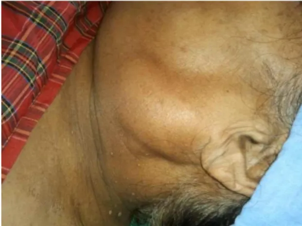
On examination, the patient’s general condition was good. Local examination revealed a mass in the left submandibular region, 7×4cm in size, smooth surface, regular margins, soft in consistency, freely mobile, lying in the subcutaneous plane. The swelling moved to the parotid region on lying down (Figure 1).
Provisional diagnosis of lipoma was made and the patient was investigated. Blood and urine investigations were normal. FNAC of the mass revealed presence of epithelial elements and the pathologist suspected a malignant secondary in the
ᴪCorrespondence at:
#525, Tank Road, Doddballapur, Bangalore Rural District -560037, Karnataka, India; Cell: +919845061813; Email:
neck. CT scan revealed a swelling arising from the left parotid extending lower down. A probable diagnosis of myxoma / adenoma was made.
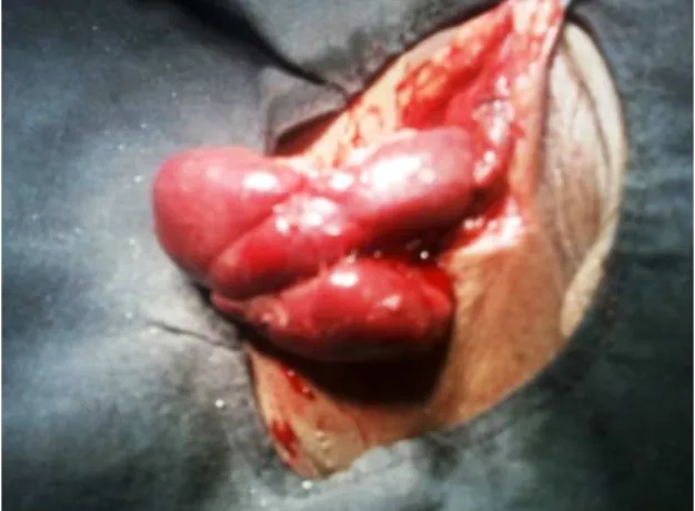
The patient was planned for excision biopsy under general anaesthesia. After intubation, the patient was positioned with face towards right, neck extended and head up position (Figure 1). A transverse incision was made over the swelling. On dissection, a well encapsulated swelling was noted in subplatysmal plane easily separable from surrounding tissue. On dissection, the upper limit was found extending close to the left parotid to which the swelling was connected by a vascular pedicle (Figure 2). The pedicle was ligated and divided. The swelling was removed (Figure 3) and sent for histopathological examination.
Kumar et al / Pedunculated parotid oncytoma
59
Copyrighted © by Dr. Arun Kumar Agnihotri. All rights reserved
Figure 2: Swelling connected to parotid gland by vascular pedicle
Figure 3: Well defined tumour
Postoperative recovery was uneventful. The patient was given oral liquids after six hours and was discharged the next day. Histopathological examination revealed presence of epithelial sheets within a well defined capsule (Figure 4). The diagnosis was oncocytosis.
Figure 4: Histological features suggesting oncocytosis
DISCUSSION
Salivary gland tumours account for 2 to 4% of head and neck tumours. 75% of these tumours arise from the parotid gland, followed by submandibular and other glands. About 80% of parotid gland tumours are benign compared to submandibular (50% benign) and minor salivary (20% benign) glands. Pleomorphic adenomas are the most common benign tumours of salivary glands.
Oncocytoma, also known as oxyphil adenoma is an uncommon benign tumour accounting for 1% of all benign tumours of the parotid.
This tumour is usually found after 50 years of age, with female and male ratio of 2:1. These tumours are mostly found in the superficial lobe of the parotid gland. Clinically they cannot be distinguished from other benign salivary gland tumours.
Histologically, they are characterised by uniformly spherical and large cells (oncocytes) arranged in solid sheets.
Excision is the treatment of choice and incomplete excision results in recurrence.2 Radioactive iodine has also been shown to have an effect on large and recurrent oncocytomas.3
All the reported cases of parotid oncocytomas had arisen from the superficial lobe. Our case was peculiar in that the swelling was in the submandibular region connected to the parotid by a vascular pedicle. Only one case of malignant pedunculated oncocytoma has been reported arising from buccal mucosa.4 No pedunculated parotid oncocytomas have been reported earlier.
REFERENCES
1. Sarma DP, Santos EE. Oncocytoma of the parotid gland. Ear, Nose Throat J. 2009 May;88(5):914.
2. Buxton RW, Maxwell JH, French AJ. Surgical treatment of epithelial tumors of the parotid gland. Surg Gynecol Obstet. Oct 1953;97(4):401-16.
3. Kosuda S, Ishikawa M, Tamura K, et al. Iodine-131 therapy for parotid oncocytoma. J Nucl Med. 1988 Jun;29(6):1126-9.