C
a s eR
e p o Rt1 2 8 Arq Bras Oftalmol. 2017;80(2):128-30 http://dx.doi.org/10.5935/0004-2749.20170031
INTRODUCTION
Oncocytes are defined as modified epithelial cells with charac te-ristic fine, granular, eosinophilic cytoplasm containing excess num-bers of mitochondria. Tumors composed of oncocytes have been des cribed in various organs, including the kidneys, adrenal gland, thyroid, parathyroid, pancreas, and respiratory tract, among others(1-3).
Oncocytomas of the lacrimal gland are extremely rare. The estimated incidence of histopathologically-proven oncocytic lesions in the ocular region is 0.3 per million per year(2). Although a MEDLINE search of the
world literature from 1960 to present found 11 such documented cases, only one case occurred in childhood(3).
Oncocytomas are generally accepted to be benign and slow-gro-wing in nature with an excellent prognosis in the majority of cases, although malignant cases have been reported(4).
Here we report a case of a proptosis caused by a lacrimal gland oncocytoma in a 4-year-old healthy girl and provide a review of lite-rature relevant to the present case.
CASE REPORT
A 4-year-old African girl was referred to the oculoplastic unit of our hospital with an asymptomatic proptosis affecting her right eye. Her mother first noticed the proptosis 4 months prior to seeking
ocu-loplastic consultation, and she reported no significant change in size over that time. The patient was otherwise healthy with no systemic symptom. She had no previous history of ocular disease, surgery, or trauma. Her general medical and family history was unremarkable.
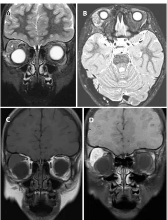
On examination at our hospital, her uncorrected visual acuity was found to be 20/20 in both eyes. Ophthalmic examination revealed a firm, painless, non-fixed, right-sided supraorbital mass causing mini-mal axial proptosis. There was no palpable lymphadenopathy. The left eye was normal. Slit lamp examination, intraocular pressures, and fundoscopy were unremarkable in both eyes. Magnetic resonance imaging (MRI) of the right orbit revealed an oval, solid, well-circums-cribed, homogeneous mass extending from the lacrimal gland and measuring 2.5 × 2.3 × 1.7 cm without any evidence of invasion into adjacent bones or paranasal sinuses (Figure 1).
The patient subsequently underwent a lateral orbitotomy via a zygomatic bony flap with an excisional biopsy of the lesion under general anesthesia. Histological examination of the lacrimal gland de monstrated a circumscribed, nodular tumor composed of acini lined with oncocytic cells without atypia and with abundant, granular, and eosinophilic cytoplasm (Figure 2). Immunohistochemical staining revealed immunoreactivity for pan-cytokeratins (CK) CK7 and CK8. Tumor cells were negative for CK20. No mitotic figures were identi-fied. In light of these findings and considering the location of the
Oncocytoma of the lacrimal gland: a case report
Oncocitoma de glândula lacrimal: relato de caso
Eduardo Muniz FEnElon1, ivElisE ThErEsa BalBy1,2, naThália TElEsdas nEvEs2, Florêncio FiguEirEdo3, Eliza carla Barros duarTE3, PaTrick FrEnsEl TzElikis1,2
Submitted for publication: August 21, 2015 Accepted for publication: December 9, 2015
1 Hospital Oftalmológico de Brasília (HOB), Brasília, DF, Brazil. 2 Hospital de Base do Distrito Federal (HBDF), Brasília, DF, Brazil. 3 Universidade de Brasília (UNB), Brasília, DF, Brazil.
Funding: No specific financial support was available for this study.
Disclosure of potential conflicts of interest: None of the authors have any potential conflict of interest to disclose.
Corresponding author: Patrick Frensel Tzelikis. SQN 203, Bl.K, ap. 502 - Brasília, DF - 70833-110 Brazil - E-mail: [email protected]
ABSTRACT
Here we describe a rare case of a benign tumor in the lacrimal gland of a healthy 4-year-old girl. Mild proptosis was the only abnormality observed on clinical exa-mination. Magnetic resonance imaging of the right orbit revealed an oval, solid, well-circumscribed, homogeneous mass extending from the lacrimal gland and measuring 2.5 × 2.3 × 1.7 cm without any evidence of invasion into adjacent bones. The lesion was surgically excised and histological analyses defined the diagnosis of oncocytoma of the lacrimal gland. Although rare, oncocytoma should be included in the differential diagnosis of lacrimal gland tumors.
Keywords: Adenoma, oxyphilic; Lacrimal gland; Exophthalmos; Orbital neoplasms; Magnetic resonance imaging; Child, preschool; Case reports
RESUMO
Nós descrevemos um raro caso de tumor benigno na glândula lacrimal em uma criança sadia de 4 anos de idade. Clinicamente, a paciente apresentava apenas uma discreta proptose. A ressonância nuclear magnética (RNM) de órbita direita revelou a presença de uma massa oval, sólida, bem-circunscrita, homogênea, se extendendo a partir da glândula lacrimal, medindo 2,5 cm x 2,3 cm x 1,7 cm, sem nenhum sinal evidente de invasão a estrutura óssea adjacente. A lesão foi cirurgicamente removida e analizada histopatologicamente, sendo estabelecido o diagnóstico de oncocitoma de glândula lacrimal. Apesar de raro, o oncocitoma deve ser incluído no diagnóstico diferencial de qualquer tumor originado da glândula lacrimal.
Fenelon eM, e ta l.
1 2 9 Arq Bras Oftalmol. 2017;80(2):128-30
tumor, the diagnosis was indicated a benign oncocytoma originating from the right lacrimal gland. At 6 months postoperatively, clinical examination and imaging studies demonstrated no evidence of residual or recurrent disease.
DISCUSSION
Herein, we describe an interesting case of benign oncocytoma of the lacrimal gland. Oncocytomas are composed of oncocytes, which are epithelial cells with a distinctive appearance characterized by a large size and abundant, granular, eosinophilic cytoplasm(5). Electron
microscopy of oncocytomas revealed a cytoplasm packed with mito-chondria of varying sizes and shapes with irregular and fragmented cristae(6). Oncocytomas occur in various organs and structures, such
as the salivary gland, thyroid, adrenal gland, kidney, liver, pharynx, larynx, trachea, etmoid sinuses, and breasts(1,5). Radnot was the first
author to describe an oncocytoma in the ocular adnexa(6). Since then,
oncocytomas have been reported in the caruncle, lacrimal gland, bulbar and forniceal conjunctiva, lacrimal sac, and eyelid margin(1-3).
The caruncle is the most frequent site of lacrimal tumors, accounting for 3%-8% of biopsied caruncular masses, followed by the lacrimal sac(1). A previous study has reported that 59% of ocular oncocytic
neoplasms involve the caruncle, 19% involve the lacrimal sac, and only 6% involve the lacrimal gland(4).
Lacrimal tumors are more commonly found in elderly patients, with a female to male ratio of 2.4:1 observed only for oncocytic lesions occurring in the caruncle(1). Only four cases of oncocytic neoplasm in
patients aged <40 years have previously been reported, the youngest being an 18-month-old girl with a lacrimal gland oncocytoma(3).
Oncocytic lesions of the lacrimal gland are rare, and do not appear to have a predisposition toward a particular sex. The present case is the second youngest patient with an oncocytic neoplasm to be reported. MRI provides information on anatomic tumoral extent, margins, angulation, and configuration of the lacrimal gland fossa mass. Benign oncocytic neoplasms of the orbit typically appear as solid, well-circumscribed, oval, round-edged, enhancing masses on MRI(7).
In contrast with benign lesions, malignant tumors demonstrate irregu-lar margins with noduirregu-larity and infiltration into adjacent orbital fat(4).
In the present case, the tumor did not produce changes in adjacent bones. The presence of bone destruction strongly favors the diagnosis of a malignant lacrimal gland tumor(7,8).
With respect to published outcomes and management of benign lacrimal gland oncocytoma, the first case report of benign oncocytoma of the lacrimal gland by Beskid and Zarycka included a patient who had an orbital mass of unknown histology that was incompletely excised 8 years prior to the reported presentation(8).
Complete or incomplete resection during the reoperation was not acknowledged in the article, and the patient was reported to be free of recurrence after a follow-up period of 20 months. Riedel et al.(3)
described two benign oncocytomas: one in a child in which the tu-mor was completely excised and followed up for 3 months without recurrence; and another in a 76-year-old female patient in which the completeness of resection was not reported and followed up for 3.5 years without recurrence. Hartman et al.(9) described the case
of a 72-year-old male with a lacrimal gland oncocytoma initially diagnosed with fine needle aspiration cytology and subsequently completely excised via a lateral orbitotomy. This patient was followed up for 18 months without recurrence. More recently, Calle et al.(10)
re-ported a case of an incompletely excised oncocytoma, due to mass extending beyond the lacrimal gland fossa and was followed up for 22 months without signs of recurrence. In the present case, the tu-mor was completely excised and followed up for 6 months without recurrence. According to previous literature, benign oncocytomas may recur or transform into malignant oncocytomas. Therefore, complete surgical resection is the treatment of choice with long-term follow-up for local recurrence(4).
A B
D C
Figure 1. Preoperative imaging of the right orbit demonstrating a soft tissue mass lesion extending from the lacrimal gland and measuring 2.5 × 2.3 × 1.7 mm. The lesion demonstrated intermediate to low signal intensity on T-2 weighted coronal (A) and axial (B) images. T1 pre- (C) and post-contrast (D) coronal MRI imaging demonstrated an enhancing mass in the superotemporal aspect of the right orbit.
Figure 2. Microscopy of the lesion demonstrating tubular and pseudoglan -du lar structures lined with large cells and with abundant eosinophilic granular cytoplasm and small nuclei (A; hematoxylin-eosin staining, ×100 magniication). Immunoreacti vity for pan-cytokeratin (B; AE1/AE3; recognizes the 58-kDa cytokeratin 5, 56-kDa cytokeratin 6, and 52-kDa cytokeratin 8).
On c O c y t O m aO ft h el a c r i m a lg l a n d: ac a s er e p O rt
1 3 0 Arq Bras Oftalmol. 2017;80(2):128-30
In conclusion, although lacrimal gland oncocytomas are very rare, they should be included in the differential diagnosis of lacrimal gland tumors. The present case is an unusual example of a rare, although be-nign, ocular lesion involving the lacrimal gland. A high degree of clinical suspicion with appropriate diagnostic tests may facilitate early diagnosis and curative surgical excision.
REFERENCES
1. Biggs SL, Font RL. Oncocytic lesions of the caruncle and other ocular adnexa. Arch Ophthalmol. 1977;95(3):474-8.
2. Østergaard J, Prause JU, Heegaard S. Oncocytic lesions of the ophthalmic region: a clini copathological study with emphasis on cytokeratin expression. Acta Ophthalmol. 2011;89(3):263-7.
3. Riedel K, Stefani FH, Kampik A. [Oncocytoma of the ocular adnexa]. Klin Monatsbl Augenheilkd. 1983;182(6):544-8. German.
4. Say EA, Shields CL, Bianciotto C, Eagle RC Jr, Shields J. Oncocytic lesions (oncocytoma) of the ocular adnexa: report of 15 cases and review of literature. Ophthal Plast Re-constr Surg. 2012:28(1):14-21.
5. Hamperl H. Benign and malignant oncocytoma. Cancer. 1962;15:1019-27. 6. Radnot M, Lapis K. Ultrastructure of the caruncular oncocytoma. Ophthalmologica.
1970;161(1):63-77.
7. Gündüz K, Shields CL, Günalp I, Shields J. Magnetic resonance imaging of unilateral lacrimal gland lesions. Graefe’s Arch Clin Exp Ophthalmol. 2003:241(11):907-13. 8. Beskid M, Zarzycka M. [A caseçof onkocytoma of the lacrimal gland]. Klin Oczna. 1959;
29:311-5. Polish.
9. Hartman LJ, Mourits MP, Canninga-van Dijk MR. An unusual tumour of the lacrimal gland. Br J Ophthalmol. 2003;87(3):363.
10. Calle CA, Castillo IG, Eagle RC Jr, Daza MT. Oncocytoma of the lacrimal gland: case report and review of the literature. Orbit. 2006;25(3):243-7.