Journal of Krishna Institute of Medical Sciences University 109
Ó
Ó
CASE REPORTAbstract:
Oncocytomas of salivary gland are benign tumors composed exclusively of oncocytic cells. Majority of them occur in parotid gland and have excellent prognosis following complete excision. They should be differentiated from other salivary gland tumors with more prominent oncocytic component and nodular oncocytic hyperplasia. Herein we present a case of oncocytoma of parotid gland in a 48 year old male patient who has no recurrence four years after excision.
Keywords: Oncocytoma, parotid gland, oncocytic hyperplasia.
Introduction:
Oncocytomas of salivary gland are benign tumors composed exclusively of oncocytes with no myoepithelial or basal cell participation [1, 2]. They constitute < 1% of salivary gland tumours [3, 4]. Majority occur in the parotid gland [4]. They can pose diagnostic problems in differentiating from other salivary gland tumours which show prominent oncocytic change.
Case Report:
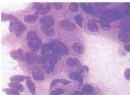
A male patient aged 48 year was admitted with a history of swelling in front of right ear lobule of two months duration. There was no history of exposure to radiation. The mass was freely mobile. No cervical lymph nodes were palpated. The general physical and systemic examination was within normal limits. The laboratory tests revealed no abnormalities. No preoperative sialogram was performed. Fine Needle Aspiration
Oncocytoma of Parotid Gland
1 1 1
Meena N. Jadhav , Rekha M. Haravi , Kittur S.K.
1
Department of Pathology, Belagavi Institute of Medical Sciences, Belagavi-590001(Karnataka) India
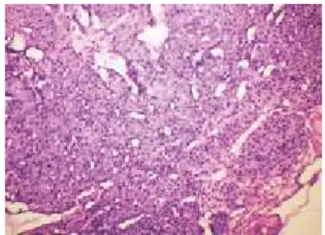
Cytology (FNAC) of mass showed feature of benign cystic neoplastic lesion (Fig.1). It was excised and sent for histopathology examination. Gross appearance of the mass was globular measured 2.5x1.5x1.5 cms with solid, lobulated cut surface and cystic spaces (Fig.2). Microscopy showed a well encapsulated tumor composed of oncocytes arranged in organoid and trabecular pattern separated by thin fibrovascular stroma (Fig.3). These cells had intensely granular eosinophilic cytoplasm with central round vesicular nuclei and indistinct nucleoli (Fig.4). Occasional cells showed clear cell change. Few cystic structures were seen at places. There was minimal cellular atypia. Abnormal mitotic figures were absent. A diagnosis of oncocytoma of right parotid gland was offered. There has been no recurrence of the tumour after four years of excision.
ISSN 2231-4261 JKIMSU, Vol. 6, No. 1, January-March 2017
Journal of Krishna Institute of Medical Sciences University 110
Ó
Ó
Discussion:
Oncocytomas are also known as oxyphilic adenomas or mitochondriomas. Oncocytic change is not limited to the salivary glands but is seen in other organs like thyroid gland, parathyroid gland, adrenal gland, kidney and pancreas. About 7% of salivary gland oncocytomas are bilateral [4]. Occasionally they th th are multifocal. Peak incidence is seen in 7 -8 decade. About 20 % of patients have a history of radiation therapy to the face [4]. Patients with previous radiation exposure are on average 20 years younger at tumour discovery than those without a documented history of radiation.
Pathogenesis of these tumours is unclear. The stimulus for induction of oncocytic change is unknown but, it is generally considered to be age related. Some theories support a neoplastic growth whereas others suggest a hyperplastic/ metaplastic phenomenon. Oncocytic tumourigenesis, secon-dary to acquired mitochondrial dysfunction has been proposed as a plausible mechanism, but few tumours harbor mtDNA alteration with control region to support this theory [5].
Grossly, oncocytomas are typically larger than four cms and less than two years in duration. Microscopically, they may show focal sebaceous, goblet cell, squamous cell differentiation. The lumen of glandular spaces may contain psammoma bodies or tyrosine rich crystals [2]. They may show cytologic atypia, infarction, necrosis, acute/chronic inflammation, fibrosis or hemorrhage. These tumour cells showed deep blue cytoplasmic granules by PTAH. PAS stain before and after diastase digestion demonstrated granules in the cytoplasm [4]. Electron microscopy shows abundant abnormally sized and shaped mitochondria. Immunohistochemical stain for AMA has been shown to be useful for identification of oncocytes [5].
Meena N. Jadhav et al. JKIMSU, Vol. 6, No. 1, January-March 2017
Fig. 2: Gross photograph showing Solid Lobulated Cut Surface with Cystic Spaces
Fig. 3: Microphotograph showing Well Encapsulated Tumor with Oncocytes Arranged in Organoid and Trabecular
Pattern. (H & E, X 100)
Fig. 4: Microphotograph showing Cells with Intensely Granular Eosinophilic Cytoplasm
Journal of Krishna Institute of Medical Sciences University 111
Ó
Ó
The differential diagnosis includes Warthin's tumor, pleomorphic adenoma, basal cell adenoma, clear cell carcinoma, mucoepidermoid carcinoma, acinic cell carcinoma, oncocytic carcinoma and metastatic renal cell carcinoma. Sometimes they can also be mistaken for oncocytic nodular metaplasia and oncocytic hyperplasia [2, 4, 6-8].
In mucoepidermoid carcinoma the tumor cells show squamous cells, mucus cells, intermediate cells with infiltrative growth pattern and the cells are PTAH negative. In acinic cell carcinoma the cell nuclei are peripherally located in contrast to central round nuclei in oncocytoma. In oncocytic carcinoma the tumor shows infiltrative growth pattern with increased mitosis. Metatstatic renal cell carcinoma shows infiltrative growth,
prominent vascularity, more cellular and nuclear pleomorphism [5]. In nodular oncocytic hyperplasia the nodules are less organized and less circumscribed than those of oncocytoma and fibrous capsule is lacking [2]. Oncocytic hyperplasia is unencapsulated and the presence of non oncocytic salivary gland parenchyma within the oncocytic nodules helps in differentiating from oncocytoma.
Prognosis is excellent. Local recurrence is rare [9] but when it occurs it may be multiple and bilateral [4]. There are no histologic features that reliably predict recurrence. The case is presented for its rarity and its importance in the differential diagnosis of tumors and tumor like conditions with oncocytic change.
Meena N. Jadhav et al. JKIMSU, Vol. 6, No. 1, January-March 2017
References
1. Thomson LD, Wenig BM, Ellis GL.Oncocytomas of the submandibular gland: A series of 22 cases and a review of the literature. Cancer 1996; 78: 2281-87. 2. Cheuk W, Chan JKC. Salivary gland tumors. In:
Fletcher CD Ed. Diagnostic histopathology of tumors. 3rd ed. China: Churchill Livingstone; 2005: 261-64. 3. Brandwein MS, Huvas AG. Oncocytic tumors of major
salivary glands. A study of 68 cases with follow-up of 44 patients. Am J Surg Pathol 1991; 15: 514-28. 4. Huvos AG, Paulino AFG. Salivary glands. In: Mills SE
ed. Sternberg's diagnostic surgical pathology. 4th ed. India: Lippincott Williams and Wilkins; 2004:942. 5. Wenig BM. Neoplasms of the salivary glands. In:
Wenig BM ed. Atlas of head and neck pathology. 2nd ed. China: Saunders Elsevier; 2008: 600-602.
* th
Author for Correspondence: Dr. Meena N. Jadhav, S-04, Sadashiva residency Near Shivalaya Temple, 11 Cross,
Sadashiva Nagar, Belagavi 590001 Karnataka, India Email: shubhamj2003@yahoo.co.in Cell: 9886042408
6. Vigliani R, Genetta C. Diffuse hyperplastic oncocytosis of parotid gland. Case report with histochemical observations. Virchows Arch (Pathol
Anat) 1982; 397: 235-40.
7. Capone RB, Ha PK, Westra WH, Pilkington TM, Sciubba JJ, Koch WM et al. Oncocytic neoplasms of the parotid gland: a 16-year institutional review.
Otolaryngol Head Neck Surg 2002; 126(6): 657-62.
8. Chakrabarti I, Basu A, Ghosh N. Oncocytic lesion of parotid gland: A dilemma for cytopathologists. J Cytol
2012; 29(1) 80-82.
9. Sepulveda I, Platin E, Spencer ML, Mucientes P, Frelinghuysen M, Ortega P et al. Oncocytoma of the parotid gland: A case report and review of literature.