J of Evolution of Med and Dent Sci/ eISSN- 2278-4802, pISSN- 2278-4748/ Vol. 4/ Issue 55/ July 09, 2015 Page 9665
RARE PRESENTATION OF BILATERAL CHOROIDAL METASTASIS FROM
PRIMARY MUCO-EPIDERMOID CARCINOMA OF THE PAROTID GLAND: A
CASE REPORT
G. Premalatha1, Ramya Seetamraju2
HOW TO CITE THIS ARTICLE:
G. Premalatha, Ramya Seetamraju. Rare Presentation of Bilateral Choroidal Metastasis from Primary Muco-Epidermoid Carcinoma of the Parotid Gland: A Case Report . Journal of Evolution of Medical and Dental Sciences 2015; Vol. 4, Issue 55, July 09; Page: 9665-9669, DOI: 10.14260/jemds/2015/1394
ABSTRACT: Choroidal metastases are the most common intraocular tumours in adults. The most common primary tumours that metastasize to eye are malignancies of the breast and lung. The authors present a case of 49 year old male; a known case of parotid gland malignancy and on treatment who presented with gradual diminution of vision in both eyes. Fundus examination showed choroidal metastases in both eyes with serous retinal detachment. Metastasis to the choroid from salivary gland malignancy is rare and mucoepidermoid carcinoma is even more rare. This report may add to the already existing reports of salivary gland metastasis to choroids.
KEYWORDS: Rare Case-Parotid Gland Carcinoma-Defective Vision-Distant Metastasis-Bilateral Choroidal Mass Lesion-Retinal Detachment-OCT, B-Scan Findings.
CASE REPORT: A 49 years old man presented with gradual onset of painless progressive visual loss in both eyes for 10 days and sudden worsening for 2 days. There was no history of redness or pain in both eyes. There was history of a mass in the parotid region for one month which had gradually increased in size and was painful. There was loss of weight and loss of appetite. He was diagnosed with Mucoepidermoid Carcinoma Right Parotid gland with right supraclavicular lymph node metastasis and was being treated for the same using radiotherapy.
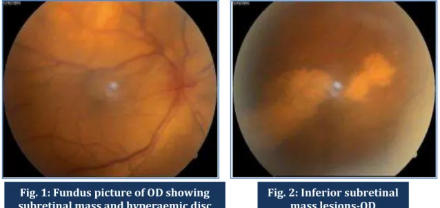
On examination, in the right eye, the anterior segment was normal. Fundus examination using indirect ophthalmoscopy revealed disc hyperaemia, an elevated choroidal mass lesion of size 3DD was seen in the superotemporal quadrant with surrounding exudative retinal detachment (Fig.1) Also there was inferior subretinal mass lesion with seedings and exudative retinal detachment (Fig.2) Visual acuity was counting fingers close to face. In the left eye, anterior segment was normal. Fundus examination showed a normal optic disc. There was an elevated mass lesion of 3DD size in superotemporal region with surrounding retinal detachment (Fig.3)
J of Evolution of Med and Dent Sci/ eISSN- 2278-4802, pISSN- 2278-4748/ Vol. 4/ Issue 55/ July 09, 2015 Page 9666
Fig. 2: Inferior subretinal mass lesions-OD Fig. 1: Fundus picture of OD showing
subretinal mass and hyperaemic disc
Fig. 3: Fundus picture of OS showing Mass lesion
J of Evolution of Med and Dent Sci/ eISSN- 2278-4802, pISSN- 2278-4748/ Vol. 4/ Issue 55/ July 09, 2015 Page 9667
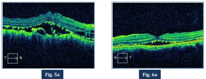
Fig. 5a & 6a: OCT of OD and OS showing multiple subretinal and choroidal deposits with serous retinal detachment.
DISCUSSION: Choroidal metastases are the most common intraocular tumor.(1) Metastatic cancer
reaches the intraocular structure through the hematogenous route and most commonly develops in the uveal tract with> 90% involving the choroid and < 10% involving the iris and /or the ciliary
Fig. 5: B scan showing choroidal mass
lesion with retinaldetachment OD
Fig. 6: B scan showing choroidal mass lesion with retina detachment OS
J of Evolution of Med and Dent Sci/ eISSN- 2278-4802, pISSN- 2278-4748/ Vol. 4/ Issue 55/ July 09, 2015 Page 9668
body.(2) Majority are from breast cancer in women and lung carcinoma in men. Less commonly they
can occur from the alimentary tract, kidney, thyroid and prostate. Of the patients who present to the ophthalmologist, most of them have no known history of primary cancer. In 10% of the cases, there is no known detectable primary cancer, it being occult.
Tumours of the salivary glands account for approximately 5% of all head and neck neoplasms. The average age of patients with malignant neoplasm is approximately 55 years of age; for benign tumours, approximately 40 years. One-quarter of parotid neoplasms are malignant. Metastases are present in 20–25% of patients at the time of diagnosis. Mucoepidermoid carcinoma is the most common carcinoma of the salivary glands. By definition it consists of mucous cells, epidermoid cells and intermediate cells. It occurs more commonly in adults. It accounts for 30% of the cancers of the salivary glands(3) and usually affects major salivary glands. Metastasis from this
tumour occurs to the lung and bone frequently apart from the lymph nodes.(4) In a large survey of
eyes with uveal metastases, it was noted that bilateral, multifocal presentation is less common than unilateral or unifocal presentation.(5) We report a rare case of choroidal metastasis from a salivary
gland tumour.
CONCLUSION: There are few reported cases of ocular metastasis from salivary gland tumours, mostly adenoid cystic carcinomas. To the best of the authors’ knowledge this is the first of such cases being reported with ocular metastasis due to mucoepidermoid carcinoma of the parotid gland. This could be due to the reason that patients with poor systemic condition may not present to the ophthalmic department for consultation.
REFERENCES:
1. Foster BS, Samy CN, Gragoudas ES: Choroidal metastasis, in Guyer, Yanuzzi, Change (eds): Retina-Vitreous-Macula, Vol. 2. Chap. 93, pp 1103-1109.
2. Jerry A. Shields, Carol L. Shields; Metastatic tumours to the intraocular structures, Intraocular tumours, an atlas and textbook, Part-1, 198-199.
3. Tang EW, Tsang CS, Li KK., Presumed bilateral choroidal metastases from mucoepidermoid carcinoma of the submandibular gland. Retin Cases Brief Rep. 2012 Summer; 6(3): 330-2. 4. Eugene Myers, Robert Erris, Salivary Gland Disorders, Chapter 3: Pathology of Salivary gland
disease, 61-65.
J of Evolution of Med and Dent Sci/ eISSN- 2278-4802, pISSN- 2278-4748/ Vol. 4/ Issue 55/ July 09, 2015 Page 9669
AUTHORS:
1. G. Premalatha 2. Ramya Seetamraju
PARTICULARS OF CONTRIBUTORS:
1. Assistant Professor, Department of Ophthalmology, Andhra Medical College, Visakhapatnam, Andhra Pradesh. 2. Senior Resident, Department of
Ophthalmology, Andhra Medical College, Visakhapatnam, Andhra Pradesh.
FINANCIAL OR OTHER
COMPETING INTERESTS: None
NAME ADDRESS EMAIL ID OF THE CORRESPONDING AUTHOR:
Dr. G. Premalatha 1-68-22/1,
Sector 2, M. V. P. Colony, Visakhapatnam-530017, Andhra Pradesh.
E-mail: [email protected]