774
Brazilian Journal of Microbiology (2011) 42: 774-777ISSN 1517-8382
ASSESSMENT OF THE QUALITY OF DNA EXTRACTED BY TWO TECHNIQUES FROM MYCOBACTERIUM
TUBERCULOSIS FOR FAST MOLECULAR IDENTIFICATION AND GENOTYPING
Marcelo Miyata1*; Adolfo Carlos Barreto Santos1; Natália Helena Mendes1; Eunice Atsuko Cunha2; Fernando Augusto Fiúza de Melo3; Clarice Queico Fujimura Leite1
¹Universidade Estadual Paulista, Faculdade de Ciências Farmacêuticas, Araraquara, SP, Brasil; ²Laboratório Central de Saúde
Pública, Campo Grande, MS, Brasil; ³Instituto Clemente Ferreira, São Paulo, SP, Brasil.
Submitted: March 31, 2010; Returned to authors for corrections: May 30, 2010; Approved: November 04, 2010.
ABSTRACT
We report a comparative study of two DNA extraction techniques, thermolysis and chemical lysis (CTAB),
for molecular identification and genotyping of M. tuberculosis. Forty DNA samples were subjected to PCR
and the results demonstrated that with thermolysis it is possible to obtain useful data that enables fast
identification and genotyping.
Key words: DNA extraction - Mycobacterium tuberculosis - thermolysis - CTAB
Tuberculosis (TB) is responsible for the death of
approximately 3 million people per year and it is estimated that
one third of the human population is infected latently with
Mycobacterium tuberculosis, Brazil occuping 16th place in the
world ranking (13). The development of fast molecular biology
techniques to identify and genotype M. tuberculosis isolates is
crucial for the prevention and control of tuberculosis. Although
techniques based on amplification of nucleic acids are highly
sensitive and reduce the time required for identification or
epidemiological study of M. tuberculosis, there are few studies
assessing the quality of M. tuberculosis DNA extracted from
cultures. Several techniques for DNA extraction have been
described (6, 9), for application in distinct diagnostic methods.
Here we aimed to find a fast and easy method to extract
DNA of sufficiently high quality to be used in the molecular
identification and genotyping of M. tuberculosis isolates. Two
methods for DNA extraction from M. tuberculosis cultures
were compared and the extracted DNA was utilized for
identification of the isolates by PCR and their genotyping by
the spoligotyping technique.
A total of 40 M. tuberculosis clinical isolates were
analyzed. The first method of extraction was a modified
thermolysis technique (10) and the second was chemical lysis
with cetyltrimethylammonium bromide (CTAB) (12). In the
thermolysis method, a loopful of mycobacteria grown on
Lowenstein-Jensen medium was suspended in 300 mL of TE
buffer (10 mM Tris, 1 mM EDTA pH 8.5) and subjected to 3
cycles of boiling and freezing for 20 minutes at -20ºC. In the
CTAB method, the samples were inactivated by heating for 18
- 24 hours at 80ºC in a dry bath. After that, proteinase K and
sodium dodecyl sulfate (SDS) were added and the samples
incubated for 10 minutes at 65ºC. 5M NaCl and CTAB solution
were then added and incubated for another 10 minutes at 65ºC.
Chloroform: isoamyl alcohol (24:1) mixture was added and
775
Miyata, M. et al.
DNA extracted from M. tuberculosis
mixed until a milky mixture was obtained, which was
centrifuged to 11,750 rcf for 5 minutes. The upper phase was
transferred to a new tube containing isopropanol and the
samples were vortexed and frozen for 18 hours. After thawing,
the samples were centrifuged for 30 minutes at 4ºC and the
supernatant was discarded. After drying, the samples were
washed with 70% ethanol and centrifuged for 20 minutes at
11,750 rcf at 4°C. The supernatant was discarded and the
pellets were dried at room temperature. The samples were
resuspended in TE buffer and incubated for one hour at 65ºC
and, finally, frozen for 2 hours at -20ºC.
For the molecular identification by PCR, a pair of
universally accepted primers were used to amplify a fragment
of the insertion sequence IS6110 (PCR-IS6110), a specific
sequence for the M. tuberculosis complex (11). For the PCR
reaction, 21.5 µL of 1x PCR-Master Mix (Fermentas™, USA),
0.5 µL of each primer (INS-1 5’-CGTGAGGGCATCGAGGT
GGC-3’ and INS-2 5’GCGTAGGCGTCGGTGACAAA-3’)
and 2.5 µL of genomic DNA were mixed. After this, the
reaction was performed in the PTC-100 Thermo cycler (MJ
Research) under the following conditions: initial cycle of 95ºC
for 10 minutes, followed for 30 cycles of 94°C for 1 minute,
56°C for 2 minutes and 72°C for 1 minute and a final cycle of
72°C for 7 minutes. Subsequently, 10 µL of amplified product
was loaded on 1% agarose gel, resolved by electrophoresis and
stained with ethidium bromide.
Strains were genotyped by the spoligotyping technique,
which is a standard method for M. tuberculosis. Spoligotyping
is a PCR-based technique that simultaneously detects and types
M. tuberculosis through patterns of multiple, well-conserved
36-bp direct repeats (DRs) with nonrepetitive spacer
sequences, 34 to 41 bp long (5).
In the spoligotyping technique, two amplifications of the
extracted DNA were performed. In the first amplification
reaction, 20 µL of 1x PCR-Master Mix (Fermentas™, USA), 1
µL of each primer at 5 µM (DRa 5’biotinylated-GGTTTTGGG
TCTGACGAC-3’, and DRb 5’-CCGAGAGGGGACGGAAA
C-3’) (5) and 1 µL of extracted DNA. In the second
amplification reaction, PCR-amplified products were used as
template. The reactions were performed in a PTC–100 Thermo
cycler (MJ Research), with an initial cycle of 15 minutes at
95ºC, followed for twenty cycles of 60 seconds at 95ºC, 60
seconds at 55ºC and 30 seconds at 72ºC and a final cycle of 5
minutes at 72ºC. Following DNA amplification, 20 µL of
amplicons were added to 150 µL of 2x SSPE/0.1% SDS
solution and heated at 100ºC for 10 minutes. Next, 150 µ L of
this mixture were applied to a membrane and incubated for 60
minutes at 60ºC for hybridization.
The membrane was washed twice with 2x SSPE/0.5%
SDS solution and incubated in 1:4,000-diluted
streptavidin-peroxidase conjugate (Zymed Laboratories, USA) for 60
minutes at 42ºC. Afterwards, the membrane was washed twice
with 2x SSPE and incubated for 1 minute with ECL™ (GE
Healthcare, UK), for chemoluminescence. As the DRa primer
is labeled with biotin, the amplified DNA could be used for
hybridization to spacer nucleotides, which were covalently
bound to a membrane (5). After 30 minutes contact with the
membrane, X-ray film was labeled and the spoligopatterns
were visualized. DNA from M. bovis BCG and M. tuberculosis
H37Rv were used as positive controls and 2x SSPE/0.1% SDS
solution as negative control. The results of spoligotyping of
DNA extracted by both lysis techniques were analyzed
comparatively, using the Fourth International Spoligotyping
Database (SpolDB4) (2).
Out of these two extraction techniques, thermolysis
proved to be easier and faster. In thermolysis, the DNA was
extracted after 2 hours, while in chemical lysis with CTAB, the
extracted DNA was obtained after 30 hours. In laboratories that
work with pathogenic bacteria such as M. tuberculosis, the
thermolysis technique is of special interest because the cyclic
procedures of heating and freezing cause the death of the
bacteria, preventing contamination. Several studies have
demonstrated the usefulness of thermolysis for inactivation and
776
Miyata, M. et al.
DNA extracted from M. tuberculosis
In the molecular identification tests, the PCR results
indicated that both techniques (thermolysis and CTAB) can be
used for DNA extraction, as illustrated in Figures 1a and
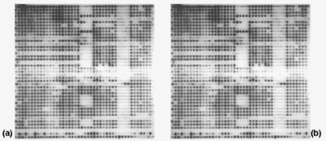
Figure 1b, respectively. Regarding genotyping by
spoligotyping, it was possible to obtain the spoligotypes with
the DNA extracted by both techniques (thermolysis and
CTAB), as illustrated in Figures 2a and 2b, respectively.
The spoligotypes found belong to 5 families and 12
sub-families: LAM2, LAM4, LAM6, LAM9, LAM3 S Convergent,
H1, H3, S, T1, T4-CEU1, U Likely T3 and U. These results
showed that the spoligotyping technique can be successfully
performed with DNA extracted by thermolysis, because in the
molecular epidemiology, the quality and the efficiency of
amplification depend on the efficiency of DNA extraction
methods (1) and the quality of Taq polymerase (3).
In conclusion, thermolysis is a simple, fast method that
can be employed in the extraction of M. tuberculosis DNA.
The quality of the DNA obtained by thermolysis was sufficient
to allow molecular identification by PCR and genotyping by
spoligotyping, contributing to the rapid diagnosis and control
of tuberculosis for molecular epidemiology.
Figure 1. Multiplex PCR amplification products from various
Mycobacterium tuberculosis DNAs extracted by (a)
thermolysis and (b) CTAB methods after electrophoresis on a
1% agarose gel and staining with ethidium bromide. Arrows
indicate the fragments with 245 bp.
Positive (+) (H37Rv) and negative (-) controls and a 100 bp
molecular ladder were used.
Figure 2. Spoligotype patterns obtained from various Mycobacterium tuberculosis DNAs extracted by (a) thermolysis and (b)
777
Miyata, M. et al.
DNA extracted from M. tuberculosis
ACKNOWLEDGEMENTS
This work was supported by Conselho Nacional de
Desenvolvimento Científico e Tecnológico (CNPq) and
Fundação de Amparo à Pesquisa do Estado de São Paulo
(FAPESP).
REFERENCES
1. Amaro, A.; Duarte, E.; Amado, A.; Ferronha, H.; Botelho, A. (2008). Comparison of three DNA extraction methods for Mycobacterium bovis,
Mycobacterium tuberculosis and Mycobacterium avium subsp. avium.
Lett. Appl. Microbiol. 47(1), 8-11.
2. Brudey, K.; Driscoll, J.R.; Rigouts, L. et al. (2006). Mycobacterium tuberculosis complex genetic diversity: mining the fourth international spoligotyping database (SpolDB4) for classification, population genetics and epidemiology. BMC Microbiol. 6, 23.
3. Cavalcanti, H.R.; Marques,E.; Fonseca L.S.; Saad, M.H.F. Do DNA extraction methods and Taq polimerase quality improve the double repetitive element (DRE) PCR typing method for Mycobacterium tuberculosis strains?. Braz. J. Microbiol. (2007). 38, 409-412.
4. Clarridge, J.E.; Shawar, R.M.; Shinnick, T.M.; Plikaytis, B.B. (1993). Large-scale use of polymerase chain reaction for detection of
Mycobacterium tuberculosis in a routine mycobacteriology laboratory. J. Clin. Microbiol. 31(8), 2049-56.
5. Kamerbeek, J.; Schouls, L.; Kolk, A.; van Agterveld, M.; van Soolingen, D.; Kuijper, S.; Bunschoten, A.; Molhuizen, H.; Shaw, R.; Goyal, M.; van Embden, J. (1997). Simultaneous detection and strain differentiation of Mycobacterium tuberculosis for diagnosis and epidemiology. J. Clin.
Microbiol. 35(4), 907-14.
6. Lee, J.H.; Park, Y.; Choi, J.R.; Lee, E.K.; Kim, H.S. (2010). Comparisons of three automated systems for genomic DNA extraction in a clinical diagnostic laboratory. Yonsei Med. J. 51(1), 104-10.
7. Ramazanzadeh, R.; Farnia, P.; Amirmozafari, N. (2009). Characterization of Mycobacterium tuberculosis Complex isolated from Iranian and Afghani patients by Spoligotyping method. Braz. J. Microbiol. 40, 314-320.
8. Shawar, R.M.; el-Zaatari, F.A.; Nataraj, A.; Clarridge, J.E. (1993). Detection of Mycobacterium tuberculosis in clinical samples using two-step PCR and nonisotopic hybridization methods. J. Clin. Microbiol. 3, 61-65.
9. Tan, S.C.; Yiap, B.C. (2009). DNA, RNA, and protein extraction: the past and the present. J. Biomed. Biotechnol. Article ID 574398. 10. Telenti, A.; Marchesi, F.; Balz, M.; Bally, F.; Böttger, E.C.; Bodmer, T.
(1993). Rapid identification of mycobacteria to the species level by polymerase chain reaction and restriction enzyme analysis. J. Clin. Microbiol. 31(2), 175-8.
11. Thierry, D.; Cave, M.D.; Eisenach, K.D.; Crawford, J.T.; Bates, J.H.; Gicquel, B.; Guesdon, J.L. (1990). IS6110, an IS-like element of
Mycobacterium tuberculosis complex. Nucleic Acids Res. 18(1), 188. 12. van Embden, J.D.; Cave, M.D.; Crawford, J.T.; Dale, J.W.; Eisenach,
K.D.; Gicquel, B.; Hermans, P.; Martin, C.; McAdam, R.; Shinnick, T.M. et al. (1993). Strain identification of Mycobacterium tuberculosis
by DNA fingerprinting: recommendations for a standardized methodology. J. Clin. Microbiol. 31(2), 406-9.
13. WHO Library Cataloguing-in-Publication Data (2008). Global tuberculosis Control: surveillance, planning, financing: WHO report 2008. [WHO/HTM/TB/2008.393]. 2008: 294 pp. http://www.who.int/tb/ publications/global_report/en/