Formyl Peptide Receptor as a Novel
Therapeutic Target for Anxiety-Related
Disorders
Irene Gallo1., Lorenza Rattazzi1., Giuseppa Piras1
, Thomas Gobbetti1, Elisabetta Panza2, Mauro Perretti1, Jeffrey W. Dalley3,4", Fulvio D’Acquisto1*"
1.William Harvey Research Institute, Barts and the London School of Medicine, Queen Mary University of London, London, United Kingdom,2.Department of Experimental Pharmacology, University of Naples ‘‘Federico II’’, Naples, Italy,3.Department of Psychology, University of Cambridge, Cambridge, United Kingdom,4.Department of Psychiatry, University of Cambridge, Cambridge, United Kingdom
.These authors contributed equally to this work. "JD and FD are joint senior authors on this work.
Abstract
Formyl peptide receptors (FPR) belong to a family of sensors of the immune system that detect microbe-associated molecules and inform various cellular and sensorial mechanisms to the presence of pathogens in the host. Here we demonstrate that Fpr2/3-deficient mice show a distinct profile of behaviour characterised by reduced anxiety in the marble burying and light-dark box paradigms, increased exploratory behaviour in an open-field, together with superior performance on a novel object recognition test. Pharmacological blockade with a formyl peptide receptor antagonist, Boc2, in wild type mice reproduced most of the behavioural changes observed in the Fpr2/3-/-mice, including a significant improvement in novel object discrimination and reduced anxiety in a light/dark shuttle test. These effects were associated with reduced FPR signalling in the gut as shown by the significant reduction in the levels of p-p38. Collectively, these findings suggest that
homeostatic FPR signalling exerts a modulatory effect on anxiety-like behaviours. These findings thus suggest that therapies targeting FPRs may be a novel approach to ameliorate behavioural abnormalities present in neuropsychiatric disorders at the cognitive-emotional interface.
OPEN ACCESS
Citation:Gallo I, Rattazzi L, Piras G, Gobbetti T, Panza E, et al. (2014) Formyl Peptide Receptor as a Novel Therapeutic Target for Anxiety-Related Disorders. PLoS ONE 9(12): e114626. doi:10.1371/ journal.pone.0114626
Editor:Tiziana Rubino, University of Insubria, Italy
Received:July 10, 2014
Accepted:November 11, 2014
Published:December 17, 2014
Copyright:ß2014 Gallo et al. This is an open-access article distributed under the terms of the
Creative Commons Attribution License, which permits unrestricted use, distribution, and repro-duction in any medium, provided the original author and source are credited.
Data Availability:The authors confirm that all data underlying the findings are fully available without restriction. All relevant data are within the paper.
Funding:The authors have no funding or support to report.
Introduction
The immune system is equipped with a vast variety of biological weapons to sense the presence of pathogens via the recognition of pathogen-associated molecular patterns (PAMPs) [1,2]; these elicit a complex series of events leading to the specialization and differentiation of the immune cells, B and T lymphocytes [3]. Formyl peptide receptors (FPRs) are G protein-coupled receptors whose main function is to sense the presence of harmful or noxious molecules such as formylated peptides and guide cells to the site where pathogen-associated molecules have been released [4]. This sensing function of FPRs is not limited to a particular pathogen and is extended to a wide range of endogenous ligands including classical biomarkers of inflammation and immune activation such as serum amyloid A (SAA) [5], formylated peptides released by mithochondria of damaged cells and tissue [6], the antimicrobial peptide LL-37 [7] and the dual pro- and anti-inflammatory protein Annexin-A1 [8].
There are currently three functional FPRs in humans as well as in mouse -FPR1, FPR2 and FPR3- which all recognise to different degrees a wide range of endogenous and exogenous ligands [6,9,10]. Activation of these receptors causes their homo- or hetero-dimerization which in turn depends on the precise ligand they bind to [11,12]. In this way FPRs are able to exert both pro- and anti-inflammatory effects on immune cells [4,8,10].
The expression of FPRs is highest in sentinel innate cells with phagocytic or chemotactic activity such as neutrophils [13,14], monocytes [13,15], macro-phages[15,16] and dendritic cells [15,17]. However, FPR are also expressed in non-phagocytic and ‘‘immobile’’ sentinel cells such as mucosal epithelial cells [18,19], endothelial cells [20–22] and glia [23–25]. In these cells, FPRs exert a genuine ‘‘sentinel role’’ by sensing pathogens present in the microenvironment as well as by favouring repair upon damage and inflammation. Recent findings show that FPRs are expressed in the vomeronasal system, where they are postulated to detect the presence of infection in the ‘‘macro environment’’ through volatile FPR ligands present in the faeces of pathogen-infected animals [26–29]. Thus, FPRs exert a unique role in the response of the host to pathogens because they signal at two levels; firstly at the level of the central nervous system to alert the host of impeding dangers and secondly at the level of the immune system by initiating a protective inflammatory response.
intraperitoneal injection of the pan-FPR inhibitor Boc2 [35,36], which was accompanied by a decreased activation of downstream FPR signalling pathways in the gut. Together these results support the hypothesis that FPRs may have an important role to play in the regulation of aversive emotional responses. Thus targeting FPRs might provide new avenues of treatment for a range of brain disorders linked to anxiety.
Materials And Methods
Mice
Four to six week old male mice were used for all experiments.Fpr2/3-/-mice have been previously described [34] and were backcrossed onto C57BL/6 for more than 8 generations. Animals were housed in groups of 4–5 under specific-pathogen-free conditions, with free access to food and water and in a room under a 12 h light/ dark cycle (light on at 7:00 am). C57BL/6 mice were purchased from Charles River (Margate, UK) and housed for at least 10 days in the same room as the Fpr2/3 -/-prior to testing to allow acclimatization.Fpr2/3+/+littermate controls and C57BL/ 6 mice were used in equal number and are collectively referred to as wild-type controls since they showed no significant differences in all the preliminary tests. All animal studies were conducted with ethical approval from the Local Ethical Review Committee. This research was carried out in accordance with the UK Animals (Scientific Procedures) Act, 1986 and under the UK Home Office project license number 70/6994.
Behavioural tests and pharmacological treatment
If not otherwise stated, tests were performed double-blind every other day during the light phase of the light-dark cycle, as previously described and recommended [37]. All the efforts were made to minimize mouse discomfort in these behavioral experiments. Mice were brought to the testing room at least 30 minutes before the start of the test session to allow habituation to the testing environment. Unless otherwise specified, standard lighting (about 50 lux) and quiet conditions were maintained throughout each experiment. FPR antagonist studies were performed with male C57BL/6 mice receiving an intraperitoneal injection of the FPR2 antagonist Boc2 (t-Boc-FLFLF; at a previously validated dose of 10 mg/animal
[38,39] or an equal volume of phosphate-buffered saline (PBS) as a control solution (200 ml), 30 minutes before the behavioural tests. This research was
carried out in accordance with the UK Animals (Scientific Procedures) Act, 1986.
Open field activity test
(50 cm630 cm620 cm) divided into 10 cm610 cm squares (n515). The 3
central squares defined the ‘‘centre’’ region (seeFig. 1). Each mouse was placed in a corner square, facing the wall, and observed and recorded for 3 minutes. The total number of squares crossed (all four paws in), total number of rears (defined as both front paws off the ground, but not as a part of grooming) and number of centre crossings was recorded. The walls and floor of the arena were thoroughly cleaned between each trial.
Climbing activity test
The climbing test is used to assess vertical activity and exploratory behaviour. The test was performed as previously described but with some modifications [41,42]. Briefly, mice were placed, one at a time, on a thin layer of fresh wood chip bedding on a laboratory bench and covered with a cylindrical climbing mesh (60 cm630 cm base diameter) (seeFig. 2). They were each observed and
recorded for 5 minutes. The number of climbing events and total duration of climbing activity was assessed. The criterion for climbing was for a mouse to have all 4 feet on the wire mesh while a climb terminated as soon as one foot touched the bench. This test was conducted in the late afternoon, when mice are known to be more active [43].
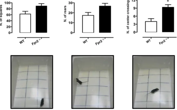
Fig. 1. Reduced anxiety-like behaviour ofFpr2/3-/-mice in the open field test.The bar graphs and images show total number of squares crossed, rears and centre crossings during a 5-minute session. Values are expressed as median¡S.E.M. and representative of four experiments, each involving 6–9 mice per group. *P,0.05 indicate significant values compared with wild-type (WT) control mice (Mann–WhitneyU-test).
Light-dark shuttle box
In this test exploratory activity reflects the combination of hazard and risk avoidance [44]. The apparatus consisted of a 45 cm620 cm621 cm box, divided
into two distinct compartments: one third (15 cm long) painted black, with a black lid on top, the remaining two thirds painted white and uncovered (see
Fig. 3). A 2.5 cm62.5 cm opening joined the two compartments. One side of the
bright box was transparent to enable behavioural assessment and the averseness of this compartment was increased by additional illumination supplied by a 50 W lamp placed 45 cm above the centre of the box floor. The test was performed in accordance with a previous published protocol [45]. Each mouse was placed in the bright compartment, facing away from the opening and allowed to explore the box for 5 minutes. Dependent variables included the time spent in the light area, latency to cross to the dark area (all four paws in) and the total number of transitions between compartments. The apparatus was cleaned after each trial.
Marble burying test
The marble-burying test (MBT) is thought to reflect repetitive and perseverative behaviour, possibly related to compulsions and/or anxiety disorders [46]. The test was carried out as described by Deacon and colleagues [47] with some
modifications. Briefly, mice were individually placed in a clear plastic box (14 cm610 cm611 cm) filled with approximately 5 cm depth of wood chip
bedding lightly pressed to give a flat surface (see Fig. 4). Fifteen 1.5 cm diameter glass marbles were placed on the surface, evenly spaced, each about 4 cm apart, so to form 5 rows of 3. The latency to start digging (defined as the mouse digging the bedding with front and hind paws for more than 1 second), the total number of digging bouts and the number of buried marbles (to 2/3 of their depth) were manually recorded during the 10 minute-test.
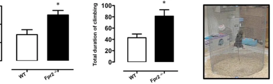
Fig. 2. Increased exploratory behaviour ofFpr2/3-/-mice in the climbing test.The bar graphs show the number of climbing events and total time (seconds) spent on the climbing mesh during a 5-minute trial. Values are expressed as median¡S.E.M. and representative of three different experiments, each involving 6–9 mice per group. *P,0.05 indicate significant values compared with wild-type (WT) control mice (Mann–WhitneyU-test).
Fig. 3. Reduced anxiety-like behaviour ofFpr2/3-/-mice in the light and dark box test.The bar graphs and images show the total time (seconds) spent in the lit area, latency (seconds) to first cross to the dark chamber and total number of transition during a 5-minute trial. Values are expressed as median¡ S.E.M. and representative of four different experiments involving 6–9 mice per group. *P,0.05 and **P,0.05 indicate significant values compared with wild-type (WT) control mice (Mann–WhitneyU-test).
doi:10.1371/journal.pone.0114626.g003
Fig. 4. Reduced digging and marble burying behaviour ofFpr2/3-/-mice in the marble burying test.The bar graphs and relative pictures show the total number of buried marbles, total duration (seconds) of digging and the latency (seconds) to the first digging bout during a 10-minute trial. Values are expressed as median¡S.E.M. and representative of four different experiments involving 6–9 mice per group. *P,0.05 and **P,0.05 indicate significant values compared with wild-type (WT) control mice (Mann–WhitneyU-test).
Novel object recognition test (NORT)
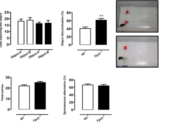
The novel object recognition test has been widely used to assess the mouse’s ability to discriminate between a previously encountered and a novel object [48]. The test relies on the idea that mice approach more frequently and spend more time exploring a novel object when previously exposed to a familiar one that they recognize as already encountered. The test was carried out as previously described [49,50]. On day one mice were firstly habituated to the open-field box for 10 minutes. On day two, mice were placed in the same arena for a 10 minute acquisition period, during which time they were exposed to two identical toys (3 cm3 non-toxic red wooden cubes (object A and B). Objects were glued to the floor 10 cm apart from each other, 8 cm away from both box edges. After being returned in their home cage, mice were given a one-hour inter-trial interval. Each subject was then placed back into the arena, where everything was the same as during the acquisition phase except that object A was replaced with a wooden, green, cylinder (4 cm height, 1 cm base diameter) (novel object). During this test phase, mice were allowed to explore both of the objects for 5 minutes. Acquisition and test phases were recorded with a video camera and time spent visiting each object (visit defined as when the animal’s nose touched the object or was pointed towards it within 1 cm radius) was manually assessed in both phases. Results were expressed as percentage of object discrimination [(Time spent exploring novel object/total time spent exploring during testing phase) 6100]. After each trial
both the arena and the objects were cleaned with 70% ethanol, in order to eliminate olfactory traces.
Y-Maze spontaneous alternation test
The Y maze was made of three enclosed transparent plastic arms (A, B, C) 29 cm68 cm619 cm each, set at an angle of 120
˚
to each other in the shape of aY. It was fixed on a white wooden board and placed on the floor of a room containing several large immovable objects to use as spatial cues. In this test for spatial memory mice tend to enter the maze arm that was explored most recently and remember the order of the arm entry, thanks to their ability to allocate the arm’s positions through spatial clues surrounding the testing apparatus. Mice were allowed to freely explore the arena for 5 minutes, during which time the total number of arm entries was recorded, along with the entering sequence, not including the initial arm. A spontaneous alternation occurred when the animal entered into all three arms of the maze on consecutive choices in overlapping triplet sets (e.g. CBABCABCBACB58 alternations) [51]. Spontaneous alternation percentage was calculated as: [Total number of actual alternations/(total arm entries –2)] 6100. The maze was thoroughly cleaned after each test.
Colon whole mount preparation
containing 0.1% Triton X-100 for 5 minutes. Thereafter, samples were washed again, then blocked in PBS containing 5% foetal bovine serum (FBS) for 1 hour. Samples were incubated alternatively with mouse monoclonal anti-phospho-p38 (#sc-7973, Santa Cruz Biotechnology) (1:100 dilutions) for 90 minutes and then all with Alexa Fluor 488 goat polyclonal anti-mouse IgG (H+L) (ab150113,
Abcam) 1:100 for 1 hour. Rinsed samples were finally mounted in Optimum Cutting Temperature (O.C.T.; Tissue-Tek) and frozen at 280
˚
C. Five mm thicksections were mounted on slides and visualized by fluorescence microscopy [52].
Plasma corticosterone and cytokine measurement
Blood was collected from untested mice by intracardiac puncture performed under anaesthesia, and all efforts were made to minimize suffering. Plasma was obtained from the clotted blood by centrifugation (8000 rpm, 5 min) and stored at 280
˚
C before the assay. Corticosterone concentrations were measured in diluted (1:32) plasma by Enzymatic Immuno Essay (EIA) assay following the manufacturer’s instructions (Enzo Life Sciences, Exeter, UK). Cytokine levels in the same samples were measured (dil. 1:500) using mouse Th1/Th2/Th17/Th22 16 plex Kit FlowCytomix and according to the manufacturer’s instructions (eBioscience).Statistical analysis
Results were analysed as previously described [53–55] using GraphPad. Unpaired Student’s t test was performed for experiments where differences between two groups needed to be analysed. For non-parametric data, the Mann–Whitney U-test was applied and results were expressed as medians (interquartile range). Statistical significance was determined at p,0.05. The results were expressed as mean ¡ S.E.M.
Results
Reduced anxiety in
Fpr2/3
null mice
Although we found no statistically significant difference between Fpr2/3-/-mice and wild-type control mice with respect to ambulation and rearing (Fig. 1,left
and middle panels, respectively), Fpr2/3-/- mice showed reduced thigmotaxis (walking along the edges) and significantly increased centre crossings (Fig. 1, right panel) indicating a reduced level of anxiety [56,57].
We further tested anxiety-related behaviour using the climbing test where vertical exploratory behaviour is assessed [41,42]. As shown in Fig. 2, Fpr2/3 -/-mice performed a greater number of climbing acts compared with wild-type (p,0.05) and spent on average more time climbing than control animals (p,0.05).
significantly more time in the aversive, brightly lit compartment compared with wild-type controls (p,0.05) and waited longer to move to the less aversive, dark side of the box (p,0.01) (Fig. 3, leftand middle panels, respectively).
Fpr2/3-/-mice also buried less marbles and spent less time in this activity compared with wild-type (Fig. 4 leftand middle panels, respectively). The latency to start this behaviour was also significantly increased in Fpr2/3-/- mice (Fig. 4 right panel) consistent with reduced anxiety.
Improved novel object recognition in
Fpr2/3
-/-mice
To investigate whether reduced anxiety of Fpr2/3-/- mice was linked to an increased preference for novelty, indicative of low anxiety, we next assessed the performance of animals on a novel object recognition task. This test has been widely used as an explicit test of novel versus familiar object discrimination and relies on the idea that animals tend to preferentially approach novel objects [48]. We found that Fpr2/3-/-mice and controls showed no difference in their
exploration of two identical objects (Fig. 5A, left panel). However, following the introduction of the novel object, wild-type mice spent about 40% of their time with the novel object as previously reported [49,50] while Fpr2/3-/-mice spent a significantly greater proportion (about 60%) (Fig. 5A, right panel).
Fpr2/3
-/-mice show no difference in the Y-maze test
We next tested theFpr2/3-/-mice in the Y maze. In this test mice tend to enter the maze arm that was explored most recently and recall the order of the arm entry. As shown inFig. 5B, there were no significant difference between wild types and Fpr2/3-/-mice in the number of arm entries or percentages of alternations in this maze. These data show that Fpr2/3-/-mice are not impaired on a spatial memory task and imply that the effects reported earlier pertain mainly to diminished anxiety and fear-related responses in this group of animals.
Higher basal corticosterone levels in
Fpr2/3
null mice
Fig. 5. Increased discriminatory activity ofFpr2/3-/-mice in the novel object recognition test.The bar graph inAshows the total time (seconds) spent exploring the objects used in the test (shown in the top picture) during the 10-minute acquisition phase (left panel) and the % of time spent on the novel object (shown in the bottom picture) in the subsequent 5-minute test phase (right panel). The bar graphs inBshow the total number of arm entries and spontaneous alternation percentage (calculated as described in material and Methods section) in the Y-maze during a 5-minute trial. Values are expressed as median¡S.E.M. and representative of n54 different experiments involving 6–9 mice per group. **P,0.05 indicates significant values compared with wild-type (WT) control mice (Mann–WhitneyU-test).
doi:10.1371/journal.pone.0114626.g005
Fig. 6. Increased level of corticosterone inFpr2/3-/-mice.Levels of corticosterone in the plasma of WT and
Fpr2/3-/-. Values are expressed as ngml21
and are representative of three experiments with 6 mice.
Administration of an FPR antagonist reduces some anxiety
behaviours
We next investigated whether the reduced anxiety of Fpr2/3-/-mice could be mimicked by administering the FPR inhibitor Boc2 in wild type animals. As shown in Fig. 7, Boc2 had no significant effect on general locomotion or explorative behaviour in the open field test (A) but did increase both the time in the brightly lit aversive compartment and the latency to cross to the ‘safe’ dark compartment (B). Moreover, Boc2-treated mice showed an increased preference for the novel object on the object recognition task compared with vehicle-treated wild-type animals (Fig. 8). These findings suggest FPR blockers may reduce some anxiety-related behaviours, including neophobia
Reduced FPR signalling in the gut of
Fpr2/3
-/-and Boc2-treated
mice
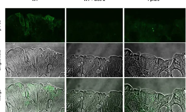
Since the behavioural phenotype of Fpr2/3-/-mice could be partly reproduced by FPR antagonism we next investigated whether these effects were related to changes in the local (peritoneal) microenvironment. Given previous findings showing a key role of FPRs in regulating gut microbiota homeostasis we measured p-p38, a widely recognised intracellular readout of FPR activation [61]. Fluorescence microscopy of colonic tissue confirmed our prediction of increased staining for p-p38 in wild-type mice compared with Fpr2/3-/- and Boc2-treated mice (Fig. 9).
Discussion
Fig. 7. Boc2-treatment reduces anxiety-like behaviour in C57BL/6 mice.The bar graphs inAshow the total number of squares crossed, rears and centre crossings of Boc2-treated mice compared to PBS vehicle-treated during a 5-minute trial in the open field test. The bar graph in B shows the total time (seconds) spent in the lit area and the latency (seconds) to first cross to the dark chamber of Boc2-treated mice compared to PBS vehicle-treated mice during a 5-minute trial. Values are expressed as median¡S.E.M. and representative of four different experiments involving 6 mice per group. *P,0.05 and **P,0.01 indicate significant values compared to PBS-vehicle treated mice (Mann–WhitneyU-test).
doi:10.1371/journal.pone.0114626.g007
Fig. 8. Boc2-treatment increases the recognition of a novel object in C57BL/6 mice.The bar graphs show the total time (seconds) spent exploring the objects used in the test during the 10-minute acquisition phase (left panel) and the % of time spent on the novel object in the subsequent 5-minute test phase (right panel) of Boc2-treated mice compared to PBS vehicle-treated mice. Values are expressed as median¡ S.E.M. and representative of four different experiments involving 6 mice per group. **P,0.01 indicates significant values compared to PBS-vehicle treated mice (Mann–WhitneyU-test).
for the enriched or ‘exercised’ mice, theFpr2/3-/-mice show both increased level of corticosterone and an overall increase in exploratory and locomotory activity as shown by the open field (Fig. 1) and climbing test (Fig. 2). In light of these findings it is tempting to speculate that the increased corticosterone levels inFpr2/ 3-/-mice might be the results of their increased ‘engagement’ with the external and social environment. Interestingly, these data contrast with those observed in Fpr1-/-mice [30] suggesting orthogonal regulation of corticosterone levels by FPR2/3 and FPR1 receptors.
Further exploration of the inquisitive and fearless nature ofFpr2/3-/-mice using the novel object test showed an almost 50% increase their discriminatory activity and no difference in spatial memory. These results suggest that the absence of homeostatic FPR2/3 signalling might induce a state of behavioural disinhibition and reduced anxiety. This conclusion is consistent with the widely recognised sensing/alerting function of FPRs in the olfactory system [29] and thus provides a further example of behavioural modulation by FPR signalling.
To support this hypothesis and to explore the therapeutic potential of our findings we investigated the effects of a FPR blocker on behaviour. Our findings reveal that administration of the pan-FPR antagonist Boc2 induced a behavioural profile that resembled, at least in part, what we observed in theFpr2/3-/-mice. We think that this is most likely due to the metabolism of this inhibitor and hence to its transient effect as previously shown [72,73]. Thus, we observed a significant increase in the number of center crossings in the open field and a significant
Fig. 9. Reduced p-p38 staining in the colon of Boc2-treated C57BL/6 andFpr2/3-/-mice.Immunofluorescence (top panel) of phospho-p38 of intestinal whole mount preparations (as described in Material and Methods) in either PBS-vehicle treated, Boc2 treated orFpr2/3-/-intestinal mucosa. The middle and
bottom panel show the bright field and the overlay pictures of the same samples.
increase in the time spent in the lit area of the light and dark box, both
observations that are indicative of reduced anxiety. We also observed a marked improvement in the ability of wild-type animals to discriminate the novel object. We found that these differences (readily detectable after as little as 2 hour post treatment) were present only after intraperitoneal but not intravenous admin-istration of Boc2 (data not shown). The lack of effect of intravenous
administration of Boc2 led us to test whether Boc2 inhibited FPR signalling in the gut or intestinal mucosa.
A number of studies have shown that the intestinal mucosa expresses receptors for formylated peptides produced by the gut microbiota [18]. These commensal bacteria are known to play important and non-detrimental roles for the host [74–
76] and have provided a perfect example of consensual interaction between microbes and immune sentinels present throughout the gut. These immune-microbiome interactions are known to be an important part of a dual circuit that controls behaviour and overall emotional wellbeing [77–79]. Indeed, one of the best examples of this system are the germ-free mice that are known to show signs of increased anxiety and reduced neurogenesis [80–83].
Our findings also show that bothFpr2/3-/-mice and Boc2-treated mice have a reduced immunostaining for p-p38 – a key FPR signalling pathway [4,11,84,85]. Similar findings have been previously reported in other studies where it has been shown that commensal bacteria such as the Lactobacillus species stimulated these pathways in gut epithelial cells [19,52,74,76]. It was recently suggested that the expression of FPR2 on the apical and lateral membrane of mouse colonic epithelial cells may have important biological significance, as it enables the epithelial cells to respond to both locally and systemically available ligands under various pathophysiological conditions [86]. Although we have not systematically explored this idea using a wider range of doses and other FPR antagonists our results show that the effects of Boc2 on behaviour occurs in parallel with a modulation of microbiota-induced FPR signalling in the gut. More specifically, the homeostatic and protective inflammatory state of the gut sustained by the commensal microbiota might contribute to a ‘‘homeostatic’’ status of focus and alertness that feature what we know as physical and mental wellbeing. Conversely, in the absence of this physiological loop a state of alertness and reduced anxiety might help the host to ‘‘focus’’ on the possible origin of ‘‘internal conflicts and dangers’’ (Fig. 10).
Author Contributions
None. Conceived and designed the experiments: FD JD. Performed the experiments: IG LR GP TG EP. Analyzed the data: IG LR FD. Contributed reagents/materials/analysis tools: MP. Wrote the paper: FD JD.
References
1. Janeway CA Jr, Medzhitov R(2002) Innate immune recognition. Annu Rev Immunol 20: 197–216.
2. Medzhitov R, Janeway C Jr(2000) Innate immune recognition: mechanisms and pathways. Immunol Rev 173: 89–97.
3. Sallusto F, Lanzavecchia A, Araki K, Ahmed R(2010) From vaccines to memory and back. Immunity 33: 451–463.
4. Ye RD, Boulay F, Wang JM, Dahlgren C, Gerard C, et al.(2009) International Union of Basic and Clinical Pharmacology. LXXIII. Nomenclature for the formyl peptide receptor (FPR) family. Pharmacol Rev 61: 119–161.
5. Su SB, Gong W, Gao JL, Shen W, Murphy PM, et al.(1999) A seven-transmembrane, G protein-coupled receptor, FPRL1, mediates the chemotactic activity of serum amyloid A for human phagocytic cells. J Exp Med 189: 395–402.
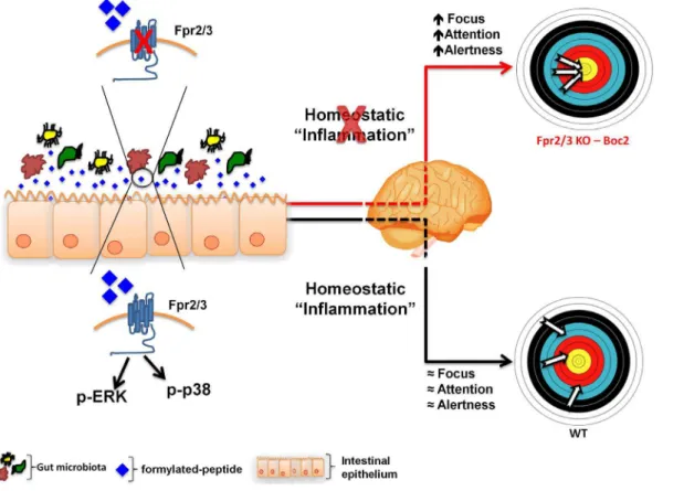
Fig. 10. Hypothetical schema of the role of Fpr2/3 at the interface of the gut-brain axis.Non-pathogenic gut microbiota releases physiological levels of formylated peptides that activate FPR signalling in the gut epithelium. This homeostatic level of protective inflammation influences brain function maintaining a physiological level of focus and attention. The blockage of FPR signalling by an antagonist or the absence of gut microbiota causes a reduction in FPR activation and a parallel increase in the state of alertness, as observed inFpr2/3-/-and Boc2-treated mice.
6. Zhang Q, Raoof M, Chen Y, Sumi Y, Sursal T, et al.(2010) Circulating mitochondrial DAMPs cause inflammatory responses to injury. Nature 464: 104–107.
7. De Y, Chen Q, Schmidt AP, Anderson GM, Wang JM, et al.(2000) LL-37, the neutrophil granule- and epithelial cell-derived cathelicidin, utilizes formyl peptide receptor-like 1 (FPRL1) as a receptor to chemoattract human peripheral blood neutrophils, monocytes, and T cells. J Exp Med 192: 1069–1074.
8. Perretti M, D’Acquisto F (2009) Annexin A1 and glucocorticoids as effectors of the resolution of inflammation. Nat Rev Immunol 9: 62–70.
9. Panaro MA, Acquafredda A, Sisto M, Lisi S, Maffione AB, et al.(2006) Biological role of the N-formyl peptide receptors. Immunopharmacol Immunotoxicol 28: 103–127.
10. Dufton N, Perretti M(2010) Therapeutic anti-inflammatory potential of formyl-peptide receptor agonists. Pharmacol Ther 127: 175–188.
11. Cooray SN, Gobbetti T, Montero-Melendez T, McArthur S, Thompson D, et al.(2013) Ligand-specific conformational change of the G-protein-coupled receptor ALX/FPR2 determines proresolving functional responses. Proc Natl Acad Sci U S A 110: 18232–18237.
12. Kasai RS, Suzuki KG, Prossnitz ER, Koyama-Honda I, Nakada C, et al.(2011) Full characterization of GPCR monomer-dimer dynamic equilibrium by single molecule imaging. J Cell Biol 192: 463–480.
13. Spurr L, Nadkarni S, Pederzoli-Ribeil M, Goulding NJ, Perretti M, et al.(2011) Comparative analysis of Annexin A1-formyl peptide receptor 2/ALX expression in human leukocyte subsets. Int
Immunopharmacol 11: 55–66.
14. El Kebir D, Jozsef L, Filep JG(2008) Opposing regulation of neutrophil apoptosis through the formyl peptide receptor-like 1/lipoxin A4 receptor: implications for resolution of inflammation. J Leukoc Biol 84: 600–606.
15. Yang D, Chen Q, Le Y, Wang JM, Oppenheim JJ (2001) Differential regulation of formyl peptide receptor-like 1 expression during the differentiation of monocytes to dendritic cells and macrophages. J Immunol 166: 4092–4098.
16. Gemperle C, Schmid M, Herova M, Marti-Jaun J, Wuest SJ, et al.(2012) Regulation of the formyl peptide receptor 1 (FPR1) gene in primary human macrophages. PLoS One 7: e50195.
17. Yang D, Chen Q, Gertz B, He R, Phulsuksombati M, et al.(2002) Human dendritic cells express functional formyl peptide receptor-like-2 (FPRL2) throughout maturation. J Leukoc Biol 72: 598–607.
18. Anton P, O’Connell J, O’Connell D, Whitaker L, O’Sullivan GC, et al.(1998) Mucosal subepithelial binding sites for the bacterial chemotactic peptide, formyl-methionyl-leucyl-phenylalanine (FMLP). Gut 42: 374–379.
19. Babbin BA, Jesaitis AJ, Ivanov AI, Kelly D, Laukoetter M, et al.(2007) Formyl peptide receptor-1 activation enhances intestinal epithelial cell restitution through phosphatidylinositol 3-kinase-dependent activation of Rac1 and Cdc42. J Immunol 179: 8112–8121.
20. Keitoku M, Kohzuki M, Katoh H, Funakoshi M, Suzuki S, et al.(1997) FMLP actions and its binding sites in isolated human coronary arteries. J Mol Cell Cardiol 29: 881–894.
21. Heo SC, Kwon YW, Jang IH, Jeong GO, Yoon JW, et al.(2014) WKYMVm-induced activation of formyl peptide receptor 2 stimulates ischemic neovasculogenesis by promoting homing of endothelial colony-forming cells. Stem Cells 32: 779–790.
22. Mou H, Li Z, Kong Y, Deng B, Qian L, et al.(2012) Proinflammatory stimulants promote the expression of a promiscuous G protein-coupled receptor, mFPR2, in microvascular endothelial cells. Inflammation 35: 656–664.
23. Chen K, Iribarren P, Huang J, Zhang L, Gong W, et al.(2007) Induction of the formyl peptide receptor 2 in microglia by IFN-gamma and synergy with CD40 ligand. J Immunol 178: 1759–1766.
24. Cui YH, Le Y, Zhang X, Gong W, Abe K, et al.(2002) Up-regulation of FPR2, a chemotactic receptor for amyloid beta 1–42 (A beta 42), in murine microglial cells by TNF alpha. Neurobiol Dis 10: 366–377.
25. Cui YH, Le Y, Gong W, Proost P, Van Damme J, et al.(2002) Bacterial lipopolysaccharide selectively up-regulates the function of the chemotactic peptide receptor formyl peptide receptor 2 in murine microglial cells. J Immunol 168: 434–442.
27. Liberles SD, Horowitz LF, Kuang D, Contos JJ, Wilson KL, et al.(2009) Formyl peptide receptors are candidate chemosensory receptors in the vomeronasal organ. Proc Natl Acad Sci U S A 106: 9842– 9847.
28. Munger SD(2009) Olfaction: Noses within noses. Nature 459: 521–522.
29. Riviere S, Challet L, Fluegge D, Spehr M, Rodriguez I(2009) Formyl peptide receptor-like proteins are a novel family of vomeronasal chemosensors. Nature 459: 574–577.
30. Gao JL, Schneider EH, Dimitrov EL, Haun F, Pham TM, et al.(2011) Reduced fear memory and anxiety-like behavior in mice lacking formylpeptide receptor 1. Behav Genet 41: 724–733.
31. Sato T, Nishio H, Iwata M, Kentotsuboi, Tamura A, et al.(2010) Postmortem molecular screening for mutations in ryanodine receptor type 1 (RYR1) gene in psychiatric patients suspected of having died of neuroleptic malignant syndrome. Forensic Sci Int 194: 77–79.
32. Saigo M, Abe K, Abe K, Aihara H, Akatsu M, et al.(2005) Study of the suppressed decays B- —
.[K+pi-](D)K- and B- —.[K+pi-]Dpi. Phys Rev Lett 94: 091601.
33. Condo GT, Handler T, Shimony J, Abe K, Armenteros R, et al.(1991) Photoproduction of an isovector rho pi state at 1775 MeV. Phys Rev D Part Fields 43: 2787–2791.
34. Dufton N, Hannon R, Brancaleone V, Dalli J, Patel HB, et al.(2010) Anti-inflammatory role of the murine formyl-peptide receptor 2: ligand-specific effects on leukocyte responses and experimental inflammation. J Immunol 184: 2611–2619.
35. Stenfeldt AL, Karlsson J, Wenneras C, Bylund J, Fu H, et al.(2007) Cyclosporin H, Boc-MLF and Boc-FLFLF are antagonists that preferentially inhibit activity triggered through the formyl peptide receptor. Inflammation 30: 224–229.
36. Derian CK, Solomon HF, Higgins JD 3rd, Beblavy MJ, Santulli RJ, et al.(1996) Selective inhibition of N-formylpeptide-induced neutrophil activation by carbamate-modified peptide analogues. Biochemistry 35: 1265–1269.
37. McIlwain KL, Merriweather MY, Yuva-Paylor LA, Paylor R(2001) The use of behavioral test batteries: effects of training history. Physiol Behav 73: 705–717.
38. Gavins FN, Hughes EL, Buss NA, Holloway PM, Getting SJ, et al.(2012) Leukocyte recruitment in the brain in sepsis: involvement of the annexin 1-FPR2/ALX anti-inflammatory system. FASEB J 26: 4977–4989.
39. Gavins FN, Yona S, Kamal AM, Flower RJ, Perretti M(2003) Leukocyte antiadhesive actions of annexin 1: ALXR- and FPR-related anti-inflammatory mechanisms. Blood 101: 4140–4147.
40. Deacon RM, Croucher A, Rawlins JN(2002) Hippocampal cytotoxic lesion effects on species-typical behaviours in mice. Behav Brain Res 132: 203–213.
41. Deacon RM, Rawlins JN(2005) Hippocampal lesions, species-typical behaviours and anxiety in mice. Behav Brain Res 156: 241–249.
42. Sutton LM, Sanders SS, Butland SL, Singaraja RR, Franciosi S, et al.(2013) Hip14l-deficient mice develop neuropathological and behavioural features of Huntington disease. Hum Mol Genet 22: 452– 465.
43. Onnela JP, Christakis NA (2012) Spreading paths in partially observed social networks. Phys Rev E Stat Nonlin Soft Matter Phys 85: 036106.
44. Barr JL, Forster GL(2011) Serotonergic neurotransmission in the ventral hippocampus is enhanced by corticosterone and altered by chronic amphetamine treatment. Neuroscience 182: 105–114.
45. Bourin M, Hascoet M(2003) The mouse light/dark box test. Eur J Pharmacol 463: 55–65.
46. Kedia S, Chattarji S (2014) Marble burying as a test of the delayed anxiogenic effects of acute immobilisation stress in mice. J Neurosci Methods.
47. Deacon RM(2006) Digging and marble burying in mice: simple methods for in vivo identification of biological impacts. Nat Protoc 1: 122–124.
48. Antunes M, Biala G(2012) The novel object recognition memory: neurobiology, test procedure, and its modifications. Cogn Process 13: 93–110.
50. Taglialatela G, Hogan D, Zhang WR, Dineley KT (2009) Intermediate- and long-term recognition memory deficits in Tg2576 mice are reversed with acute calcineurin inhibition. Behav Brain Res 200: 95– 99.
51. Carpenter AC, Saborido TP, Stanwood GD (2012) Development of hyperactivity and anxiety responses in dopamine transporter-deficient mice. Dev Neurosci 34: 250–257.
52. Wentworth CC, Jones RM, Kwon YM, Nusrat A, Neish AS(2010) Commensal-epithelial signaling mediated via formyl peptide receptors. Am J Pathol 177: 2782–2790.
53. van Gaalen MM, Reul JH, Gesing A, Stenzel-Poore MP, Holsboer F, et al. (2002) Mice overexpressing CRH show reduced responsiveness in plasma corticosterone after a5-HT1A receptor challenge. Genes Brain Behav 1: 174–177.
54. Pellow S, File SE(1985) The effects of putative anxiogenic compounds (FG 7142, CGS 8216 and Ro 15-1788) on the rat corticosterone response. Physiol Behav 35: 587–590.
55. Jahng JW(2011) An animal model of eating disorders associated with stressful experience in early life. Horm Behav 59: 213–220.
56. Bourin M, Petit-Demouliere B, Dhonnchadha BN, Hascoet M(2007) Animal models of anxiety in mice. Fundam Clin Pharmacol 21: 567–574.
57. Walsh RN, Cummins RA(1976) The Open-Field Test: a critical review. Psychol Bull 83: 482–504.
58. Aoki M, Shimozuru M, Kikusui T, Takeuchi Y, Mori Y (2010) Sex differences in behavioral and corticosterone responses to mild stressors in ICR mice are altered by ovariectomy in peripubertal period. Zoolog Sci 27: 783–789.
59. Marquez C, Nadal R, Armario A(2006) Influence of reactivity to novelty and anxiety on hypothalamic-pituitary-adrenal and prolactin responses to two different novel environments in adult male rats. Behav Brain Res 168: 13–22.
60. Touma C, Bunck M, Glasl L, Nussbaumer M, Palme R, et al.(2008) Mice selected for high versus low stress reactivity: a new animal model for affective disorders. Psychoneuroendocrinology 33: 839–862.
61. Cattaneo F, Guerra G, Ammendola R(2010) Expression and signaling of formyl-peptide receptors in the brain. Neurochem Res 35: 2018–2026.
62. Strohle A, Holsboer F (2003) Stress responsive neurohormones in depression and anxiety. Pharmacopsychiatry 36 Suppl 3: S207–214.
63. Ganella DE, Kim JH(2014) Developmental rodent models of fear and anxiety: from neurobiology to pharmacology. Br J Pharmacol 171: 4556–4574.
64. Schoenfeld TJ, Gould E(2012) Stress, stress hormones, and adult neurogenesis. Exp Neurol 233: 12– 21.
65. Costa-Pinto FA, Palermo-Neto J(2010) Neuroimmune interactions in stress. Neuroimmunomodulation 17: 196–199.
66. Leasure JL, Jones M(2008) Forced and voluntary exercise differentially affect brain and behavior. Neuroscience 156: 456–465.
67. Santos-Soto IJ, Chorna N, Carballeira NM, Velez-Bartolomei JG, Mendez-Merced AT, et al.(2013) Voluntary running in young adult mice reduces anxiety-like behavior and increases the accumulation of bioactive lipids in the cerebral cortex. PLoS One 8: e81459.
68. Fediuc S, Campbell JE, Riddell MC (2006) Effect of voluntary wheel running on circadian corticosterone release and on HPA axis responsiveness to restraint stress in Sprague-Dawley rats. J Appl Physiol (1985) 100: 1867–1875.
69. Benaroya-Milshtein N, Hollander N, Apter A, Kukulansky T, Raz N, et al.(2004) Environmental enrichment in mice decreases anxiety, attenuates stress responses and enhances natural killer cell activity. Eur J Neurosci 20: 1341–1347.
70. Marashi V, Barnekow A, Ossendorf E, Sachser N(2003) Effects of different forms of environmental enrichment on behavioral, endocrinological, and immunological parameters in male mice. Horm Behav 43: 281–292.
72. Sato AK, Viswanathan M, Kent RB, Wood CR(2006) Therapeutic peptides: technological advances driving peptides into development. Curr Opin Biotechnol 17: 638–642.
73. Park S, Kim SD, Lee HY, Hwang D, Park JS, et al.(2014) A novel delivery platform for therapeutic peptides. Biochem Biophys Res Commun 450: 13–18.
74. Alam A, Leoni G, Wentworth CC, Kwal JM, Wu H, et al.(2014) Redox signaling regulates commensal-mediated mucosal homeostasis and restitution and requires formyl peptide receptor 1. Mucosal Immunol 7: 645–655.
75. Molloy MJ, Grainger JR, Bouladoux N, Hand TW, Koo LY, et al.(2013) Intraluminal containment of commensal outgrowth in the gut during infection-induced dysbiosis. Cell Host Microbe 14: 318–328.
76. Wentworth CC, Alam A, Jones RM, Nusrat A, Neish AS(2011) Enteric commensal bacteria induce extracellular signal-regulated kinase pathway signaling via formyl peptide receptor-dependent redox modulation of dual specific phosphatase 3. J Biol Chem 286: 38448–38455.
77. De Palma G, Collins SM, Bercik P, Verdu EF(2014) The Microbiota-Gut-Brain axis in gastrointestinal disorders: Stressed bugs, stressed brain or both? J Physiol.
78. Foster JA, McVey Neufeld KA (2013) Gut-brain axis: how the microbiome influences anxiety and depression. Trends Neurosci 36: 305–312.
79. Cryan JF, Dinan TG(2012) Mind-altering microorganisms: the impact of the gut microbiota on brain and behaviour. Nat Rev Neurosci 13: 701–712.
80. Cryan JF, O’Mahony SM (2011) The microbiome-gut-brain axis: from bowel to behavior. Neurogastroenterol Motil 23: 187–192.
81. Diaz Heijtz R, Wang S, Anuar F, Qian Y, Bjorkholm B, et al.(2011) Normal gut microbiota modulates brain development and behavior. Proc Natl Acad Sci U S A 108: 3047–3052.
82. Neufeld KM, Kang N, Bienenstock J, Foster JA (2011) Reduced anxiety-like behavior and central neurochemical change in germ-free mice. Neurogastroenterol Motil 23: 255-264, e119.
83. Gareau MG, Wine E, Rodrigues DM, Cho JH, Whary MT, et al. (2011) Bacterial infection causes stress-induced memory dysfunction in mice. Gut 60: 307–317.
84. Krump E, Sanghera JS, Pelech SL, Furuya W, Grinstein S(1997) Chemotactic peptide N-formyl-met-leu-phe activation of p38 mitogen-activated protein kinase (MAPK) and MAPK-activated protein kinase-2 in human neutrophils. J Biol Chem 272: 937–944.
85. Rane MJ, Carrithers SL, Arthur JM, Klein JB, McLeish KR (1997) Formyl peptide receptors are coupled to multiple mitogen-activated protein kinase cascades by distinct signal transduction pathways: role in activation of reduced nicotinamide adenine dinucleotide oxidase. J Immunol 159: 5070–5078.
86. Chen K, Liu M, Liu Y, Yoshimura T, Shen W, et al.(2013) Formylpeptide receptor-2 contributes to colonic epithelial homeostasis, inflammation, and tumorigenesis. J Clin Invest 123: 1694–1704.
87. Miceli F, Soldovieri MV, Lugli L, Bellini G, Ambrosino P, et al.(2009) Neutralization of a unique, negatively-charged residue in the voltage sensor of K V 7.2 subunits in a sporadic case of benign familial neonatal seizures. Neurobiol Dis 34: 501–510.
88. Luisi R, Panza E, Barrese V, Iannotti FA, Viggiano D, et al.(2009) Activation of pre-synaptic M-type K+channels inhibits [3H]D-aspartate release by reducing Ca2+entry through P/Q-type voltage-gated Ca2+channels. J Neurochem 109: 168–181.
89. Lim H, Jang S, Lee Y, Moon S, Kim J, et al.(2012) Enhancement of Anxiety and Modulation of TH and pERK Expressions in Amygdala by Repeated Injections of Corticosterone. Biomol Ther (Seoul) 20: 418– 424.