TLR4 Induced Inflammation and Death in Mice
Rabindra N. Bhattacharjee1,2,3*, Birendranath Banerjee4, Shizuo Akira1,2,5, M. Prakash Hande4*
1Akira Innate Immunity Project, Exploratory Research for Advanced Technology (ERATO), Osaka University, Osaka, Japan,2Department of Host Defense, Research Institute for Microbial Diseases, Osaka University, Osaka, Japan,3Department of Anatomy and Cell Biology, University of Western Ontario, London, Canada,4Department of Physiology, Yong Loo Lin School of Medicine, National University of Singapore, Singapore, Singapore,5World Premier International Immunology Frontier Research Center, Osaka University, Osaka, Japan
Abstract
Background:Telomeres are essential to maintain chromosomal stability. Cells derived from mice lacking telomerase RNA component (mTERC2/2 mice) display elevated telomere-mediated chromosome instability. Age-dependent telomere shortening and associated chromosome instability reduce the capacity to respond to cellular stress occurring during inflammation and cancer. Inflammation is one of the important risk factors in cancer progression. Controlled innate immune responses mediated by Toll-like receptors (TLR) are required for host defense against infection. Our aim was to understand the role of chromosome/genome instability in the initiation and maintenance of inflammation.
Methodology/Principal Findings:We examined the function of TLR4 in telomerase deficientmTERC2/2mice harbouring chromosome instability which did not develop any overt immunological disorder in pathogen-free condition or any form of cancers at this stage. Chromosome instability was measured in metaphase spreads prepared from wildtype(mTERC+/+),
mTERC+/2andmTERC2/2mouse splenocytes. Peritoneal and/or bone marrow-derived macrophages were used to examine the responses of TLR4 by their ability to produce inflammatory mediators TNFaand IL6. Our results demonstrate that TLR4 is highly up-regulated in the immune cells derived from telomerase-null(mTERC2/2)mice and lipopolysaccharide, a natural ligand for TLR4 stabilises NF-kB binding to its promoter by down-regulating ATF-3 inmTERC2/2macrophages.
Conclusions/Significance:Our findings implied that background chromosome instability in the cellular level stabilises the action of TLR4-induced NF-kB action and sensitises cells to produce excess pro-inflammatory mediators. Chromosome/ genomic instability data raises optimism for controlling inflammation by non-toxic TLR antagonists among high-risk groups.
Citation:Bhattacharjee RN, Banerjee B, Akira S, Hande MP (2010) Telomere-Mediated Chromosomal Instability Triggers TLR4 Induced Inflammation and Death in Mice. PLoS ONE 5(7): e11873. doi:10.1371/journal.pone.0011873
Editor:Mikhail V. Blagosklonny, Roswell Park Cancer Institute, United States of America
ReceivedApril 21, 2010;AcceptedJune 18, 2010;PublishedJuly 29, 2010
Copyright:ß2010 Bhattacharjee et al. This is an open-access article distributed under the terms of the Creative Commons Attribution License, which permits unrestricted use, distribution, and reproduction in any medium, provided the original author and source are credited.
Funding:MPH acknowledges the support from Academic Research Fund, Ministry of Education, Singapore (T206B3108) for funding. This work was supported in part by Exploratory Research for Advanced Technology of Japan Science and Technology Agency, grants of the Japanese Ministry of Education, Culture, Sports, Science and Technology, and from the 21st Century Center of Excellence Program of Osaka University to SA. The funders had no role in study design, data collection and analysis, decision to publish, or preparation of the manuscript.
Competing Interests:The authors have declared that no competing interests exist.
* E-mail: [email protected] (RNB); [email protected] (MPH)
Introduction
Telomeres are specialised DNA structures consisting of tandem arrays of short, repetitive G-rich sequences that are oriented in 59-39 direction towards the end of the chromosome. They preserve chromosome integrity and protect chromosomes from being recognised as damaged DNA. The end replication problem results in a gradual loss of telomeric sequence under normal conditions in cells lacking the enzyme telomerase [1,2,3,4]. Activation of telome-rase, a ribonucleoprotein, has been shown to be essential for the stable maintenance of telomere lengthin vitroandin vivo. Telomerase consists of an essential RNA component (in mouse called mTR or mTERC) which serves as a template for telomeric DNA synthesis and a catalytic protein (mTERT) component. This enzyme protects against chromosome end-to-end fusions not only by lengthening the telomere repeats but also by providing a cap at the end of the chromosome [4]. Telomere shortening, telomerase reverse transcriptase and telomerase hold important implications for ageing and oncogenesis.
to negatively modulate the senescence-associated inflammatory mediators (IL-6 and IL-8) [15].
Infection and autoimmune disorders are commonly associated with inflammation and evidences support the notion that the presence of microbes can be a cofactor in tumour promotion and progression [16]. For example, there is a strong association between inflammatory bowel disease and colon cancer; infection with Helicobacter pylori and gastric cancer, chronic viral hepatitis and liver cancer [17]. Data from mouse models of human cancer have established that chronic inflammation creates a pro-tumour microenvironment that is an essential component of neoplastic processes [18,19,20]. The soluble inflammatory molecules secreted by tumour associated immune cells promote cell motility; induce angiogenesis and extravasation of tumour cells [21]. Interestingly, metastatic carcinoma cells secrete macrophage-activating factors leading to production of IL-6 and TNF-athrough activation of TLR2 and 6 [20,22]. However, till to date the precise mechanisms that link inflammation with tumour progression and metastasis remain to be elucidated.
Innate immune system is the body’s first line of defense against infection. In vertebrates, the main function of innate immune receptors (Toll-like receptors or TLR) is to recognise the presence of foreign invaders in the form of pathogen associated molecular patterns (PAMPs,) on invading microorganisms and initiate downstream signals to produce inflammatory mediators as protective measures to defend hosts. The members of the TLR
family are type 1 transmembrane proteins expressed primarily by certain cells of the innate immune system (e.g., macrophages, dendritic cells and neutrophils). TLRs and interleukin 1 receptors (IL-1Rs) share significant similarity in their cytoplasmic domain (called Toll-IL-1R or TIR domain. Typical ligands for the TLRs are microbial products such as bacterial unmethylated deoxycy-tidyl-deoxyguanosine (CpG) DNA, and lipopolysaccarides or viral RNA or nucleic acids. They mediate signals through several adaptor proteins such as MyD (myeloid differentiation factor) 88, MAL/TIRAP (MyD88-adaptor-like/TIR-associated protein), and TRIF (Toll-receptor-associated activator of interferon) to liberate pro- inflammatory transcription factor NF-kB [23,24] which initiates robust transcription of most of the genes responsible for inflammation such as interleukin (IL)-6, cyclooxygenase (COX)-2, reactive oxygen species (ROS), TNF-a, etc as part of the tumour supportive micro-environment. Besides TLR’s ability to recognise PAMPs, they also play an essential role in non-infectious sterile inflammation by recognising endogenous ligands from the damaged cells such as b-defensins, and oxidised lipids [25] or protein molecules modified by oxidation and nitration [23].
While innate immune responses via TLRs are absolutely required for host defence against infections, uncontrolled reaction from a TLR can also cause chronic inflammation eventually leading to cancer. Our aim was to understand the role of chromosome instability in the initiation and maintenance of
Figure 1. Genomic instability in mTERC2/2 MEFs with dysfunctional telomeres. (A) Left arrow points a binucleated cell with two
micronuclei, a marker for chromosomal instability. Right arrow points to a normal binucleated cell. (B) Telomere PNA-FISH on metaphase spreads
frommTERC2/2MEFs. Chromosomal DNA was stained with DAPI (blue) and telomeres were hybridised with Cy3-labeled telomere probe (pink). Left
arrows point Robertsonian fusion like configuration (RLC) and down arrow indicates a dicentric chromosome (DC) representing chromosomal aberrations.C). Histogram showing number of micronuclei measured by the CBMN assay D) Histogram showing total chromosome aberrations detected per cell in different cell types.
inflammation. We examined the function of TLR4 in telomerase deficientmTERC2/2mice harbouring chromosome instability. At this stage, these mice did not exhibit any overt immunological disorder in pathogen-free condition or development of cancer. Genomic instability was measured in metaphase spreads obtained from wildtype (mTERC+/+
), mTERC+/2 and mTERC2/2 mouse splenocytes. Peritoneal and/or bone marrow-derived macrophag-es were used to examine the rmacrophag-esponsmacrophag-es of TLR4 by their ability to produce inflammatory mediators TNFaand IL6.
Results and Discussion
Absence of active telomerase in mice resulted in progressive telomere shortening both in vivo and in vitro [5,6,26]. Short or dysfunctional telomeres produced massive chromosome/genomic instability in these cells [5,6]. In our earlier study, we have observed that the cells with dysfunctional telomeres are specifically sensitive to oxidative damage [7]. A similar response to oxidative stress was detected in mouse cells lacking a DNA break sensing molecule, PARP-1 [13] and PARP-12/2MEFs display abnormal telomeres and chromosome instability. Microarray analysis on
mTERC2/2and PARP-12/2MEFs revealed differential expres-sion of genes involved in immune response (unpublished). In the present study, we sought to determine the link, if any, between telomere mediated chromosome/genomic instability and initiation and maintenance of inflammation. Genomic instability, quantified following telomere staining, includes micronuclei induction (Fig. 1A), dicentric chromosomes and Robertsonian-fusion-like configurations (Fig. 1B). Percentage of micronuclei (Fig. 1C) and chromosomal aberrations (Fig. 1D) in the splenocytes were three times higher inmTERC2/2 mice compared tomTERC+/+
mice. Although mTERC+/+
and mTERC+/2 macrophages produced normal level of TNFa-IL6 in response to lipopolysaccharide (LPS), a 6–12 fold induction of IL6 and TNFahave been observed in mTERC2/2 macrophages (both in peritoneal exudate cells and bone marrow-derived macrophages) (Fig. 2A). Progressive telo-mere shortening was seen inmTERC2/2cells in culture [6,7] and inmTERC2/2mice [26]. To find out any contribution of telomere shortening in TLR4 action, we incubated PD80 (,37 kb; PD
-Population Doubling) and PD200 (,16 kb) MEFs with LPS and
measured TNF-aand IL6. Consistent with themTERC2/2bone marrow-derived macrophages, PD 200 MEFs produced 4–7 fold
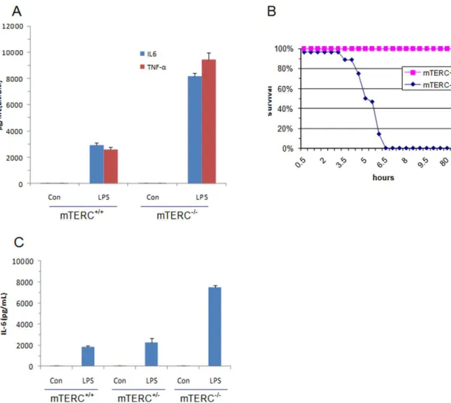
Figure 2. TLR4 induced inflammation inmTERC+/+and
mTERC2/2mice.(A) Upon LPS stimulation ofmTERC+/+
andmTREC2/2BMDMs, IL6 and
TNFalevels were determined by ELISA (right) (B) Age-matchedmTERC+/+andmTERC2/2littermates (n = 8) were administered intraperitoneally with
LPS (1mg/g body weight) and monitored for survival. (C) Sera were collected after 2 h of LPS injection from different group of mice (n = 3) and
increase of IL6 and TNF-a as compared to PD 80 (data not shown). Using microarray analysis, Lou et al.[11] demonstrated that upregulation of ISG15, an interferon stimulated gene which has cytokine-like immunomodulatory properties [27], correlates with telomere shortening in human cells. This increase in the level of ISG15 was not genotoxic stress dependent but correlated with telomere length [11].
To clarify the in vivo function of TLR4 in chromosome instability, we produced systemic inflammation in age-matched WT and mTERC2/2 mice by intra-peritoneal LPS injection. Notably, mTERC2/2 mice exhibited significant reduction of survival (succumbed between 3.5 h to 6.5 h after LPS adminis-tration) than WT littermate (none expired within 100 h period) (Fig. 2B). Death of mTERC2/2 mice was relevant to severe endotoxin shock and subsequent systemic inflammatory reaction as their serum circulating levels of IL6 and TNF-areached greater than 4-fold within 2 h of LPS injection and 1.5 h before the first death (Fig. 2C).
TLR4-induced transcription of IL-6 is closely controlled by the nuclear factor (NF)-kB [28]. To determine the specific role of NF-kB in the differential immune regulation in WT andmTERC2/2bone marrow-derived macrophages, we performed NF-kB DNA binding assays with nuclear extract from these cells at several time points. NF-kB binds with equal intensity to the TNF-a promoter probe within 1 h of LPS stimulation of the WT andmTERC2/2 bone marrow-derived macrophages. In mTERC+/+
cells, this binding disappears within 2.5 h. In contrast, binding ofmTERC2/2NF-kB remains constant for at least 4 h and only started declining after 5.3 h of LPS stimulation (Fig. 3A and B). We have identified the mechanism of this longer half-life of NF-kB in chromosome instability as the function of activating transcription factor (ATF)-3.
We measured the relative nuclear concentration of Rel A (p65 subunit of NF-kB) and ATF-3 in the nuclear extracts, which we used for NF-kB DNA binding assays. While LPS-induced augmentation of Rel A persisted up to 4 h in WT cells, that inmTERC2/2cells remained steady until 5.3 h (Fig. 3C). An increasing amount of ATF-3 was consistently correlated with the decreasing amount of Rel A at all time points, indicative of negative regulatory function of ATF-3 in LPS-induced inflammation and chromosomal instability.
Many cancer cells do not metastasise because successful metastasis requires cancer cell specific intrinsic factors as well as extrinsic factors derived from tumour microenvironment. In these cases, we believe that mutational events during the process of carcinogenesis select out cells with activated TLRs that can orchestrate the production of inflammatory mediators (IL-1, IL-6, TNF-aetc.) and recruits immune cells overloaded with TLRs. In combination, they orchestrate a signalling cascade leading to overproduction of many molecules such as angiogenic factors that facilitate cancer progression and metastasis. While exploring the connection between chromosomal instability and immune re-sponses, we have now established the obligatory role of toll-like receptors. Our findings implicate that background chromosome instability in the cellular level stabilises the action of TLR4-induced NF-kB action and sensitises cells to produce excess pro-inflammatory mediators. Chromosome instability status may be used as a prognostic indicator because it has potential to orchestrate an inflammatory microenvironment for many pro-gressive diseases including cancers. The finding that TLR4 action correlates to the telomere length and associated chromosome instability in mouse cells suggests that telomeres are involved in broader immune functions beyond the protection of chromo-somes. We explained one of the root causes of the inflammation by linking host’s telomere-dependent chromosomal status and deregulation of immunity. It raises optimism for controlling inflammation by non-toxic TLR antagonists among high-risk groups.
Materials and Methods
Ethics Statement
The animal breeding protocol as well as the experiments conducted in this study are approved by the Institutional Animal Care and Use Committee (IACUC) of the National University of Singapore (IACUC Protocol 755/05 and 828/05).
Reagents and cells
LPS from E.coli serotype 055:B5 (phenol extracted and then chromatograpically purified by gel filtration) was purchased from Sigma. LPS was solubilised in distilled water by sonication. Bone marrow was collected from femurs in complete RPMI with 10% heat-inactivated FBS, 2 mM glutamine, 100 U/ml penicillin, 100 ug/ml streptomycin and 50 ng/ml rmM-CSF. Resident peritoneal exudate cells from wild type and mTERC2/2 mice were isolated from the peritoneal cavity of mice 3 d after injection with 2 ml of 4% thioglycollate medium (Sigma) and were cultured in RPMI1640 supplemented with 10% foetal bovine serum. Both peritoneal exudate cells and bone marrow-derived macrophages were stimulated with LPS (10 ng/ml) for the indicated times and production of IL6 and TNFa were measured by enzyme-linked immunosorbent assay (ELISA).
Florescencein SituHybridisation (FISH) with Peptide Nucleic Acid-telomere Probe
Plates containing primary MEFs or established cell lines were treated with colcemid (0.1mg/ml) for 4 - 5 h and subsequently
Figure 3. TLR4 induced inflammation inmTERC+/+and
mTERC2/2
mice. (A) and (B) BMDMs frommTERC+/+
and mTERC2/2 mice were
stimulated with 10 ng/mL LPS for the indicated times. Nuclear extracts were subjected to EMSA using NF-kB probe. (C) Nuclear extracts above were also subjected to western blot for ATF3 and Rel (p65 subunit of NF-kB). Histone H1 was used as a loading control (right).
trypsinised and spun for 8 min at 120 g. After hypotonic swelling in sodium citrate (0.03 M) for 20 min at 37uC, cells were fixed in methanol/acetic acid (3:1). After 2–3 additional changes of fixative, cell suspensions were dropped on wet, clean slides and dried overnight. FISH with Cy-3 labelled (CCCTAA)3 peptide
nucleic acid, and subsequent quantitative analysis of digital images were performed [6].
Cytokinesis-blocked micronucleus assay
Chromosome instability was also measured by cytokinesis-blocked micronucleus assay [29,30]. Cells were incubated with cytochalasin B (Sigma, 5mg/ml) for 22 h. The cells were then trypsinised and subsequently fixed using a combination of both Carnoy’s fixative (acetic acid/methanol 1:3) and three to four drops of formaldehyde (to fix the cytoplasm). Fixed cells were dropped onto clean slides and stained with 3mg/ml of acridine orange, which differentially stains cytoplasm and nucleus [31,32]. One thousand binucleated cells were scored for each sample.
Measurement of proinflammatory cytokine concentrations
Primary peritoneal macrophages, bone marrow-derived mac-rophages, and MEFs were cultured with 50 ng/ml ofEscherichia coli LPS for 4–8 h. To produce systemic inflammation in mouse, LPS (1mg/g body weight) was given by intraperitoneal injection. Blood was obtained by retro-orbital puncture after anaesthesia with methoxyflurane, and the mice were asphyxiated with carbon
dioxide. Survival of the mouse was monitored for one week post injection of LPS. Concentration of TNFa and IL6 in culture supernatants and serum were measured by ELISA according to manufacturer’s instructions (R & D Research systems).
Electrophoretic Mobility shift assay
The nuclear extracts of bone marrow derived macrophages (56106) were purified after LPS stimulation. The extract was incubated with a specific probe for NF-kB DNA binding site, electrophoresed, and visualised by autoradiography.
Western Blot analysis
Purified nuclear extracts used in electrophoretic mobility shift assay were resolved by SDS-PAGE (40mg total protein) and transferred onto a polyvinylidene fluoride membrane. The membrane was blotted with the specific antibodies to indicated proteins and visualised with an enhanced chemiluminescence system (NEN life science).
Acknowledgments
Dr. Veena Hande is thanked for critically reading the manuscript.
Author Contributions
Conceived and designed the experiments: RNB SA MPH. Performed the experiments: RNB BB. Analyzed the data: RNB MPH. Contributed reagents/materials/analysis tools: BB SA MPH. Wrote the paper: RNB.
References
1. Blackburn EH (1991) Telomeres. Trends Biochem Sci 16: 378–381. 2. Blackburn EH (1992) Telomerases. Annu Rev Biochem 61: 113–129. 3. Blackburn EH (2001) Switching and signaling at the telomere. Cell 106:
661–673.
4. Blackburn EH, Greider CW, Szostak JW (2006) Telomeres and telomerase: the path from maize, Tetrahymena and yeast to human cancer and aging. Nat Med 12: 1133–1138.
5. Blasco MA, Lee HW, Hande MP, Samper E, Lansdorp PM, et al. (1997) Telomere shortening and tumor formation by mouse cells lacking telomerase RNA. Cell 91: 25–34.
6. Hande MP, Samper E, Lansdorp P, Blasco MA (1999) Telomere length dynamics and chromosomal instability in cells derived from telomerase null mice. J Cell Biol 144: 589–601.
7. Newman JP, Banerjee B, Fang W, Poonepalli A, Balakrishnan L, et al. (2008) Short dysfunctional telomeres impair the repair of arsenite-induced oxidative damage in mouse cells. J Cell Physiol 214: 796–809.
8. Nijnik A, Woodbine L, Marchetti C, Dawson S, Lambe T, et al. (2007) DNA repair is limiting for haematopoietic stem cells during ageing. Nature 447: 686–690.
9. Rudolph KL, Chang S, Lee HW, Blasco M, Gottlieb GJ, et al. (1999) Longevity, stress response, and cancer in aging telomerase-deficient mice. Cell 96: 701–712. 10. Wiemann SU, Satyanarayana A, Buer J, Kamino K, Manns MP, et al. (2005) Contrasting effects of telomere shortening on organ homeostasis, tumor suppression, and survival during chronic liver damage. Oncogene 24: 1501–1509.
11. Lou Z, Wei J, Riethman H, Baur JA, Voglauer R, et al. (2009) Telomere length regulates ISG15 expression in human cells. Aging (Albany NY) 1: 608–621. 12. Poonepalli A, Balakrishnan L, Khaw AK, Low GK, Jayapal M, et al. (2005)
Lack of poly(ADP-ribose) polymerase-1 gene product enhances cellular sensitivity to arsenite. Cancer Res 65: 10977–10983.
13. d’Adda di Fagagna F, Hande MP, Tong WM, Lansdorp PM, Wang ZQ, et al. (1999) Functions of poly(ADP-ribose) polymerase in controlling telomere length and chromosomal stability. Nat Genet 23: 76–80.
14. Altmeyer M, Hottiger MO (2009) Poly(ADP-ribose) polymerase 1 at the crossroad of metabolic stress and inflammation in aging. Aging (Albany NY) 1: 458–469.
15. Bhaumik D, Scott GK, Schokrpur S, Patil CK, Orjalo AV, et al. (2009) MicroRNAs miR-146a/b negatively modulate the senescence-associated inflammatory mediators IL-6 and IL. Aging (Albany NY) 1: 402–411. 16. Coussens LM, Werb Z (2002) Inflammation and cancer. Nature 420: 860–867.
17. Shacter E, Weitzman SA (2002) Chronic inflammation and cancer. Oncology (Williston Park) 16: 217–226, 229; discussion 230–212.
18. Karin M (2006) Nuclear factor-kappaB in cancer development and progression. Nature 441: 431–436.
19. Karin M, Greten FR (2005) NF-kappaB: linking inflammation and immunity to cancer development and progression. Nat Rev Immunol 5: 749–759. 20. Park EJ, Lee JH, Yu GY, He G, Ali SR, et al. (2010) Dietary and genetic obesity
promote liver inflammation and tumorigenesis by enhancing IL-6 and TNF expression. Cell 140: 197–208.
21. Forst B, Hansen MT, Klingelhofer J, Moller HD, Nielsen GH, et al. (2010) Metastasis-inducing S100A4 and RANTES cooperate in promoting tumor progression in mice. PLoS One 5: e10374.
22. Kim S, Takahashi H, Lin WW, Descargues P, Grivennikov S, et al. (2009) Carcinoma-produced factors activate myeloid cells through TLR2 to stimulate metastasis. Nature 457: 102–106.
23. Akira S, Takeda K (2004) Toll-like receptor signalling. Nat Rev Immunol 4: 499–511.
24. Takeda K, Kaisho T, Akira S (2003) Toll-like receptors. Annu Rev Immunol 21: 335–376.
25. Ulevitch RJ (2004) Therapeutics targeting the innate immune system. Nat Rev Immunol 4: 512–520.
26. Guachalla LM, Ju Z, Koziel R, von Figura G, Song Z, et al. (2009) Sod2 haploinsufficiency does not accelerate aging of telomere dysfunctional mice. Aging (Albany NY) 1: 303–315.
27. D’Cunha J, Knight E Jr, Haas AL, Truitt RL, Borden EC (1996) Immunoregulatory properties of ISG15, an interferon-induced cytokine. Proc Natl Acad Sci U S A 93: 211–215.
28. Kawai T, Akira S (2007) Signaling to NF-kappaB by Toll-like receptors. Trends Mol Med 13: 460–469.
29. Fenech M (2007) Cytokinesis-block micronucleus cytome assay. Nat Protoc 2: 1084–1104.
30. Fenech M (2008) The micronucleus assay determination of chromosomal level DNA damage. Methods Mol Biol 410: 185–216.
31. Hande MP, Boei JJ, Natarajan AT (1996) Induction and persistence of cytogenetic damage in mouse splenocytes following whole-body X-irradiation analysed by fluorescence in situ hybridization. II. Micronuclei. Int J Radiat Biol 70: 375–383.