Recebido em 03.10.2003. / Received in October, 03rdof 2003.
Aprovado pelo Conselho Consultivo e aceito para publicação em 08.10.2003. / Approved by the Consultive Council and accepted for publication in October 08thof 2003. * Trabalho realizado no Ambulatório de Dermatologia Pediátrica do Serviço de Dermatologia do Hospital das Clínicas da Faculdade de Medicina da UFMG. / Work done at the Pediatric
Dermatology Outpatient Clinic, Dermatology Service of the Hospital das Clínicas, Faculty of Medicine, UFMG.
1Professor Adjunto - Doutor em Dermatologia da Faculdade de Medicina da Universidade Federal de Minas Gerais; Coordenador do Ambulatório de Dermatologia Pediátrica do Serviço de Dermatologia do Hospital das Clínicas da Faculdade de Medicina da UFMG. / Adjunct Professor- Ph.D. in Dermatology, Faculty of Medicine, Federal University of Minas Gerais (UFMG); Coordinator of the Pediatric Dermatology Outpatient Clinic, Dermatology Service, Hospital das Clínicas, Faculty of Medicine, UFMG.
2Mestre em Dermatologia pela UFMG. Médica do Ambulatório de Dermatologia Pediátrica do Serviço de Dermatologia do Hospital das Clínicas da Faculdade de Medicina da UFMG. / Masters in Dermatology, UFMG. M.D. at the Pediatric Dermatology Outpatient Clinic, Dermatology Service, Hospital das Clínicas, Faculty of Medicine, UFMG.
3Professora Assistente de Dermatologia da Faculdade de Medicina da UFMG. Mestre em Dermatologia pela UFMG. Docente do Ambulatório de Dermatologia Pediátrica do Serviço de Dermatologia do Hospital das Clínicas da Faculdade de Medicina da UFMG./ Assistant Professor of Dermatology, Faculty of Medicine, UFMG. Masters in Dermatology, UFMG. Member of teaching staff at the Pediatric Dermatology Outpatient Clinic, Dermatology Service, Hospital das Clínicas, Faculty of Medicine, UFMG.
©2003by Anais Brasileiros de Dermatologia
Hemangioma da infância
*
Hemangioma of infancy
*
Bernardo Gontijo
1Cláudia Márcia Resende Silva
2Luciana Baptista Pereira
3Resumo:As novas classificações disponíveis e os modernos recursos diagnósticos por imagem não só per-mitiram a diferenciação entre os tumores e as malformações vasculares, mas também modificaram de forma substancial a abordagem e o tratamento dessas anomalias. O hemangioma da infância, o mais comum dos tumores vasculares dessa faixa etária e objeto deste trabalho, é revisto do ponto de vista de suas caracterís -ticas clínicas e laboratoriais, diagnóstico diferencial e opções terapêu-ticas. Embora a conduta expectante permaneça como o tratamento de escolha para a maioria dos casos, o julgamento crítico é crucial para o emprego de outras modalidades terapêuticas.
Palavras-chave: hemangioma; hemangioma capilar; literatura de revisão.
Summary:New classifications and availability of modern radiologic diagnostic tools have not only
allowed a precise distinction between vascular tumors and vascular malformations but have also sig nificantly modified management and treatment of these vascular anomalies. Hemangioma of infan cy, the most common vascular tumor of this age and subject of this review, is approached from its clin ical and laboratory features, differential diagnosis and therapeutic options. Although noninterven -tion remains the treatment of choice for the majority of cases, critical judgement is mandatory to decide whether other therapeutic modalities should be used.
Key words: hemangioma; hemangioma, capillary; review literature.
INTRODUÇÃO
Por muitos séculos as lesões vasculares congênitas foram denominadas nevus maternus, refletindo a crença
popular da participação da mãe nas lesões de seus filhos. Acreditava-se que emoções e desejos maternos poderiam imprimir uma marca nos recém-nascidos, e, dependendo da cultura de cada povoado, as mães eram culpadas por inge-rir ou não determinadas frutas vermelhas ou outros tipos de alimento durante a gravidez, advindo daí os termos que adjetivavam as lesões como morango, framboesa, vinho do Porto e salmão.1
Virchow, em 1863, foi quem classificou pela pri-meira vez as anomalias vasculares, com base em seu qua-dro microscópico, em angioma simples, cavernoso e race-moso.2Acreditava que cada um desses tipos poderia
trans-formar-se em outro por proliferação celular ou dilatação de
INTRODUCTION
For many centuries congenital vascular lesions were denominated nevus maternus, reflecting a popular belief that mothers were responsible for their children's lesions. It was believed that emotions and maternal desires could print a mark on the newly born, and, depending on the culture of each region, the mothers were blamed for ingesting or not ingesting certain red fruits or other food types during the pregnancy, hence the terms describing the lesions as strawberry, raspberry, port wine and salmon.1
Virchow, in 1863, classified the vascular anomalies for the first time, on the basis of their microscopic aspect, into simple, cavernous and racemosum angioma.2He
vasos. Wegener, discípulo de Virchow, com base nos estu-dos deste, propôs em 1877 uma classificação semelhante para as alterações linfáticas que persistiu até fins do século XX: linfangioma simples, cavernoso e cistóide. Atribuía a gênese dessas lesões à inflamação e dilatação linfática, mal-formação ou proliferação endotelial.3
O termo hemangioma foi empregado durante anos, de forma ampla e indiscriminada, para designar anomalias vasculares totalmente distintas quanto a sua gênese, carac-terísticas clínicas e histopatológicas, evolução e prognósti-co. Assim, hemangioma capilar ou em morango eram as expressões utilizadas para designar o que atualmente se conhece como a forma superficial do hemangioma da infância: um tumor, na acepção da palavra, provocado pela multiplicação das células endoteliais, presente ou não ao nascimento, com fases consecutivas de crescimento, para-lisação e regressão, geralmente desprovido de maior signi-ficado clínico e de conseqüências na maioria das vezes cos-méticas. Ao mesmo tempo, hemangioma plano era a deno-minação do que hoje é classificado como mancha em vinho do Porto: uma malformação vascular, na quase-totalidade dos casos presente ao nascimento, com crescimento pro-porcional ao desenvolvimento da criança, de caráter per-manente, podendo associar-se a síndromes diversas. Além disso, adjetivos como cavernoso, um termo descritivo his-tológico e que deve ser preservado com esse fim, referiam-se a características clínicas como a cor azulada sugestiva de lesões profundas.1 Dessa forma, lesões radicalmente
diversas, como a forma profunda do hemangioma da infân-cia (que regride espontaneamente) e as malformações vas-culares do subcutâneo (que são permanentes), que apresen-tam em comum apenas a coloração azulada, eram igual-mente caracterizadas como cavernosas. A nomenclatura confusa e a ausência de uma classificação adequada foram os principais responsáveis pelas dificuldades diagnósticas e terapêuticas das anomalias vasculares.
Em 1982, Mulliken e Glowacki propuseram uma nova classificação das lesões vasculares baseada nas mani-festações clínicas, quadro histopatológico e história natural (Quadro 1), distinguindo-as em hemangiomas e malforma-ções vasculares.4,5
O hemagioma da infância é o tumor vascular mais comum nessa faixa etária. Entretanto, outros tumores vas-culares, mais raros, têm sido descritos. Em função disso, em 1996, a classificação de Mulliken e Glowacki foi revista, e a nova classificação, adotada como oficial pela Sociedade Internacional para o Estudo de Anomalias Vasculares - ISSVA, divide as lesões vasculares em dois grupos: tumores e malformações vasculares.6Os tumores
vasculares são neoplasias da vasculatura (proliferação celular) e incluem hemangioma da infância, hemangioma congênito rapidamente involutivo, hemangioma congênito não involutivo, angioma em tufos, hemangioendotelioma kaposiforme e granuloma piogênico. As malformações vasculares consistem em erros da morfogênese e são
clas-these studies, proposed a similar classification for the lym-phatic alterations that was used until the end of the 20th Century: lymphangioma simplex, cavernosum and cys-ticum. It attributed the genesis of these lesions to lymphatic inflammation and dilation, and to endothelial malformation or proliferation.3
The term hemangioma was used for many years, in a wide and indiscriminate manner, to designate vascular anomalies that were totally different in terms of their gene-sis, clinical and histopathological characteristics, clinical course and prognostic. Hence, capillary or strawberry hemangioma were the expressions used to designate what is now known as the superficial form of hemangioma of child-hood: a tumor, in the meaning of the word, caused by the multiplication of endothelial cells, that may or may not be present at birth, with consecutive phases of growth, stop-page and regression. They are usually of little clinical sig-nificance and in the majority of cases the consequences are only cosmetic. At the same time, hemangioma planum was the denomination that today is classified as port wine stain: a vascular malformation, almost always present at birth, with growth proportional to the child's development and with a permanent character that can be associated to sev-eral syndromes. Furthermore, adjectives such as cav-ernous, a histological descriptive term that should be reserved for this finality, referred to clinical characteristics such as the blue coloration suggestive of deep lesions.1in
this way, lesions that are radically different, as in the case of the deep form of hemangioma of childhood (with sponta-neous regression) and the subcutasponta-neous vascular malfor-mations (that are permanent), each presenting no more than a bluish coloration in common, were characterized equally as cavernous. The confusing nomenclature and absence of an appropriate classification were the main cause of the diagnostic and therapeutic difficulties surrounding vascu-lar anomalies.
In 1982, Mulliken and Glowacki proposed a new classification for the vascular lesions based on the clinical manifestations, histopathological picture and natural histo-ry (Chart 1), distinguishing them into hemangiomas and vascular malformations.4,5
classified according to the predominant vein (Chart 2).6,7,8
As in all classifications, it is not absolute. However, its sim-plicity and clinical relevance enabled an important advance in the management of vascular anomalies.
This EMC-D (Continuing Medical Education
-Dermatology) article specifically approaches hemangioma of childhood. The other vascular tumors will be commented on in summarized form in the differential diagnosis, and the vascular malformations will be the subject of the next chap-ter of EMC-Din this Journal.
HEMANGIOMA OF CHILDHOOD
sificadas de acordo com o vaso predominante (Quadro 2).6,7,8Assim como todas as classificações, essa não é
abso-luta. Porém, sua simplicidade e relevância clínica possibi-litaram um avanço importante na abordagem das anoma-lias vasculares.
Este artigo de EMC-D aborda especificamente o hemangioma da infância. Os demais tumores vasculares serão comentados de forma sumária no diagnóstico diferen-cial, e as malformações vasculares serão objeto do próximo capítulo da EMC-Dneste periódico.
HEMANGIOMA DA INFÂNCIA
Hemangiomas / Hemangiomas Malformações / Malformations
Apresentam fase proliferativa e involutiva Malformações capilares, venosas, arteriais, linfáticas e fístulas sem fase proliferativa e involutiva
Presenting proliferative and involution phases Malformations of capillaries, veins, arteries, lymphs and fistulae, without a proliferative and involution phase
Proliferação das células endoteliais Ciclo das células endoteliais normal Proliferation of the endothelial cells Normal cycle of endothelial cells 40% presentes ao nascimento, usualmente como 90% reconhecidas ao nascimento uma mancha vermelha
Present at birth in 40%, usually with a red patch Recognized at birth in 90%
Crescimento pós-natal rápido e involução lenta Crescem proporcionalmente à criança Rapid postnatal growth and slow involution Grow proportionally with the child Razão sexo feminino:masculino - 5:1 Razão sexo feminino:masculino - 1:1 Female:male ratio - 5:1 Female:male ratio - 1:1
Fonte: Mulliken JB, Gloawacki, J, 1982 / Source: Mulliken JB, Gloawacki, J, 1982
Fonte: Enjolras O,1997; Bruckner AL, Frieden IJ, 2003 / Source: Enjolras O,1997; Bruckner AL, Frieden IJ, 2003 Quadro 1: Classificação das lesões vasculares segundo Mulliken e Glowacki (1982).
Chart 1: Classification according to Mulliken and Glowacki (1982).
Quadro 2: Tumores e malformações vasculares. / Chart 2: Tumors and vascular malformations
Tumores vasculares / Vascular tumors Malformações vasculares / Vascular malformations Hemangioma Simples Combinadas Hemangioma Simple Combined Hemangima congênito rapidamente involutivo Capilar (MC) Fístula AV Congenital hemangioma rapid involuting-type Capillary (MC) Fistula AV Hemangioma congênito não involutivo Linfática (ML) MAV, MCV Non-involuting congenital hemangioma Lymphatic (ML) MAV, MCV Granuloma piogênico Venosa (MV) MCLV, MLV Pyogenic granuloma Venous (MV) MCLV, MLV Hemangioendotelioma kaposiforme Arterial (MA) MCAV, MCLA Hemangioendothelioma kaposiform Arterial (MA) MCAV, MCLAV Angioma em tufos
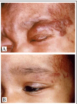
Figura 1: Lesão precurso-ra: mácula eritematosa com telangiectasias
Figure 1: Precursory lesion: erythematous
macule with telangiectasia
É o tumor mais comum da infância, ocorrendo em 10 a 12% das crianças com um ano de idade e atingindo até 30% daquelas com peso muito baixo ao nascimento (menos de 1.000g).9O risco de hemangioma é 10 vezes maior nas
crian-ças cujas mães foram submetidas à biópsia de vilo coriônico durante a gravidez,10e é nítida a predileção pelo sexo
femini-no em razão que varia de 3:1 até 7:1.11,12 Cerca de 80% dos
pacientes apresentam lesões únicas, sendo rara a presença de quatro ou mais lesões. A pele é o órgão mais comumente aco-metido, e as regiões da cabeça e do pescoço (60%), e o tron-co (25%) são as mais afetadas.12,13,14O tamanho pode variar de
poucos milímetros até vários centímetros.
QUADRO CLÍNICO
A apresentação clínica varia de acordo com tamanho da lesão, local acometido, profundidade e estágio de evolução. Em geral ausente ao nascimento, o hemangioma da infância torna-se aparente já nos primeiros dias de vida. Uma lesão inicial, conhecida como lesão precursora (Figura 1), está presente em percentual que varia de 30% a 50% dos casos ao nascimento e pode apresentar-se clinicamente sob a forma de uma mancha anêmica, mancha eritematosa e/ou equimótica, pequeno agru-pamento de pápulas vermelho-vivo ou ainda telangiectasias cir-cundadas ou não por um halo anêmico.15O crescimento é
rápi-do, e mais de 90% dos hemangiomas estão bem evidentes ao término do primeiro mês de vida.1,15,16,17Hemangiomas mais
pro-fundos (subcutâneo) podem tornar-se aparentes mais tardia-mente, alguns meses após o nascimento.8,12,13 Em raras
situa-ções, ao nascimento, o hemangioma apresenta-se como lesão ulcerada cujo componente angiomatoso aparece posteriormen-te ou como lesão tumoral parcialmenposteriormen-te desenvolvida e que tam-bém apresenta crescimento pós-natal rápido.16,18,19,20
Podem ser divididos em superficiais, profundos ou combinados de acordo com sua aparência clínica.21,22Os
super-ficiais (50-60% dos casos)15,23são bem delimitados, de cor
ver-melho-vivo, nodulares ou em placa, com pele normal ao redor, por vezes de aspecto semelhante a um morango (Figura 2). Normalmente restritos à derme papilar e reticular, revelam-se à palpação como uma massa firme, pouco compressível, podendo apresentar aumento da temperatura no local. Com a regressão da lesão, a cor
aver-melhada vai clareando, do cen-tro para a periferia, adquirindo um tom azul-acinzentado.9,24
Eventualmente podem ter apa-rência macular, com a pele levemente elevada, sendo con-fundidos com a malformação
This is the most common tumor of childhood, occur-ring in 10 to 12% of children with one year of age and involving up to 30% of those with very low birth weight (less than 1,000 g).9 The risk of hemangioma is 10 times
higher in children whose mothers were submitted to biopsy of the chorionic villi during pregnancy,10and there is a clear
feminine bias with a ratio that varies from 3:1 to 7:1.11,12
About 80% of the patients present single lesions and the presence of four or more lesions is rare. The skin is the organ most commonly involved, and particularly in areas of the head and neck (60%), and the trunk (25%).12,13,14The size
can vary from a few millimeters to several centimeters.
CLINICAL PICTURE
The clinical presentation varies according to the size of the lesion, location, depth and stage of clinical course. In general absent at birth, hemangioma of the childhood becomes apparent within the first days of life. An initial lesion, known as a precursory lesion (Figure 1), is present in a percentage that varies from 30 to 50% of the cases at birth and can present clinically in the form of an anemic stain, erythematous and/or ecchymotic patch, a small group of bright-red papules or even telangiectasia that may or may not be surrounded by an anemic halo.15The growth is fast,
and over 90% of the hemangiomas are very evident by the end of the first month of life.1,15,16,17More profound
heman-giomas (subcutaneous) become apparent later, generally several months after birth.8,12,13In rare situations, the
heman-gioma presents at birth as an ulcerated lesion whose angiomatous component appears later or as a partially developed tumoral lesion and that also presents fast post-natal growth.16,18,19,20
They can be divided into superficial, profound or combined according to their clinical appearance.21,22 The
superficial form (50-60% of the cases)15,23 have
well-defined borders, with a bright red color and nodular or in plaques, with normal surrounding skin, often with an aspect similar to a strawberry (Figure 2). They are usual-ly restricted to the papillary and reticular dermis, on pal-pation they have a firm, only slightly compressible mass that can present a localized increase in temperature. With regression of the lesion, the red color becomes lighter, from the center to the edges and acquires a gray-ish-blue tone.9,24 They can
Figure 2: Hemangioma of infancy - frontal region (superficial form)
Figura 2: Hemangioma da infância - região frontal (forma superficial)
Figura 3: Hemangioma da infância (forma combinada)
Figure 3: Hemangioma of infancy (combined form)
appearance, with slightly raised skin, that can be mistaken by capil-lary malformation. The deep hemangiomas (15% of cases),15,23
previously denominated cavernous, are nodular lesions, either present-ing the skin color or of a bluish tone, sometimes with telangiectasia in the surface, it is occasionally possible to observe drainage veins in the periphery. They involve the lower dermis and subcutaneous tis -sue.8,25 Mixed or combined lesions
(Figure 3) account for the remain-ing 25 to 35% of cases.15,23
The rapid phase of growth usually lasts from six to 10 months of life. It can be a little more prolonged in the deep hemangioma, however it rarely exceeds two years of age.26In general the maximum size is reached by the end of
the first year of life, although a few cases present in a fray form, with very discreet growth, and remain almost indis -tinguishable from the precursory lesion.8Later the
heman-gioma enters a quiescent phase that persists for several months, followed by a slow involution. It is considered that 50% of the lesions regress either totally or partially by up to five years of age and 90% by 10 years of age.13,23
Involution and proliferation can be observed simultaneous-ly in different parts of the same lesion or in different heman-giomas of a child with multiple lesions.9The regression is
considered esthetically acceptable - with discreet altera-tions of the skin - in more than 50% of the cases. Residual lesions observed include telangiectasia, atrophy, redundant skin, retraction of the skin and hypopigmentation, among others.1,8,9,12,13,16The complete resolution of the hemangioma
is not influenced by size of the lesion, ulceration, depth, the child's sex or age at onset, but appears to be related to an early beginning of the involution process.9,15
ETIOPATHOGENESIS
Although the
phys-capilar. Os hemangiomas profundos (15% dos casos),15,23 anteriormente
denominados cavernosos, são lesões nodulares, da cor da pele ou de tom azulado, algumas vezes com telan-giectasias na superfície, sendo ocasio-nalmente possível observar vasos de drenagem na periferia. Acometem a derme profunda e o tecido subcutâ-neo.8,25As lesões mistas ou
combina-das (Figura 3) respondem pelos 25 a 35% dos casos restantes.15,23
A fase de crescimento rápi-do normalmente dura até o períorápi-do de seis a 10 meses de vida. Pode
ser um pouco mais prolongada no hemangioma profundo, porém raramente excede os dois anos de idade.26Em geral
o tamanho máximo é atingido ao final do primeiro ano de vida, embora alguns poucos casos se apresentem como forma frusta, de crescimento muito discreto, e permane-çam quase indistinguíveis da lesão precursora.8
Posteriormente o hemangioma entra em uma fase quies-cente, que persiste por alguns meses, e então involui len-tamente. Estima-se que 50% das lesões regridam total ou parcialmente até os cinco anos de idade e 90% até os 10 anos de idade.13,23Involução e proliferação podem ser
vis-tas simultaneamente em partes distinvis-tas de uma mesma lesão ou em diferentes hemangiomas de uma criança com múltiplas lesões.9A regressão é considerada
esteticamen-te aceitável - com alesteticamen-terações discretas da pele - em mais de 50% dos casos. Lesões residuais observadas incluem telangiectasias, atrofia, pele redundante, retração da pele e hipopigmentação, entre outras.1,8,9,12,13,16A completa
reso-lução do hemangioma não é influenciada por tamanho da lesão, ulceração,
profundi-dade, sexo da criança ou idade de apresentação, mas parece estar relacionada ao início precoce da involu-ção.9,15
ETIOPATOGENIA
fisiopato-genia não ser bem conhecida, considera-se atualmente que os hemangiomas são o resultado de um desequilíbrio na angio-gênese que permite a proliferação descontrolada de elementos vasculares.1,8,27,28 Durante a fase proliferativa do tumor, vários
marcadores da angiogênese estão aumentados, como o fator de crescimento básico de fibroblasto (bFGF), fator de cresci-mento do endotélio vascular (VEGF), metaloproteinases, antí-geno de proliferação celular nuclear (PCNA), colagenase tipo IV, uroquinases, proteases e E-selectina. Esses fatores dimi-nuem quando da involução do tumor, com exceção do bFGF, que permanece elevado no início da regressão, mas atinge níveis normais quando o hemangioma completa sua involu-ção. O nível urinário de bFGFpode ser empregado como parâ-metro da eficácia do tratamento dos hemangiomas. Os fatores bFGFe VEGFtêm um importante papel na diferenciação das células endoteliais na fase de proliferação rápida do tumor. Essas células endoteliais diferenciadas, por sua vez, além de atraírem um grande número de mastócitos, expressam o inibi-dor tecidual da metaloproteinase tipo 1 (TIMP-1), um potente bloqueador da formação de novos vasos sangüíneos. Especula-se sobre a possibilidade de que os mastócitos secre-tem substâncias como interferons, que reduziriam a angiogê-nese e promoveriam também a involução. Outros marcadores da fase involutiva dos hemangiomas são a angiostatina, endostatina, fator 4 plaquetário, interleucina-12, interferon-α
e glicocorticóides.8,29-33
Um erro no desenvolvimento fetal no primeiro tri-mestre da gravidez ou uma alteração gênica são algumas das teorias aventadas para explicar esse desequilíbrio da angiogênese.27,28
A hipótese de uma alteração genética é baseada na existência de irmãos afetados e em relatos de maior incidên-cia de hemangiomas em alguns grupos familiares. Especula-se a possibilidade de uma herança autossômica dominante na transmissão desses tumores.8,24,27,28
HISTOPATOLOGIA
As biópsias de lesões superficiais e profundas apre-sentam quadros histopatológicos semelhantes. Na fase de crescimento do hemangioma observam-se agregados de células endoteliais proliferativas constituindo cordões sóli-dos e massas, por vezes com formação de lúmen. Essas célu-las tendem a se agrupar formando delicados lóbulos separa-dos por finos feixes de tecido conjuntivo. Nenhum separa-dos lóbu-los é encapsulado ou fibrótico, e muitos contêm tecido nor-mal e uma artéria alimentadora. A trombose e os depósitos de hemossiderina são raros e estão limitados às áreas de ulceração ou inflamação aguda. Perícitos, fibroblastos e par-ticularmente mastócitos são numerosos na fase tardia da pro-liferação. Na fase involutiva as células endoteliais achatam-se, há formação de canais progressivamente proeminentes e ectásicos, levando à formação de grandes vasos de paredes delgadas. As ilhas de tecido adiposo e fibroso vão substituin-do gradativamente as células tumorais.1,15,34,35
A imuno-histoquímica é positiva para o antígeno
iopathogenesis is not well understood, it is now considered that the hemangiomas are the result of an imbalance in the angiogenesis that allows an uncontrolled proliferation of vas-cular elements.1,8,27,28 During the proliferative phase of the
tumor, several markers of angiogenesis are increased, such as the basic fibroblast growth factor (bFGF), vascular endotheli-um growth factor (VEGF), metalloproteinase, proliferating cell nuclear antigen (PCNA), collagenase type IV, urokinase, proteases and E-selectin. These factors decrease when there is involution of the tumor, except for bFGFthat remains high in the beginning of the regression, but eventually reaches normal levels when the hemangioma completes its involution. The urinary level of bFGF can be used as a parameter for the effectiveness of the treatment of hemangiomas. Factors bFGF
and VEGFhave an important role in the differentiation of the endothelial cells in the phase of rapid proliferation of the tumor. These differentiated endothelial cells, in turn, besides attracting a great number of mastocytes, express the tissular inhibitor of metalloproteinase type 1 (TIMP-1), a potent block-er of the formation of new blood vessels. It has been speculat-ed that mastocytes possibility secrete substances such as interferon, that reduce the angiogenesis and also promote the involution. Other markers of the involution phase of heman-giomas are angiostatin, endostatin, platelet factor 4, inter-leukin-12, interferon-αand glycocorticoid.8,29-33
An anomaly in the fetal development in the first three months of pregnancy or a genetic alteration are just two of the theories proposed to explain this imbalance in the angiogenesis.27,28
The hypothesis of a genetic alteration is based on the existence of affected siblings and in reports of a higher inci-dence of hemangiomas in certain family groups. The possi-bility of an autosomal dominant inheritance is speculated in the transmission of these tumors.8,24,27,28
HISTOPATHOLOGY
The biopsies of superficial and deep lesions present similar histopathological pictures. In the growth phase of the hemangioma, aggregations of endothelial proliferative cells are observed constituting solid strings and masses, sometimes with a lumen formation. These cells tend to group together forming delicate lobules separated by fine bands of connective tissue. None of the lobules are encap-sulated or fibrotic, and many contain normal tissue and an alimentary artery. The thrombosis and hemosiderin deposits are rare and limited to areas of ulceration or acute inflammation. Pericytes, fibroblasts and especially masto-cytes are numerous in the late phase of the proliferation. In the involution phase the endothelial cells become flattened and there is a formation of progressively prominent and ectatic channels, leading to the formation of major veins with thin walls. The islands of fatty and fibrous tissue gra-dually substitute the tumoral cells.1,15,34,35
vascular origin.8,32,33 The antigen erythrocyte-type glucose
transporter protein (GLUT1), a glucose transporter usually expressed in the endothelium of the microvasculature of the brain, retina, placenta and endoneurium, but not in the nor-mal skin, was described recently as a specific marker of hemangioma of childhood, being present in all of the clini-cal phases.34 Hemangioma of childhood also presents
intense immunoreactivity with several vascular markers of the placenta, such as merosina and Lewis Y (LeY).8,36,37
DIAGNOSIS
In almost all cases, diagnosis can be reached exclusively on the basis of the physical findings and clinical his -tory. However, some hemangiomas can be mistaken by vas-cular malformations or with other types of tumors.1,8,16,23
The radiological evaluation can be useful in the dif-ferential diagnosis and in the evaluation of the size, type and extension of the lesion. It can also be used in the fol-low-up of the response to a systemic treatment.1,38,39
The Doppler ultrasonography (US) continues to be used as an exam for screening because of its low cost, easy availability and lack of risk, and since it can be performed with little or no sedation. In the proliferative phase, a well-defined homogeneous solid mass is observed, with high flow blood vessels (low arterial resistance with increased arterial and venal speeds). These characteristics enable the differenti-ation between hemangiomas and those due to low flow such as the venous capillary and lymphatic malformations, but they do not allow its distinction from the malformations of high flow, for example when arteriovenous. USis also useful in the differential diagnosis with other common tumors of child-hood, such as dermal cyst, lipoma or meningocele. The limi-tations of USare its small field of vision, restricted visibility in deep lesions, difficulty in evaluating superficial flat lesions and in detecting very small vessels with low flow.8,39,40,41
The magnetic resonance test (MR) is considered the best exam to confirm the tissular characteristics of the lesion, its extension into the deeper anatomical levels and to evaluate associated adjacent anomalies. Computed tomography (CT) scans with contrast can be substituted for
MR, but it is less precise in the evaluation of tissular char-acteristics and of blood flow. In the proliferative phase, both CT and MR reveal well-defined lesions that are uni-form, densely lobulated, with alimentary and drainage ves-sels in the center or in the periphery. The MR test shows evi-dence of an isointense or hypointense homogeneous mass in relation to the muscle tissue in the weighted sequence in T1 and hyperintense when weighted in T2. An intense and uni-form condition is also brought out by using the gadolinium contrast. The use of the contrast is an important aid in the differential diagnosis between the hemangiomas and malig-nant tumors or other masses that may resemble a heman-gioma.8,38,42,43
The angiography is not used for diagnosis, but it can be useful for atypical lesions in which surgery or
emboliza-CD31, fator Von Willebrand e uroquinase, confirmando sua origem vascular.8,32,33 O antígeno erythrocyte-type glucose
transporter protein (GLUT1), um transportador de glicose normalmente expresso no endotélio da microvasculatura do cérebro, retina, placenta e endoneuro, mas não na pele normal, foi descrito recentemente como um marcardor específico do hemangioma da infância, estando presente em todas as suas fases evolutivas.34 Além disso, o hemangioma da infância
apresenta imunorreatividade intensa para vários marcadores vasculares da placenta, como merosina e Lewis Y (LeY).8,36,37
DIAGNÓSTICO
Na quase-totalidade dos casos o diagnóstico pode ser realizado com base exclusivamente nos achados físicos e história clínica. Entretanto, alguns hemangiomas podem ser confundidos com malformações vasculares ou com outros tipos de tumores.1,8,16,23
A avaliação radiológica pode ser útil tanto no diag-nóstico diferencial quanto na avaliação do tamanho, tipo e extensão da lesão. Pode ser utilizada também no acompa-nhamento da resposta a um tratamento sistêmico.1,38,39
A ultra-sonografia (US) com Doppler vem sendo empregada como um exame de screening por ser de baixo custo, fácil acesso e desprovida de risco, podendo ser realiza-da com pouca ou nenhuma serealiza-dação. Na fase proliferativa observa-se uma massa sólida homogênea, bem delimitada, com vasos de alto fluxo (baixa resistência arterial e velocida-des arterial e venosa aumentadas). Essas características possi-bilitam a diferenciação entre os hemangiomas e as malforma-ções de baixo fluxo, como as venosas, capilares e linfáticas, mas não permitem sua distinção das malformações de alto fluxo, como as arteriovenosas. A UStambém é útil no diagnós-tico diferencial com outros tumores comuns na infância, como cisto dermóide, lipoma ou meningocele. As limitações da US
são o pequeno campo de visão, restrição de visibilidade nas lesões profundas, dificuldade em avaliar lesões planas superfi-ciais e detectar vasos muito pequenos e com baixo fluxo.8,39,40,41
A ressonância magnética (RM) é considerada o melhor exame para confirmar as características teciduais da lesão, sua extensão nos diversos planos anatômicos e para avaliar anomalias adjacentes associadas. A tomografia com-putadorizada (TC) com contraste pode substituir a RM, mas é mais imprecisa na avaliação das características teciduais e do fluxo sangüíneo. Na fase proliferativa, a TCe a RM mos-tram lesões bem delimitadas, uniformes, densamente lobula-das, com vasos de alimentação e drenagem no centro ou na periferia. A RMevidencia massa homogênea isointensa ou hipointensa em relação ao músculo na seqüência ponderada em T1 e hiperintensa quando ponderada em T2, além do realce intenso e uniforme com o contraste gadolinium. O uso de contraste é importante no auxílio do diagnóstico diferen-cial entre os hemangiomas e tumores malignos ou outras massas que se assemelham ao hemangioma.8,38,42,43
cirurgias ou embolização.8,9,13
A biópsia é recomendada nos casos em que há incer-teza diagnóstica ou quando se faz necessário afastar a pos-sibilidade de um tumor maligno. A realização de estudo imuno-histoquímico da lesão facilita sobremaneira o diag-nóstico.1,8,11
DIAGNÓSTICO DIFERENCIAL
Os hemangiomas devem ser distinguidos das man-chas vasculares ou manman-chas salmão, das malformações vas-culares e de outros tumores vasvas-culares da infância cujas características são descritas a seguir. Essa diferenciação pode ser realizada na maioria dos casos baseando-se na his-tória clínica e no exame físico.
Manchas vasculares ou manchas salmão são a ano-malia vascular mais comum ao nascimento. São lesões pla-nas, rosadas ou avermelhadas, vistas em 40% dos recém-nascidos, principalmente na glabela, pálpebras, lábio supe-rior, fronte e região da nuca. Apesar de a etiologia ser des-conhecida, considera-se alteração fisiológica que desapare-ce espontaneamente na maioria dos casos. Cerca de 50% das manchas salmão localizadas na nuca permanecem inde-finidamente.9,24
As malformações vasculares apresentam história natural totalmente distinta da dos hemangiomas. São ano-malias congênitas, embora algumas delas possam não ser imediatamente reconhecidas ao nascimento, que crescem proporcionalmente ao desenvolvimento da criança e não involuem. São sensíveis à modulação hormonal e exibem crescimento na puberdade e gravidez, bem como após epi-sódios de infecção e trauma.8,9
Hemangiomas congênitos são tumores vasculares raros, anteriormente considerados variantes do hemangio-ma da infância por compartilhar várias características clíni-cas, radiológicas e histológicas. O avanço da microscopia e da imuno-histoquímica permitiu distingui-los do hemangio-ma da infância. As lesões, completamente desenvolvidas ao nascimento, são ovais ou redondas, em placa, exofíticas ou lobuladas, com tonalidades que vão do rosa ao purpúrico. A pele envolvida pode apresentar telangiectasias e palidez central ou periférica. Um ou mais vasos de drenagem podem ser observados na periferia e há aumento de tempe-ratura no local. São divididos em dois tumores distintos: hemangioma congênito com involução rápida (ou heman-gioma congênito não progressivo) e hemanheman-gioma congêni-to não involutivo.8O primeiro não apresenta crescimento
após o nascimento e regride rapidamente nos primeiros meses de vida (de seis a 14 meses).6À histologia
observam-se lobularidade com estroma fibrótico, depósitos de hemos-siderina, trombose focal e esclerose dos capilares lobulares, número reduzido de mastócitos e capilares em proliferação com paredes espessas. A imuno-histoquímica revela ausên-cia de reatividade aos antígenos GLUT1e LeY.8,19A
condu-ta conservadora é a modalidade terapêutica indicada, uma vez que a regressão ocorre nos primeiros meses de vida.6,19
tion is planned.8,9,13
A biopsy is recommended in those cases in which there is a diagnostic uncertainty or when it is necessary to rule out the possibility of a malignant tumor. An immunohistochemical study of the lesion facilitates the diagnosis considerably.1,8,11
DIFFERENTIAL DIAGNOSIS
Hemangiomas should be distinguished from the vas-cular stains or salmon patch, from vasvas-cular malformations and from other vascular tumors of childhood, a description of the characteristics of which is provided below. The dis-tinction can be done in most cases based upon the clinical history and on the physical examination.
Vascular stains or salmon patches are the most com-mon vascular anomalies at birth. They are pink or red, flat lesions, seen in 40% of newborns, mainly on the glabella, eyelids, upper lip, forehead and area of the nape. In spite of the unknown etiology, they are considered to be a physio-logical alteration that disappears spontaneously in most cases. But about 50% of the salmon patches located on the nape persist indefinitely.9,24
Vascular malformations present a natural history totally different from that of the hemangiomas. They are congenital anomalies, although some of them cannot be recognized immediately at birth. They grow in proportion to the child's physical development and without involution. They are sensitive to hormonal modulations and present growth during puberty and pregnancy, as well as after episodes of infection or trauma.8,9
Congenital hemangiomas are rare vascular tumors, previously considered variants of the hemangioma of child-hood because they share several clinical, radiological and histological characteristics. Advances in the science of microscopy and immunohistochemistry now permit distin-guishing them from the hemangioma of childhood. The lesions, completely developed at birth, are oval or round, in plaques, exophytic or lobulated, with shades of color that run from pink to purple. The skin involved may present telangiectasia with a central or peripheral paleness. One or more drainage vessels may be observed in the periphery and there is increased temperature in that location. They are classed as two distinct tumors: congenital hemangioma with rapid involution (or non-progressive congenital heman-gioma) and congenital hemangioma without involution.8The
first does not present growth after birth and recedes rapidly in the first months of life (from six to 14 months).6In the
his-tology, lobulation is observed along with fibrotic stroma, hemosiderin deposits, focal thrombosis and sclerosis of the capillary lobules, a reduced number of mastocytes and a proliferation of capillaries with thick walls. The immunohis-tochemistry shows an absence of reactivity to the antigens
GLUT1and LeY.8,19A conservative approach is the most
suit-able therapeutic modality, since the regression begins to occur in the first months of life.6,19On the other hand,
as the child grows, without any tendency toward regression. Its treatment is surgical excision. Histologically, it differs from the hemangioma of childhood by presenting the follow-ing characteristics: large lobules of small vessels separated by fibrous tissue, containing abnormal veins and arteries. The basal membranes of the channels are thin, not hyalin-ized or multilamellated; the endothelial cells have large, dark, round nuclei; the intralobular blood vessels are large with a starlike appearance and fine walls with microscopic arteriovenous fistulae. The immunohistochemistry does not present reactivity to GLUT1.8,44
The pyogenic granuloma or capillary lobular hemangioma is an acquired vascular lesion, it is clinically and histologically similar to the hemangioma of childhood, however, in general, with smaller dimensions. It tends to occur in the mucous membranes and skin of children and young adults. The mean age affected is six years, but 12% of the cases occur in infants less than a year old. It may appear suddenly and usually without any previous history of trau-ma. It is located mainly in the cheeks, eyelids, extremities and within capillary malformations. It may be sessile or pedunculated and presents repeated episodes of bleeding with the formation of superficial ulceration. The treatment is the removal of the lesion by surgical excision, electrocauter -ization, chemical cauterization or cryotherapy.1,9
Kaposiform hemangioendothelioma (Figure 4) is an aggressive vascular tumor of infancy, frequently associated with serious thrombocytopenia (Kasabach-Merritt syn-drome). Present at birth or appearing in early infancy, with-out sex bias, it involves other areas besides the cervicofa-cial segment, such as the extremities, the trunk and the retroperitoneal region. The lesion is hardened, red-violet and grows rapidly. Histologically it is a more invasive tumor than the common hemangioma. Lobules and bands of cells that infiltrate the dermis and the subcutaneous fatty tissue may be observed, as well as fusiform endothelial cells, microthrombi, hemosiderin deposits and blood vessels or lymphatic channels (Chart 3).1,9,21,35,45
Another benign vascular tumor that may appear in infancy or is sometimes present at birth is tufted angioma (angioblastoma or Nakagawa's angioblastoma). It presents a slow growth but in some patients it can affect large areas of the trunk and neck. The principal histolog-ical characteristic is the presence of aggregated, cir -cumscribed angiomatoses,
O hemangioma congênito não involutivo cresce proporcio-nalmente à criança, sem qualquer tendência à regressão. Seu tratamento é a excisão cirúrgica, e histologicamente difere do hemangioma da infância por apresentar as seguin-tes características: lóbulos grandes de pequenos vasos sepa-rados por tecido fibroso, contendo veias e artérias anormais; as membranas basais dos canais são finas, não hialinizadas ou multilameladas; células endoteliais com núcleos gran-des, escuros e redondos; vasos intralobulares grandes com aspecto estrelado e paredes finas e fístulas arteriovenosas microscópicas. A imuno-histoquímica não apresenta reati-vidade ao GLUT1.8,44
O granuloma piogênico ou hemangioma lobular capilar é uma lesão vascular adquirida, clinica e histologi-camente semelhante ao hemangioma da infância, porém, em geral, de dimensões menores. Tende a ocorrer na muco-sa e pele de crianças e adultos jovens. A média de idade acometida é de seis anos, mas 12% dos casos ocorrem em crianças abaixo de um ano. Aparece súbita e usualmente sem história prévia de trauma, e localiza-se preponderante-mente nas bochechas, pálpebras, extremidades e dentro de malformações capilares. Pode ser séssil ou pedunculado e apresenta episódios repetidos de sangramento com forma-ção de ulceraforma-ção superficial. O tratamento é a remoforma-ção da lesão por excisão cirúrgica, eletrocauterização, cauterização química ou crioterapia.1,9
O hemangioendotelioma kaposiforme (Figura 4) é tumor vascular agressivo da infância, sendo freqüentemente associado à trombocitopenia grave (fenônemo de Kasabach-Merritt). Presente ao nascimento ou na infância precoce e sem predileção por sexo, acomete outras áreas além do seg-mento cervicofacial, tais como extremidades, tronco e região retroperitoneal. A lesão é endurada, vermelho-violácea e de crescimento rápido. Histologicamente é tumor mais invasi-vo do que o hemangioma comum, e podem ser observados lóbulos e feixes de células que infiltram a derme e o tecido adiposo subcutâneo, células endoteliais fusiformes, micro-trombos, depósitos de hemossiderina e vasos ou canais lin-fáticos (Quadro 3).1,9,21,35,45
Outro tumor vascular benigno que pode surgir na infância ou algumas vezes
estar presente ao nascimento é o angioma em tufos (angio-blastoma ou angio(angio-blastoma de Nakagawa). Apresenta crescimento lento e em alguns pacientes pode atingir gran-des áreas do tronco e pescoço. Compõem sua principal
Figura 4: Hemangioendotelioma kaposiforme
Figure 4: Kaposiform
Incidência Localização Quadro clínico Histologia Comentários Incidence Localization Clinical Picture Histology Comments Hemangioma Muito comum Cutânea, eventu- Superficial: nó- Células endo- Crescimento rápido
almente visceral dulo ou placa ver- teliais formando inicial (5 a 9 meses) (20% têm lesões melho-vivo. Pro- capilares e sinu- e involução espontânea múltiplas) fundo: nódulo sóides; fases proli- lenta; algumas vezes
claro com telan- ferativa e involutiva associado a condições giectasias ou de risco
azulado
Hemangioma Very common Cutaneous, some- Superficial: nodule Endothelial Cells Rapid initial growth times visceral (20% or bright red plaque. forming capillaries (5 to 9 months) and with multiple Deep: clear nodule and sinusoids; Phases spontaneous slow invo- lesions) with telangiectasia of proliferation and lution; sometimes
or bluish involution associated with condi tions of risk
Hemangioen- Incomum Cutâneo e subcutâ- Grande, purpúrico, Denso infiltrado de Tipicamente associado dotelioma neo profundo, retro- bordas irregulares células fusiformes; ao FKM; Crescimento kaposiforme peritônio; solitário e nodularidade firme componente linfático rápido (semanas) e
no tumor regressão lenta Kaposiform Not common Cutaneous and deep, Large, purpuric, Dense infiltration of Typically associated hemangioendo- retroperitoneal; irregular borders fusiform cells; lym- with KMS; Rapid thelioma solitary and firm nodules phatic component growth (weeks) and
in the tumor slow regression Angioma em Raro Região cervical e Placa ou nódulo Pequenos nódulos Crescimento lento, tufos tronco superior; subcutâneo; geral- capilares em tufos raramente regride (angioblastoma) Solitário mente anular com (exceto forma
congêni-borda palpável e ta); algumas vezes centro deprimido associado ao FKM Tufted angioma Rare Cervical region Plaque or nodule Small capillary Slow growth, rarely (angioblastoma) and upper trunk; subcutaneous; nodules in tufts regresses (except
con-Solitary generally annular genital form); some-with palpable bor- times associated with der and depressed KMS
center
Quadro 3: Comparação entre hemangioma da infância, hemangioendotelioma kaposiforme e angioma em tufos /
Chart 3: Comparison between hemangioma of infancy, kaposiform hemangioendothelioma and tufted angioma
Fonte: Metry DW, Hebert AA, 2000; Mueller BU, Mulliken JB, 1999
Source: Metry DW, Hebert AA, 2000; Mueller BU, Mulliken JB, 1999
característica histológica os agregados angiomatosos cir-cunscritos, alguns dos quais são pequenos e alongados, denominados tufos, enquanto outros formam lóbulos maio-res, redondos ou irregulares. Pode apresentar fenômeno de Kasabach-Merritt, especialmente quando associado com canais linfáticos (Quadro 3).9,21,35
Outras massas e neoplasias podem simular um hemangioma, especialmente o hemangioma profundo ou combinado: angiossarcoma, miofibromatose infantil, hamartoma angiomatoso écrino, fibrossarcoma, rabdo-miossarcoma, neurofibroma, teratoma, glioma nasal e cisto dermóide. Exames como US, TC, RMou biópsia auxiliam no diagnóstico diferencial.8,9,16,35
COMPLICAÇÕES
some of which are small and elongated, forming tufts, while others form larger, round or irregular lobules. It may pres-ent the Kasabach-Merritt syndrome, especially when asso-ciated with lymphatic channels (Chart 3).9,21,35
Other masses and neoplasias can simulate a heman-gioma, especially the deep or combined hemangioma: angiosarcoma, infantile myofibromatosis, eccrine angiomatous hamartoma, fibrosarcoma, rhabdomyosarco-ma, neurofibrorhabdomyosarco-ma, teratorhabdomyosarco-ma, nasal glyoma and dermoid cyst. Exams such as US, TC, RMor biopsy aid in the differ-ential diagnosis.8,9,16,35
COMPLICATIONS
occurring in 5 to 13% of cases.9,23,46It is more common in the
phase of rapid proliferation and usually causes pain and dis-comfort, especially when it involves the lips, the perianal, gen-ital or flexural areas.8,23,46 Once established, resolution may
require a period between one week to 12 months, the mean time for cicatrization being 86 days.46The area most
frequent-ly affected is the perineum, evidence to the fact that maceration and friction are contributory factors in its formation. On rare occasions the ulceration may be present even before the angiomatous component of the lesion is clinically visible.18,20
Secondary infection is common and usually restricted to the skin, but it may involve deeper structures, leading to conditions such as erysipelas, cellulitis or septicemia. Bleeding of low intensity may occur but this usually responds well to direct compression. More serious cases that might demand surgical intervention and/or transfusions are rare.15There is no
evi-dence that the ulceration in itself propitiates a faster involution of the hemangioma.
Congestive Heart Failure (CHF): is a rare compli-cation and may be associated with the hemangiomas of greater dimensions or multiple hemangiomas. There is a fundamental need for investigation into lesions occurring in other organs, principally the liver, since they represent the most common cause of CHF.13,15It would be thoughtful to
remember that hepatic hemangioma may occur alone, with no association with cutaneous hemangiomas.8Infants
pres-ent CHFand hepatomegaly already in the first weeks of life, and this could coexist with anemia and thrombocytopenia.
CHFis caused by high deficits and therefore may respond to treatment with digitalis and diuretics. However, it is funda-mental that the blood volume be reduced, thus promoting the regression of the hemangioma, whether by use of med-ications or methods of ablation.8,9,15
Hypothyroidism:it may be associated with the larger hemangiomas. The enzyme 3-iodothyronine deiodinase, usu-ally present in the brain and placenta, can also be found in the formative tissues of the hemangioma. This enzyme catalyzes the conversion of the thyroxin into reverse triiodothyronine and the conversion of triiodothyronine into 3.3'diodothyro -nine, both biologically inactive. It is believed that in a very large hemangioma, due to its high vascularity and size, there may be excessive inactivation of the thyroid hormone by the enzyme 3-iodothyronine deiodinase, it cannot then be suffi-ciently supplanted by synthesis of the hormone through the infant's thyroid gland. However in smaller hemangiomas, which represent the majority of cases, the infant's thyroid gland compensates for that degradation by producing a larg -er quantity of hormones. Until prospective studies are devel-oped to evaluate the real risk of hypothyroidism, investigation of the thyroid function is recommended for all of the children with large and/or hepatic hemangiomas, together with peri-odic check-ups during the entire proliferation phase.8,47
Visual alterations: this may occur when a heman-gioma in the periorbital area causes obstruction of the visu-al axis, compression of the eyebvisu-all or if it should expand
Ulceração: é a complicação mais freqüente,
ocorren-do em percentual que varia de cinco a 13% ocorren-dos casos.9,23,46É
mais comum na fase de proliferação rápida e normalmente ocasiona dor e desconforto, sobretudo quando acomete lábios, região perianal, genital ou flexuras.8,23,46 Uma vez
ins-talada, a resolução ocorre no prazo de uma semana a 12 meses, com tempo médio de cicatrização de 86 dias.46O local
mais acometido é o períneo, evidenciando o fato de que a maceração e fricção são fatores contribuintes em sua forma-ção. Em raras ocasiões a ulceração pode estar presente antes mesmo de o componente angiomatoso da lesão estar clinica-mente visível.18,20 A infecção secundária é comum e
geral-mente restrita à pele, mas pode acometer estruturas mais pro-fundas, levando a quadros de erisipela, celulite ou septice-mia. Sangramentos de pouca intensidade podem ocorrer e respondem bem à compressão direta, sendo raros os casos mais graves, que demandam intervenção cirúrgica e/ou trans-fusões.15Não existem evidências de que a ulceração
isolada-mente propicie involução mais rápida do hemangioma.
Insuficiência cardíaca congestiva (ICC): é
compli-cação rara e pode estar associada a hemangiomas de gran-des dimensões ou a hemangiomas múltiplos. É fundamental a pesquisa de lesões em outros órgãos, principalmente no fígado, já que elas representam a causa mais comum da
ICC.13,15 É prudente lembrar que o hemangioma hepático
pode ocorrer isoladamente, sem associação com hemangio-mas cutâneos.8A criança apresenta ICCe hepatomegalia já
nas primeiras semanas de vida, podendo coexistirem ane-mia e trombocitopenia. A ICCé por alto débito e pode res -ponder ao uso de digitálicos e diuréticos. Entretanto, é fun-damental que se reduza o volume sangüíneo, promovendo a regressão do hemangioma quer pelo uso de medicamentos, quer por métodos ablativos.8,9,15
Hipotireoidismo:pode estar associado a
hemangio-mas grandes. A enzima 3-iodotironina deiodinase, normal-mente presente no cérebro e placenta, pode também ser encontrada nos tecidos formadores do hemangioma. Essa enzima cataliza a conversão da tiroxina em triiodotironina reversa e a conversão da triiodotironina em 3,3'-diodotiro-nina, ambas biologicamente inativas. Acredita-se que em um hemangioma gigante, devido a sua alta vascularidade e seu tamanho, haja excessiva inativação do hormônio tireoi-diano pela enzima 3-iodotironina deiodinase, incapaz de ser suplantada pela síntese do hormônio por meio da tireóide da criança. Já nos hemangiomas menores, que representam a maioria dos casos, a glândula tireóide da criança compensa essa degradação produzindo maior quantidade de hormô-nios. Até que estudos prospectivos sejam desenvolvidos para avaliar o risco real de hipotireoidismo, recomenda-se a pesquisa de função tireoidiana em todas as crianças com hemangiomas grandes e/ou hepáticos e o controle periódico durante toda a fase de proliferação.8,47
Alteração da visão: pode ocorrer quando um
o espaço retrobulbar.8Estima-se que 80% dos hemangiomas
periorbitais, principalmente os localizados na região da pál-pebra superior, apresentam complicações visuais. A altera-ção mais comum é o astigmatismo, mas outros distúrbios, como ambliopia, ptose, erros de refração, estrabismo e cera-tites, podem ocorrer. A obstrução prolongada da visão ou a compressão do nervo óptico podem levar à cegueira. A ava-liação oftalmológica precoce e periódica é fundamental nes-ses casos. Tanto a TCquanto a RMestão indicadas quando há suspeita de acometimento do espaço retrobulbar.8,9,17,23
Comprometimento da respiração:é resultante da
obstrução das vias áreas pelo hemangioma localizado nas fossas nasais, orofaringe ou na região traqueolaríngea.9,15 O
local mais acometido é a região subglótica. Considera-se que percentual variável de 10 a 20% dos casos são sintomá-ticos, podendo colocar em risco a vida da criança. Normalmente o quadro clínico é de uma criança afebril, entre seis e 12 semanas de vida e com estridor bifásico (ins-piração e ex(ins-piração).1,16,17A voz é normal e geralmente não
há dificuldade na deglutição, podendo ocorrer tosse seme-lhante à observada nos quadros de laringite.8Os sintomas
são progressivos. O diagnóstico é obtido pela história clíni-ca e visualização de uma lesão avermelhada ou azulada pela endoscopia. As crianças com maior risco de acometimento subglótico são as que apresentam hemangiomas na área da barba, principalmente se a lesão localizar-se em quatro ou mais das seguintes regiões: região pré-auricular direita e esquerda, lábio inferior, mento e pescoço anterior.48
Crianças assintomáticas devem ser monitorizadas com cau-tela, já que a obstrução se desenvolve em mais de 60% dos casos.1,17,26,49
Comprometimento de audição: pode instalar-se
quando há obstrução do conduto auditivo externo, o que usualmente ocorre com o hemangioma da glândula paróti-da. Há redução na condução auditiva que pode levar a atra-so no desenvolvimento da linguagem, atra-sobretudo se o aco-metimento for bilateral. Não há relatos de surdez. Outra complicação é a ocorrência de otites de repetição.9,13,17
Desfiguração: pode ser uma conseqüência dos
hemangiomas grandes, principalmente os que acometem face e pescoço. Alguns locais são mais críticos, como nariz, boca e pavilhão auricular. As lesões residuais podem deixar deformidades de difícil correção, e há evidências de que nessas localizações os hemangiomas regridem lenta e incompletamente.11,15,23A região mamária no sexo feminino
é local de risco estético e funcional.11
MANIFESTAÇÕES CLÍNICAS ESPECIAIS
Hemangiomatose:em raras ocasiões a criança pode
apresentar múltiplos hemangiomas cutâneos, com ou sem acometimento visceral. Quando as lesões se restringem à pele, o quadro é conhecido como hemangiomatose neonatal benigna (HNB). Quando há acometimento visceral, a expressão aplicada é hemangiomatose neonatal dissemina-da ou difusa (HND). Crianças com HNB (Figura 5)
apresen-within the retrobulbar space.8It is estimated that 80% of the
periorbital hemangiomas, mainly those located in the area of the upper eyelid, present visual complications. The most common alteration is astigmatism, but other disturbances may occur, such as amblyopia, ptosis, imperfections in refraction, squint and keratitis. A prolonged obstruction of the vision or compression of the optical nerve can lead to blindness. Early and periodic ophthalmologic evaluation is fundamental in these cases. Either CTor MR are indicated when involvement is suspected of the retrobulbar space.8,9,17,23 Compromising of the breathing:it is the result of the obstruction of the airways due to a hemangioma locat-ed in the nasal passages, in the oropharynx or in the tra-cheolaryngeal area.9,15The area most frequently involved is
the subglottis. It is estimated that from 10 to 20% of the cases are symptomatic and put the infant's life at risk. Usually the clinical situation is of an afebrile infant, with between six and 12 weeks of life and presenting two-phase strident breathing (inspiration and expiration).1,16,17 The
voice is normal and there is usually no difficulty in degluti-tion. A cough may occur similar to that observed in laryn-gitis.8 The symptoms are progressive. The diagnosis is
obtained by the clinical history and by the finding of a red or bluish lesion by endoscopy. The infants with the greatest risk of subglottic attack are those that present hemangiomas in the "beard" area, especially if the lesion is located in four or more of the following areas: right or left preauricular, inferior lip, chin and the front of the neck.48Asymptomatic
infants should be monitored with caution, since the obstruc-tion continues to grow in more than 60% of the cases.1,17,26,49 Impairment of audition: this can take place when there is an obstruction in the external hearing conduit, which usually occurs when there is a hemangioma of the parotid gland. There is a reduction in the auditory conduc-tion that can in turn lead to delay in the development of lan-guage, above all if there is bilateral involvement. There are no reports of deafness. Another complication is the occur-rence of repetitive otitis.9,13,17
Disfigurement: this complication can be a conse-quence of large hemangiomas, principally those that involve the face and neck. Some places are more critical, such as the nose, mouth and external ears. Residual lesions can leave deformities that are difficult to correct, and there is evidence that it is in these very locations that the hemangiomas regress slowly and incompletely.11,15,23 The mammary area in the
female sex is a locus of both aesthetic and functional risk.11
SPECIAL CLINICAL MANIFESTATIONS
rapid involution of their lesions, usually within the first two years of life, with an excellent prognosis. However, DNH
presents an unfavorable prognosis, with a mortality rate between 29% and 81%.8,15,17,23Three criteria are necessary
for the diagnosis of DNH: onset in the neonatal period, absence of malignancy in the hemangiomas and involve-ment of three or more organs.50At the present time, the
ten-dency is to consider BNHand DNHas two extremes of the same disease. Hemangiomatosis of the liver, composed of hepatic hemangioma, cutaneous manifestations and CCI, is an intermediate position in that spectrum.
The typical clinical picture of DNHis comprised of multiple hemangiomas, varying from 2mm to 2cm in diam-eter, bright red or bluish in color, these are present at birth or develop during the neonatal period.1,17About 83% of the
children with hemangiomatosis present more than five cuta-neous lesions, but visceral involvement can occur with a low number of cutaneous hemangiomas or even in their absence.23 The most frequently involved extracutaneous
organs are the liver (64%), central nervous system (52%), gastrointestinal tract (52%), lungs (52%), eyes (32%), mouth and tongue (44%).15,51 In 2001, Herszkowicz et al.52
reported the first case of BNH with lesions of the oral mucous membrane, demonstrating that such involvement is not exclusive to DNH. Most of the infants with significant hepatic involvement will present hepatomegaly, CHF and anemia, usually beginning in the first 16 weeks of life. CHF
is the most common cause of mortality. Other complications vary in seriousness relative to the location of involvement (gastrointestinal hemorrhage, obstructive jaundice and hydrocephaly).17
Early and periodic clinical attendance of all of the patients with hemangiomatosis is essential due to the possi-bility of DNH. Infants less than three months old with multi-ple hemangiomas are considered at risk.1In that group, the routine
per-formance of an abdominal US exam is recommended in order to evaluate the possibility of hepatic involve-ment. Other exams should be done according to the clinical history and to the physical findings: a blood count with a screening for coagula-tion for research of anemia and coagulopathy, routine exam of urine for the detection of hematuria, test-ing for traces of blood in the feces,
tam involução rápida de suas lesões, habitualmente nos pri-meiros dois anos de vida, e ótimo prognóstico. Já a HND
apresenta prognóstico reservado, com taxa de mortalidade entre 29% e 81%.8,15,17,23Três critérios são necessários para o
diagnóstico da HND: início no período neonatal, ausência de malignidade nos hemangiomas e envolvimento de três ou mais órgãos.50Atualmente a tendência é de se considerar
a HNBe a HNDextremos de uma mesma doença. A heman-giomatose do fígado, composta por hemangioma hepático, manifestações cutâneas e ICC, estaria em posição interme-diária nesse espectro.
O quadro clínico típico é composto por múltiplos hemangiomas, variando de 2mm a 2cm de diâmetro, de cor vermelho-viva ou azulada, presentes ao nascimento ou que se desenvolvem durante o período neonatal.1,17 Cerca de
83% das crianças com hemangiomatose possuem mais de cinco lesões cutâneas, mas o envolvimento visceral pode ocorrer com número reduzido de hemangiomas cutâneos ou até mesmo na ausência deles.23 Os órgãos extracutâneos
mais acometidos são fígado (64%), sistema nervoso central (52%), aparelho gastrointestinal (52%), pulmões (52%), olhos (32%), boca e língua (44%).15,51 Em 2001,
Herszkowicz et al.52relataram o primeiro caso de HNBcom lesões da mucosa oral, evidenciando que este acometimen-to não é exclusivo da HND. A maioria das crianças com aco-metimento hepático significativo irá apresentar hepatome-galia, ICCe anemia, geralmente com início nas primeiras 16 semanas de vida. A ICCé a causa mais comum de óbito, e as demais complicações variam de acordo com o local aco-metido (hemorragia gastrointestinal, icterícia obstrutiva e hidrocefalia).17
É essencial o acompanhamento clínico precoce e periódico de todos os portadores de hemangiomatose devi-do à possibilidade de HND. As crianças com menos de três meses e múltiplos hemangiomas
são consideradas de risco.1 Nesse
grupo, recomenda-se a realização rotineira de US abdominal para avaliar a possibilidade de acometi-mento hepático. Outros exames serão realizados de acordo com a história clínica e com os achados físicos; são eles: hemograma com coagulograma para pesquisa de anemia e coagulopatias, exame de urina de rotina para detecção de hematúria, pesquisa de sangue
Figure 5: Benign neonatal hemangiomatosis (courtesy of the Dermatology Service of the Hospital das Clínicas, USP)