Sao Paulo Med J. 2014; 132(4):249-52 249
CASE REPORT
DOI: 10.1590/1516-3180.2014.1324715Rare cavernous hemangioma of adrenal gland: case report
Hemangioma cavernoso raro da glândula adrenal: relato de caso
Li Wang
I, Yiwu Dang
II, Rukun He
III, Gang Chen
IVHospital of Guangxi Medical University, Nanning, Guangxi, China
ABSTRACT
CONTEXT: Cavernous hemangiomas of the adrenal gland are rare benign neoplastic tumors. The clinical presentation of adrenal hemangiomas is usually vague, and they are often discovered incidentally through imaging examinations performed for other reasons.
CASE REPORT: We report the case of a non-functional adrenal hemangioma found incidentally in a 37-year-old man with a one-year history of headache and hypertension. A right adrenal mass was detect-ed by means of magnetic resonance imaging. Physical examination and all laboratory values were unre-markable. The patient underwent laparoscopic right adrenal gland resection. Histopathological evaluation conirmed adrenal cavernous hemangioma.
CONCLUSIONS: Most occurrences of cavernous hemangiomas of the adrenal gland are non-functional and often discovered incidentally. Although rare, these unusual benign adrenal masses should form part of the diferential diagnosis of adrenal neoplasms. The proper treatment for adrenal cavernous heman-gioma is surgical removal.
RESUMO
CONTEXTO: Hemangiomas cavernosos da glândula adrenal são tumores neoplásicos raros, benignos. A apresentação clínica dos hemangiomas adrenais é geralmente vaga e muitas vezes eles são descobertos acidentalmente por exame imagiológico realizado por outras razões.
RELATO DE CASO: Nós relatamos um caso de hemangioma adrenal não funcional encontrado por acaso em um homem de 37 anos de idade com histórico de um ano de dor de cabeça e hipertensão. Foi detec-tada massa adrenal direita por meio de ressonância magnética. Exame físico e todos os valores de labo-ratoriais estavam normais. O paciente foi submetido a cirurgia laparoscópica para ressecção da glândula adrenal direita. Avaliação histopatológica conirmou hemangioma adrenal cavernoso.
CONCLUSÃO: A maioria dos hemangiomas cavernosos de glândula adrenal são não funcionais e mui-tas vezes descobertos por acaso. Embora raras, essas massas adrenais benignas devem ser parte de um diagnóstico diferencial de neoplasias suprarrenais. O tratamento adequado para o hemangioma adrenal cavernoso é a remoção cirúrgica.
IMSc. Postgraduate Student, Department of
Pathology, First Ailiated Hospital of Guangxi Medical University, Nanning, Guangxi, China.
IIMSc. Technician, Department of Pathology, First
Ailiated Hospital of Guangxi Medical University, Nanning, Guangxi, China.
IIIMD. Professor, Department of Pathology, First
Ailiated Hospital of Guangxi Medical University, Nanning, Guangxi, China.
IVMD, PhD. Associate Professor, Department of
Pathology, First Ailiated Hospital of Guangxi Medical University, Nanning, Guangxi, China.
KEY WORDS:
Hemangioma, cavernous. Adrenal glands. Hemangioma. Immunohistochemistry. Pathology, clinical.
PALAVRAS-CHAVE:
CASE REPORT | Wang L, Dang Y, He R, Chen G
250 Sao Paulo Med J. 2014; 132(4):249-52 INTRODUCTION
Adrenal hemangiomas are extremely rare. Since the irst case in 1955, 63 cases of adrenal hemangioma have been reported, as seen through searching the literature. he clinical presenta-tion of adrenal hemangiomas is usually vague, and non-speciic abdominal pain is the predominant symptom.1-3 Frequently, they
are discovered incidentally either during imaging or in autopsies. he development of various diagnostic tools, such as computed tomography and magnetic resonance imaging make it feasible to predict a tumor’s nature more precisely.4 We report the case of
a non-functional adrenal hemangioma found incidentally in a 37-year-old man.
CASE REPORT
A 37-year-old Chinese male was referred to our teaching hospital on December 4th, 2012, with an incidental
find-ing of a right adrenal mass by magnetic resonance imagfind-ing. The patient had a one-year history of headache and hyper-tension. On admission, all vital signs were normal, except blood pressure, reaching 145/101 mmHg. No abnormali-ties were found in physical examination. All laboratory val-ues were within the normal range. His endocrinology tests were performed in such a way that they would rule out a func-tional tumor: all the parameters were normal. There were no clinical signs of Cushing’s disease or adrenogenital syn-drome. An abdominal computed tomography scan revealed
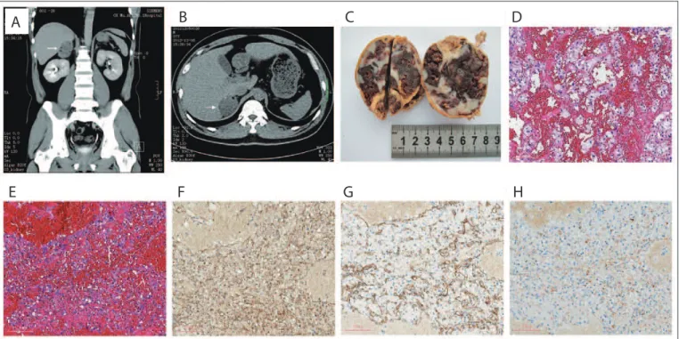
a well-defined, heterogeneous, ovoid mass with fat com-ponent and peripheral speckled calcifications, which mea-sured 5.3 × 4.6 × 6 cm, located at the upper pole of the right kidney (Figures 1A and 1B). Contrast-enhanced computed tomography showed slight enhancement of the solid compo-nent of the mass in the portal venous phase. On December 12, 2012, the patient underwent laparoscopic right adrenal gland resection because the possibility of malignant tumors could not be ruled out clinically. Pathological examination revealed a smoothly and completely encapsulated and mod-erately firm oval mass, measuring 6 × 5 × 4.5 cm and weigh-ing 150 g, with a cross-section of reddish-brown and ash-gray organized hematoma (Figure 1C). The opened mass con-tained some areas of necrosis, and the tissue was extremely heterogeneous. Histopathological evaluation showed a cav-ernous hemangioma with erythrocytes filling the lacu-nae, which was lined with a single layer of endothelial cells
(Figures 1D and 1E). Areas of hemorrhage and small focal
calcifications were observed. Atrophy of the adrenal cortex was present under the tumor capsule. There was no evidence of malignancy. Immunohistochemical examination revealed vessels lined with vascular endothelial cells that were spe-cifically positive for CD31, CD34 and blood coagulation factor VIII (Figures 1F, 1G and 1H), demonstrating their endothelial nature. On December 17, 2012, the patient was discharged from the hospital.
Figure 1. A case of cavernous hemangioma of the adrenal gland. A, B: Computed tomography scan (right adrenal mass, arrows). C: Gross pathology of adrenal mass. D, E: Hematoxylin and eosin staining (× 40). F, G, H: Immunohistochemistry for CD31, CD34 and blood coagulation factor VIII (diaminobenzidine staining, × 40).
A
B
C
D
Rare cavernous hemangioma of adrenal gland: case report | CASE REPORT
Sao Paulo Med J. 2014; 132(4):249-52 251 DISCUSSION
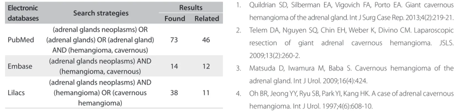
Adrenal hemangiomas occur very infrequently. Since the irst case in 1955, only 63 cases of adrenal hemangioma have been reported in diferent databases (Table 1). Adrenal hemangiomas are benign tumors that arise from endothelial cells that line blood vessels. hey consist of multiple, large vascular channels lined by a sin-gle layer of endothelial cells and supported by collagenous walls. he cause of adrenal hemangioma is not completely understood. Hemangiomas are probably congenital, and hereditary factors may play a role in their pathogenesis. Ectasia, rather than growth, is believed to contribute to hemangioma enlargement.2 Adrenal
hemangiomas are mostly cavernous, unilateral lesions of the adre-nal glands, which appear between the ages of 50 and 70 years, with a 2:1 female-to-male ratio.1,5-6 Our patient was younger than the
average age reported. However, adrenal hemangioma in a 19-year-old Saudi female was reported in 2011.5 he tumor size has ranged
from 2 cm to 25 cm in diameter, and the weight has ranged from a few grams to 5 kg. he majority have measured more than 10 cm, probably because most of these tumors are incidental indings and are usually asymptomatic, unless pain is caused by hemorrhage or mechanical mass efects of the tumor on associated structures.4-6
he size in our case was 6 × 5 × 4.5 cm and the weight was 150 g, i.e. consistent with the range reported.
Adrenal hemangiomas are most commonly non-functional tumors, and only three cases of hormone-secreting adrenal hem-angiomas have been reported to date.7-9 hese unusual benign
adrenal masses are oten discovered incidentally during imaging studies performed for other reasons. Because they do not show any symptoms of adrenal hemangiomas, they are frequently diagnosed clinically ater reaching a size of 10 cm in diameter. his was similarly observed in our patient, with no associated symptoms of either adrenalism or hypoadrenalism.
Computed tomography and magnetic resonance imaging are the most frequently used methods for diagnosing and char-acterizing adrenal masses. he typical indings of adrenal hem-angioma on computed tomography include a heterogeneous, hypodense lesion with calciications, as was seen in our patient.
Calciication characteristically appears speckled throughout the entire mass. However, it may be diicult to distinguish these lesions from carcinomas or cysts. Magnetic resonance imaging may also show homogeneous masses with central hyperintense signal on T1-weighted images and heterogeneous masses with hyperintense signal on T2-weighted images.1 he major tumors
that should be diferentiated are renal tumors, right-lobe liver tumors and other types of adrenal tumors, as well as the metasta-ses of other carcinomas: melanomas and lung, breast, renal and gastrointestinal cancers.6
hese lesions are typically well encapsulated and located in the adrenal cortex. On histopathological inspection, most of the tumors reported were cavernous and rarely capillary in type, and they may undergo degenerative changes like thrombosis, hem-orrhage, necrosis and calciication. Cavernous hemangiomas are enlarged masses of blood-illed sinusoidal channels that have eroded and displaced normal tissues. Furthermore, the presence of multiple vascular cavities at the periphery is an important fea-ture, which accounts for the characteristic peripheral nodular contrast enhancement pattern seen on imaging.7
Surgical resection remains necessary for larger adrenal masses exceeding 3.5 cm,10 even when suspected to be of an
angi-omatous nature, due to their propensity to bleed and the inabil-ity to rule out malignant elements. Several open techniques have been described, including transabdominal, lank and posterior approaches. While laparoscopic adrenalectomy has become the procedure of choice for adrenal masses, the majority of these lesions tend to be small. Although it has been reported that adre-nalectomy can be performed by means of laparoscopy for lesions measuring less than 6 cm,6 laparoscopic resection for larger
adre-nal hemangiomas of up to 12 cm in diameter has been reported.2
CONCLUSION
In summary, we reported a case of non-functional adrenal cavern-ous hemangioma discovered by imaging studies. Although rare, cavernous hemangiomas of the adrenal gland should be part of the diferential diagnosis for adrenal neoplasms. he proper treatment for adrenal cavernous hemangioma is surgical removal.
REFERENCES
1. Quildrian SD, Silberman EA, Vigovich FA, Porto EA. Giant cavernous
hemangioma of the adrenal gland. Int J Surg Case Rep. 2013;4(2):219-21.
2. Telem DA, Nguyen SQ, Chin EH, Weber K, Divino CM. Laparoscopic
resection of giant adrenal cavernous hemangioma. JSLS.
2009;13(2):260-2.
3. Matsuda D, Iwamura M, Baba S. Cavernous hemangioma of the
adrenal gland. Int J Urol. 2009;16(4):424.
4. Oh BR, Jeong YY, Ryu SB, Park YI, Kang HK. A case of adrenal cavernous
hemangioma. Int J Urol. 1997;4(6):608-10.
Table 1. Review of the literature on adrenal cavernous hemangioma
Electronic
databases Search strategies
Results Found Related
PubMed
(adrenal glands neoplasms) OR (adrenal glands) OR (adrenal gland)
AND (hemangioma, cavernous)
73 46
Embase (adrenal glands neoplasms) AND
(hemangioma, cavernous) 14 12 Lilacs
(adrenal glands neoplasms) AND (hemangioma) OR (cavernous
hemangioma)
CASE REPORT | Wang L, Dang Y, He R, Chen G
252 Sao Paulo Med J. 2014; 132(4):249-52
5. Aljabri KS, Bokhari SA, Alkeraithi M. Adrenal hemangioma in a
19-year-old female. Ann Saudi Med. 2011;31(4):421-3.
6. Arkadopoulos N, Kyriazi M, Yiallourou AI, et al. A rare coexistence of adrenal
cavernous hemangioma with extramedullar hemopoietic tissue: a case
report and brief review of the literature. World J Surg Oncol. 2009;7:13.
7. Oishi M, Ueda S, Honjo S, et al. Adrenal cavernous hemangioma
with subclinical Cushing’s syndrome: report of a case. Surg Today.
2012;42(10):973-7.
8. Ng AC, Loh HL, Shum CF, Yip SK. A case of adrenal cavernous
hemangioma presenting with progressive enlargement and
apparent hormonal hypersecretion. Endocr Pract. 2008;14(1):104-8.
9. Stumvoll M, Fritsche A, Wehrmann M, et al. A functioning
adrenocortical hemangioma. J Urol. 1996;155(2):638.
10. Deckers F, De Schepper A, Shamsi K, et al. Cavernous hemangioma
of the adrenal gland: CT appearance. J Comput Assist Tomogr.
1993;17(3):506-7.
Sources of funding: None
Conlict of interest: None
Date of irst submission: May 12, 2013
Last received: September 21, 2013
Accepted: November 19, 2013
Address for correspondence: Gang Chen
Department of Pathology
Hospital of Guangxi Medical University
Shuangyong Road, 530021
China
Tel. 008615277192143