Imaging findings and therapeutic alternatives
for peripheral vascular malformations*
Achados de imagem e alternativas terapêuticas das malformações vasculares periféricas
Lucas Moretti Monsignore1, Guilherme Seizem Nakiri1, Daniela dos Santos1, Thiago Giansante Abud2, Daniel Giansante Abud3
Peripheral vascular malformations represent a spectrum of lesions that appear through the lifetime and can be found in the whole body. Such lesions are uncommon and are frequently confounded with infantile hemangioma, a common benign neoplastic lesion. In the presence of such lesions, the correlation between the clinical and radiological findings is extremely important to achieve a correct diagnosis, which will guide the best therapeutic approach. The most recent classifications for peripheral vascular malformations are based on the blood flow (low or high) and on the main vascular components (arterial, capillary, lymphatic or venous). Peripheral vascular malformations represent a diagnostic and therapeutic challenge, and complementary methods such as computed tomography, Doppler ultrasonography and magnetic resonance imaging, in association with clinical findings can provide information regarding blood flow characteristics and lesions extent. Arteriography and venography confirm the diagnosis, evaluate the lesions extent and guide the therapeutic decision making. Generally, low flow vascular malformations are percutaneously treated with sclerosing agents injection, while in high flow lesions the approach is endovascular, with permanent liquid or solid embolization agents.
Keywords: Vascular malformations; Therapeutic embolization; Sclerotherapy; Interventional radiology.
As malformações vasculares periféricas compreendem um espectro de lesões que se tornam aparentes no decorrer da vida e podem ser encontradas em praticamente todo o corpo. São pouco comuns e frequente-mente confundidas com o hemangioma infantil. Estas doenças são completafrequente-mente distintas tanto em rela-ção à história clínica como ao prognóstico e às formas de tratamento. Nestas lesões, a história evolutiva e as características do exame físico são de extrema importância para o adequado diagnóstico clinicorradioló-gico, que guiará a melhor alternativa terapêutica. As classificações mais recentes dividem as malformações vasculares periféricas levando em consideração o fluxo sanguíneo (alto e baixo) e os componentes vasculares envolvidos (arteriais, capilares, linfáticos e venosos). As malformações vasculares periféricas representam um desafio diagnóstico e terapêutico, e exames complementares como tomografia computadorizada, ultrasso-nografia com Doppler e ressonância magnética, em conjunto com a história clínica, podem trazer informações quanto às características de fluxo e à extensão das lesões. Arteriografia e flebografia confirmam o diagnós-tico, avaliam a sua extensão e orientam a decisão terapêutica. Malformações de baixo fluxo geralmente são tratadas por abordagem percutânea e injeção de agente esclerosante, enquanto para as malformações de alto fluxo o acesso é endovascular com uso de agentes embolizantes permanentes líquidos ou sólidos.
Unitermos: Malformações vasculares; Embolização terapêutica; Escleroterapia; Radiologia intervencionista. Abstract
Resumo
* Study developed at Centro de Ciências das Imagens e Física Médica do Hospital das Clínicas da Faculdade de Medicina de Ribeirão Preto da Universidade de São Paulo (CCIFM/HC-FMRP-USP), Ribeirão Preto, SP, Brazil.
1. MDs, Residents in Therapeutic Neuroradiology and Inter-ventional Radiology at Hospital das Clínicas da Faculdade de Medicina de Ribeirão Preto da Universidade de São Paulo (HC-FMRPUSP), Ribeirão Preto, SP, Brazil.
2. Master, Interventional Radiologist at Clínica Documenta de Ribeirão Preto and Hospital das Clínicas da Faculdade de Medi-cina de Ribeirão Preto da Universidade de São Paulo (HC-FMR-PUSP), Ribeirão Preto, SP, Brazil.
3. Doctor Professor, Docent Responsible for the Service of Therapeutic Neuroradiology and Interventional Radiology at Hos-pital das Clínicas da Faculdade de Medicina de Ribeirão Preto da Universidade de São Paulo (HC-FMRPUSP), Ribeirão Preto, SP, Brazil.
Mailing address: Dr. Lucas Moretti Monsignore. CCIFM/HC-FMRPUSP. Avenida Bandeirantes, 3900, Monte Alegre. Ribeirão
abnormalities are visible at birth(1). In most
of cases, the diagnosis can be achieved with the clinical history and physical examina-tion, and imaging methods such as Doppler ultrasonography, computed tomography and magnetic resonance imaging are use-ful in dubious cases, besides being impor-tant for an appropriate therapy planning.
The correct classification of PVMs, with the use of updated terminology, guides the treatment and avoids semantic confu-sion which may result in wrong therapeu-tic indications. Some types of PVMs are Monsignore LM, Nakiri GS, Santos D, Abud TG, Abud DG. Imaging findings and therapeutic alternatives for peripheral vas-cular malformations. Radiol Bras. 2010;43(3):185–194.
INTRODUCTION
Peripheral vascular malformations (PVMs) are characterized by abnormal development of vascular structures (arte-rial, capillary, venous and lymphatic) in different proportions. Of congenital etiol-ogy, they may present along the life course and in approximately 90% of cases these
Preto, SP, Brazil, 14048-900. E-mail: lucasmonsignore@ gmail.com
erroneously denominated “hemangiomas” and are frequently confounded with infan-tile hemangioma, a benign neoplasia char-acteristically found in newborns, usually self-limited and with spontaneous regres-sion in most cases.
The PVMs’ treatment with the support of different imaging methods has been highlighted over the last years, mainly due to excellent outcomes, low risk for the pa-tient, and development of materials and techniques involved. In some situations, the endovascular treatment may be associ-ated with surgical approach.
CLASSIFICATION
From the diagnostic and therapeutic points of view, as well as in relation to fol-low-up type and requirements, PVMs and vascular tumors are completely different diseases, but both can be denominated vas-cular anomalies. One of the reasons for confusion is the terminology utilized for describing vascular anomalies by the dif-ferent practitioners involved in the treat-ment of patients(2).
Radiologists, pathologists, pediatri-cians, dermatologists, vascular surgeons, plastic surgeons, head & neck surgeons, otolaryngologists and ophthalmologists represent some of the specialists involved in the primary assistance, diagnosis, treat-ment and follow-up of such patients, in general children, teenagers and young adults. Dubious terminology for different diseases frequently leads to iatrogenic dis-orders, sometimes with a catastrophic out-come for the patient(3). Treating an
infan-tile hemangioma (a benign infaninfan-tile tumor with spontaneous involution in most cases) as if it were a low-flow venous malforma-tion (many times denominated “cavernous hemangioma”) may lead to unnecessary mutilation. On the other hand, treating a venous malformation as if it were an infan-tile hemangioma also leads to therapeutic failure, with unnecessary expenses, with-out resolution of the patient’s problem.
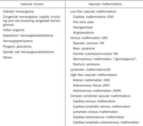
In order to solve this problem, in 1992, after many international meetings to dis-cuss this subject over several years, the International Society for the Study of Vas-cular Anomalies (ISSVA) was created. In 1996, during one of its meetings, the
cur-rent classification of disorders was created (Table 1).
Such classification breaks down the vascular abnormalities into vascular tumors (which include the infantile hemangiomas and other rare vascular tumors that may occur both in children and in adults) and vascular malformations (sub-classified ac-cording to the characteristics of blood flow characteristics – high- and low-flow – and according to histological components com-prising the lesion – arterial, capillary, venous and lymphatic). The classification was based on pathological findings re-ported by Mulliken & Glowacki in a study published in 1982(4). Vascular tumors are
differentiated from vascular malformations with basis on their clinical and radiologi-cal appearance, histopathologiradiologi-cal findings and biological behavior. The “oma” suffix means neoplastic proliferation, therefore terms such as “angioma”, “hemangioma” and “lymphangioma” are erroneous when attributed to vascular malformations, and so have been abolished(3).
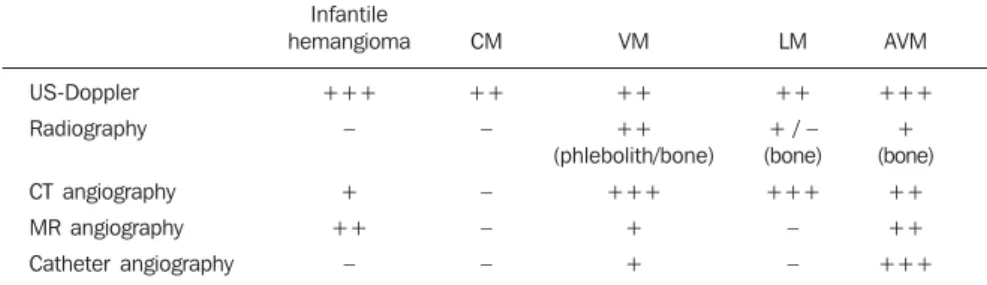
Vascular anomalies are noninvasively classified, with basis on clinical findings, developmental history and findings at
dif-ferent imaging studies(5,6): color and
spec-tral Doppler ultrasonography, plain radiog-raphy, computed tomography angiogradiog-raphy, magnetic resonance angiography(7),
phle-bography, and catheter angiography, each one of these methods with a different im-portance in the several sub-types of the disorder (Table 2).
CLINICAL CONDITION AND DIFFERENT IMAGING FINDINGS
The clinical condition is extremely vari-able and may present as: asymptomatic; small maculae at birth; expansile lesions with variable colors, associated or not with pain, and functional limitation; extensive deformities with aesthetic impact; and high-output heart failure. These findings may present isolatedly or as part of a ge-netic syndrome(8) and may be distributed
throughout the body. Male/female pre-dominance is not observed.
These malformations are always present at birth, although not always visible, pre-senting a progressive growth that occurs at different phases of life depending on the lesion components. Spontaneous
involu-Table 1 Classification of vascular abnormalities considering their vascular components (ISSVA / Mulliken, 1996). Adapted from Enjolras et al.(3).
Vascular tumors
Infantile hemangioma
Congenital hemangioma (rapidly involut-ing and non-involutinvolut-ing congenital heman-giomas)
Tufted angioms
Kaposiform hemangioendothelioma
Hemangiopericytoma
Pyogenic granuloma
Spindle cell Hemangioendothelioma
Others
Vascular malformations
Low-flow vascular malformations
Capillary malformations (CM)
Port-wine stain
Telangiectasia
Angiokeratoma
Venous malformation (VM)
Sporadic common VM
Bean syndrome
Familial cutaneous/mucosal VM
Glomuvenous malformation (“glomangioma”)
Marfucci syndrome
Lymphatic malformation(LM) High-flow vascular malformations
Arterial malformation (AM)
Arteriovenous fistula (AVF)
Arteriovenous malformation (AVM)
Complex combined vascular malformations
Capillary-venous malformation
Capillary-lymphatic-venous malformation
Lymphatic-venous malformation
Capillary-arteriovenous malformation
tion is never observed (a characteristic that is present in most infantile hemangiomas).
Capillary malformations (CMs)
Capillary malformations are the most commonly diagnosed vascular malforma-tions and include telangiectasias and Port-wine stains(9). CMs are low-flow lesions
usually visible at birth and present as red-dish patches of variable dimensions (small maculae to large lesions that may cover an entire limb)(10). These abnormalities are
most frequently found in the head and neck. In general, changes in color are not observed along life and may be confused with precursors to venous malformations, and sometimes a reevaluation of the lesion may be necessary months after birth to con-firm the diagnosis.
Doppler ultrasonography is the method of choice for evaluating such lesions when-ever there is a suspicion for other type of vascular lesion, generally deep, mimicking CM. In the case of CM, Doppler ultra-sonography will not present a specific find-ing, while in the case of other deep vascu-lar lesions mimicking CM, the findings will be specific for the disorder(6).
Venous malformations (VMs)
Venous malformations are present at birth and may develop at any period of the childhood and adolescence. Usually, VMs present as compressible bluish masses, sometimes associated with small hard nod-ules, which correspond to phleboliths. There is no alteration in temperature of the lesion or presence of fremitus at palpation. Dimensions may vary depending upon position or Valsalva maneuver(2).
Magnetic resonance imaging is the im-aging method that provides more
informa-tion regarding the actual lesion extent and its relation with adjacent structures (Fig-ures 1 and 2). These lesions are iso- or hypointense at T1-weighted sequences, and may be subtly hyperintense in case of pres-ence of fat inside the lesion. Small areas of signal absence on all sequences correspond to dystrophic calcifications or phleboliths. Classically, hyper intensity is observed on T2-weighted sequences, the most sensitive ones for demonstrating the actual lesion extent. After contrast injection, the lesion
will be homogeneous or heterogeneously enhanced, allowing accurate differentiation of the lesion in relation to perilesional tis-sue. It is the method of choice for follow-up of results following sclerotherapy(11)
(Figure 2). Such areas are shown as hetero-geneous both on T1- and T2-weighted se-quences, presenting lower enhancement in relation to non treated areas of the lesion(7).
Spectral and color Doppler US evalua-tion demonstrates monophasic or biphasic flow in about 80% of lesions. Arteriogra-phy does not play a relevant role in the evaluation of VMs, considering that the greatest majority of them will not present any specific finding, or only a subtle late venous or capillary blush. The arteries that supply the malformation may be normal or present a subtle ectasia, and the draining veins may be dilated.
Ultrasonography is the first method uti-lized for the evaluation of VMs, because of its noninvasiveness and wide availability. At ultrasonography VMs are characterized by the presence of heterogeneous,
predomi-Table 2 Relevant of the different imaging methods in the diagnosis of some vascular abnormalities. (Adapted from Enjolras et al.(3)).
US-Doppler
Radiography
CT angiography
MR angiography
Catheter angiography
Infantile hemangioma
+++
–
+
++
–
CM
++
–
–
–
–
VM
++
++ (phlebolith/bone)
+++
+
+
LM
++
+ / – (bone)
+++
–
–
AVM
+++
+ (bone)
++
++
+++
CM, capillary malformation; VM, venous malformation; LM, lymphatic malformation; AVM, arteriovenous malfor-mation.
information on the lesion, usually underes-timating its dimensions, besides exposing the patient to ionizing radiation. In this method, as well as in conventional radiog-raphy, a frequent finding is the presence of phleboliths (Figure 1), and thickening of bone cortex, in cases where an intimate relation is observed between the bone and the lesion.
Direct puncture phlebography is consid-ered the gold standard and is indicated in situations where a diagnosis confirmation or therapeutic planning is required. Differ-ent phlebographic patterns have been de-scribed, considering morphology (spongi-form, cavitary and dysmorphic) and the venous return (type I: isolated from the normal venous drainage; type II: normal venous drainage, with competent valves; type III: ectatic drainage veins with inpetent valves; type IV: completely com-posed of ectatic and dysplastic veins)(2).
This classification is useful in the therapeu-tic planning and suggests the prognosis. Cavitary and dysmorphic lesions are tech-nically easier to be treated, in spite of dysmorphic lesions presenting higher re-currence rates. Spongiform lesions require multiple sclerotherapy sessions and mul-tiple punctures at each session.
Lymphatic malformations (LMs)
Approximately 80% of LMs are visible before one year of age, predominantly in the region of the head, neck and axillas (95%), and present as masses, without skin color alteration, when superficial, and (dif-ferently from VMs) not compressible at maneuvers. These lesions are classified as follows: macrocystic, when presenting with cystic areas of variable dimensions; microcystic, when presenting with areas < 2 mm in a solid matrix; or mixed, when presenting with findings of the two previ-ous sub-types(6). The lesions may have
di-mensions that vary in accordance with re-gional or systemic inflammatory processes. Magnetic resonance imaging is the best imaging method to determine the lesion extent and therapy follow-up (Figure 3). They are characterized by lobulated masses, with iso- or hyposignal on T1-weighted sequences, and hypersignal on T2-weighted sequences. After contrast administration, a subtle halo may be observed around the nantly hypoechogenic masses, with
hypoechoic tubular structures inside corre-sponding to the vascular channels of the lesion. When detected, the phleboliths are
shown as hyperechogenic structures with posterior acoustic shadowing.
Computed tomography, even with the use of contrast agents, provides limited
septa of macrocystic lesions, and absence or minimum enhancement of microcystic lesions. Perilesional lymphedema may be observed(7). As in the case of other
vascu-lar malformations, ultrasonography is the method utilized for initial evaluation. The macrocystic sub-type is characterized by a multiloculated cystic mass without flow at the Doppler study, while the microcystic sub-type is hyperechogenic(12). Computed
tomography demonstrates low attenuation masses, occasionally presenting with fluid level, with minimum septal and peripheral enhancement. Angiography is of no diag-nostic value for this type of vascular mal-formation (Figure 4).
Arteriovenous malformations (AVMs)
Arteriovenous malformations are char-acterized by abnormal communication be-tween arteries and veins, with interposition of malformed tissue denominated nidus.
These lesions are characterized by a mass
covered by normal color or angiomatous skin, generally smooth and shinny(13), with
increased local temperature and may present fremitus and murmur. The drainage veins may become evident and tortuous, and local traumas may lead to substantial, life threatening bleeding. Ultrasonography is the method of choice for initial evalua-tion and demonstrates anechoic tubular structures, without well-defined soft tissues mass. At Doppler ultrasonography, the le-sion presents with areas of arteriovenous shunt with increased peak systolic veloc-ity and ectatic veins with arterialized flow. Magnetic resonance imaging (Figure 5) demonstrates several areas with signal ab-sence (flow-void) on T1- and T2-weighted sequences, corresponding to the supplying arteries and to the malformation nidus.
Generally no mass is observed adjacent to the pathological vessels, which facilitates the differentiation between AVMs and hypervascular tumors(7). At computed
to-mography the aspect of multiple ectatic supplying arteries is also observed, with early contrast-enhancement of the drainage veins, with no significant interposed mass. Arteriography (Figures 5 and 6) is the gold standard for the diagnosis of AVMs, char-acterized by early contrast-enhancement of
Figure 3. Macrocystic-type lym-phatic malformation. A female, six-year-old patient with a painless bulging without skin lesion at the right axillary region observed at her six months of age. Magnetic reso-nance imaging, (a) T2-weighted, (b) T1-weighted and (c) T1-weighted sequence with fat satu-ration after contrast agent injec-tion, demonstrating a well-delim-ited lesion with some thin septa inside, at the right axillary region, presenting hyper signal on the T2-weighted, hypo signal on the T1-weighted sequence and subtle enhancement of some septa after contrast agent injection.
venous structures and by the presence of malformative nidus.
Arteriovenous fistulas (AVFs)
Arteriovenous fistulas, on their turn, differentiate from AVMs for presenting with a direct communication between the artery and the vein. Differently from
intrac-ranial AVFs, peripheral AVFs are generally secondary to post-traumatic or post-infec-tious processes(14). The skin on the affected
region generally presents with normal color. It may present with fremitus and murmur at physical examination. Doppler ultrasonography demonstrates increased peak systolic velocity in the arterial system
which may be ectatic, with also ectatic drainage veins with arterialized flow. The magnetic resonance imaging findings of arteriovenous fistulas are similar to those of AVMs, except for the absence of mal-formative nidus. Arteriography is also of diagnostic value and is the gold standard for the diagnosis of AVFs, with the
entiation from AVMs being the absence of malformative nidus(Figure 7).
Arterial malformations (AMs)
Arterial malformations with relevant clinical symptoms affect mainly the aorta, its branches and limbs arteries, comprising aneurysms, coarctations and vascular hypo-plasias. The clinical status is variable and depends on the location and type of the abnormality. Aortic coarctation is the most frequent and most known AM. It may be associated with heart malformations in 5% to 8% of cases, and present with hyperten-sion, heart failure, low perfusion of the lower extremities and head ache. Although
in most cases the treatment is surgical, it may be also performed by means of interventional radiology, with balloon or stent angioplasty. Other AMs include per-sistence of fetal internal iliac artery and persistence of the sciatic artery, which pre-sents with claudication and lower limbs ischemia. Aberrant right subclavian artery may present with dilation or aneurysm at its origin (Kommerell’s diverticulum) and may present with dysphagia lusoria(1) or
asymptomatic dysphagia. These malforma-tions may be easily diagnosed by means of computed tomography angiography, mag-netic resonance angiography and catheter angiography.
Complex malformations
These malformations present with two or more different histological types (capil-lary-venous, capillary-lymphatic-venous, lymphatic-venous, capillary-arterial-ve-nous and capillary-lymphatic-arterial-venous).
Syndromes associated with vascular malformations
Many syndromes present in association with vascular malformations. Some ex-amples are Sturge-Weber syndrome, Klip-pel-Trenaunay syndrome, cutis marmorata teleangiectatica congenita, Parkes-Weber
Figure 7. Arteriovenous fistula. A male, 31-year-old patient, victim of automobile accident. Increase in volume of the left thigh after femur fracture fixation. (a,b) Selective catheter angiography of left lower limb demonstrated arteriovenous fistula between the ipsilateral branch of the deep femoral artery and the superficial femoral vein. (c) The fistula was percutaneously treated with selective microcatheterization and NBCA injection. (d) Fluoroscopic aspect of the NBCA occupying the arteriovenous fistula following the procedure completion. (e) Selective catheter angiography performed upon the procedure completion demon-strates resolution of the fistula.
syndrome, Proteus syndrome, Maffucci syndrome, among others(8).
THERAPEUTIC ALTERNATIVES AND INDICATIONS
The treatment of vascular malforma-tions is extremely varied(5,6) and comprises
surgical resection, laser therapy(15), direct
puncture sclerotherapy(16,17) and arterial
embolization(18,19), with specific
indica-tions for each lesion subgroup, according to location, extent and classification. The correct classification is imperative to deter-mine the therapeutic choice for each case, and a wrong choice of embolization mate-rials can be disastrous. Generally, low-flow malformations are percutaneously treated with sclerosing agent injection, while for high-flow malformations the approach is endovascular with the use of solid or liq-uid permanent embolizing agents.
Currently, the method indicated for the treatment of capillary malformations is pulsed dye laser with a specific wavelength for oxyhemoglobin, which promotes blood coagulation inside the lesion, with clearing along the treatment(15). When the
wave-length is determined for oxyhemoglobin, the destruction occurs almost selectively in the pathological vessels, with preservation of adjacent tissues, and absence of scarring with excellent aesthetic results. Occasion-ally, the laser energy absorption by the epi-dermal melanin may cause thermal injury, and the association with cutaneous cooling is necessary during laser application.
It is recommended that outpatient fol-low-up and therapeutic decisions should be multidisciplinary with a team including pediatricians, pediatric surgeons, derma-tologists, radiologists, interventional radi-ologists, vascular or plastic surgeons, otolaryngologists, ophthalmologists, head and neck surgeons, oncologic orthopedists, anesthetists, psychologists, physical thera-pists and occupational therathera-pists(6).
At the moment of the therapeutic deci-sion making, the risks of the proposed treat-ment in relation to the psychological im-pact and morbidities associated with the natural course of the disease should be taken in consideration. Absolute indica-tions for treatment are the following: hem-orrhage, high-output heart failure,
compli-cations secondary to venous hypertension, and presence of life threatening lesions, such as those located in the airways. Rela-tive indications include progressive pain or discomfort, functional alterations impair-ing daily activities or the quality of life, severe deformity, vascular bone syndrome (extremity length discrepancy), lesion lo-cation with high risk for complilo-cations and infection or recurrent sepsis(6).
The decision between surgical approach and interventional radiology is based on the experience of involved professionals, tak-ing into consideration the patients or caregivers option. Under certain condi-tions, both methods can be associated.
Image-guided percutaneous treatment
The percutaneous treatment can be per-formed by different access pathways, uti-lizing a range of embouti-lizing agents. The determining characteristic for the choice of the most appropriate method is the classi-fication of lesions based on blood flow velocity. Transarterial or transvenous em-bolization is preferred for treatment of high-flow malformations, and can be per-formed by means of solid embolizing agents, such as platinum metal coils with controlled detachment, eventually with polyvinyl alcohol (PVA) microparticles, Gelfoam, and others(18–20), or adhesive
liq-uid agents, such as cyanoacrylate, and non-adhesive liquid agents such as Onyx (ev3 Neurovascular, Inc.; Irvine, CA, USA)(21).
For the treatment of low-flow lesions, the choice is percutaneous sclerotherapy.
Sclerotherapy involves direct percuta-neous puncture of the lesion with a fine needle attached to a small extension tube, through which different substances are in-jected, promoting blood coagulation, pro-tein denaturation and a severe inflamma-tory process that will lead to the oblitera-tion of the malformed cavities. Most of times, the sclerosing agent injection is per-formed under fluoroscopic guidance. Punc-ture of deep lesions can be performed un-der ultrasonography or computed tomogra-phy guidance. Several sclerosing agents have been utilized(22–24), however the most
widely used and lowest-cost agent is abso-lute alcohol in association with other sub-stances to make it radiopaque and less fluid(5,6,9,12,17,25).
In the transarterial embolization, the lesion is accessed by means of remote ar-terial puncture, with the use of catheters, guide catheters and microcatheters, with the application of liquid or solid permanent material, with the purpose of filling the malformed vessels for resolution of the abnormal communications. Eventually, depending on the location of the lesion, the approach can be made by direct percutane-ous access(18,21,26,27).
The procedures are performed under general anesthesia or sedation followed by regional anesthetic blockade, depending on the lesion location. Some preoperative pre-cautions, such as evaluation of renal func-tion, blood coagulability, and platelet count, must be adopted, and identified al-terations must be corrected.
Absolute alcohol
Absolute alcohol is the most widely utilized agent for percutaneous, intrave-nous or intra-arterial sclerotherapy, depend-ing on the hemodynamic characteristics of the lesion. It is indicated for low-flow mal-formations (Figures 1, 2, and 4). It can be utilized mixed with iodinated or poppy oil-based contrast medium – Lipiodol or Ethio-dol (Savage; Melville, NY, USA). Its action occurs by injury of the vessel epithelium, blood coagulation, thrombosis and occlu-sion, with therapeutic response around 64% to 96%(5,17,28). The rate of complications
originating from the use of absolute alco-hol range from 7.5% to 27.95% and include cutaneous necrosis, transitory pain, muscu-lar contraction, neurologic lesion (generally transitory), cellulites, deep venous thrombo-sis, pulmonary embolism, and even circula-tory shock(29). The maximum safe dose is
1 mL/kg of weight per session(16);
depend-ing on the extent and location of the lesion, multiple sessions may be required. An in-tense local inflammatory reaction is ex-pected following the procedure and anal-gesic and anti-inflammatory drugs may be utilized as necessary. Anti-inflammatory drugs should be avoided as much as pos-sible, since the inflammatory activity is as-sociated with better therapeutic responses.
Bleomycin
vascu-lar epithelium, presents a sclerosing action, a collateral effect that was first utilized for therapeutic purposes in 1977 in the treat-ment of LMs of macrocystic type. Its utili-zation in cases of PVMs is more frequent for LMs, but other reports on its use for VMs and AMs are found in the litera-ture(30). It should be diluted at a
concentra-tion of 1 mg/mL, and is applied by direct puncture into the lesion. The most common complications are cellulitis, ulcerations, hair loss and cold-like symptoms.
OK-432 (Picibanil) (Chugai Pharmaceutical Co.; Tokyo, Japan)
This drug is a solution prepared with lyophilized Streptococcus pyogenes cells, treated with benzylpenicillin. It is utilized for the treatment of LMs, with good results in the macrocystic sub-type. It is injected by direct puncture into the lesion, after as-piration of the serous or whitish contents, usually without any image-guidance. Low complication rates are observed.
Ethanolamine oleate
This is an agent that is not frequently utilized for treatment of PVMs. It is more commonly employed for esophageal va-rices. It must be mixed with non-ionic io-dinated contrast agent to maintain its sur-factant effect and to be visualized at fluo-roscopy(5). Few reports on complications
are found in the literature, but ethanola-mine oleate presents the lowest therapeu-tic responses.
Polidocanol
Polidocanol is most frequently used for sclerotherapy of esophageal and lower limb varices. Because of the anesthetic action of polidocanol, no analgesia is required dur-ing procedure.
N-butyl-cyanoacrylate (NBCA)
A liquid adhesive embolizing agent that it is utilized for treatment of high-flow vascular malformations, particularly AMs, by means of direct or transarterial puncture, with injection directly into the malfor-mative nidus(31) (Figures 5 and 7). It is used
in solution with lipiodol, in concentrations that vary according to the blood flow ve-locity. Lipiodol retards the polymerization of NBCA and makes it visible at
fluoros-copy. NBCA acts by polymerizing when in contact with blood, permanently occupying the vascular space, since it is not an absorb-able agent. Experience is required from the practitioner in the handling of this sub-stance to reduce procedural risks and side effects.
Ethylene vinyl alcohol copolymer (Onyx)
This is a non-adhesive biocompatible polymer, soluble in dimethyl sulfoxide (DMSO), with a permanent action that does not act by polymerization, but by precipi-tation, causing the occlusion of the vessel into which it is injected. It is a solution comprising ethylene vinyl alcohol copoly-mer, DMSO and tantalum salts, which makes the solution radiopaque. It should be kept on a shaker for at least 20 minutes before use, so that the components are ho-mogeneously distributed in the solution at the moment of injection. It was primarily utilized in embolization of intracranial AVMs(32) and intracranial dural AVFs(33).
Because of its non-adhesive nature, its procedural time is long, with better results by session as compared with NBCA. It can be used for the treatment of high- and low-flow malformations, generally by intra-ar-terial or intravenous approach(21). This
sub-stance plays an increasing role in the treat-ment of high-flow malformations due to better control during administration, with lower rates of distant complications, par-ticularly in the case of AVMs (Figure 6).
Platinum metal coils
Platinum metal coils were primarily uti-lized in the treatment of cerebral aneurysms early in the nineties(34). Platinum coils with
controlled detachment have been in use for the treatment of other cerebral vascular le-sions such as dural and pial AVFs, as well as in peripheral AVFs, as well as in visceral aneurysms and high-flow vascular malfor-mations(35).
These coils are frequently utilized in the treatment of AVFs by means of endovas-cular access with arterial approach. They may also be utilized in cases of other vas-cular malformations reduce the blood flow velocity, allowing a safer utilization of scle-rosing agents.
Indications for AVMs are very limited, as they usually cause proximal occlusion of
the pedicles, leading to a recruitment of collateral vessels for the malformative ni-dus, which makes a later approach more difficult, and does not result in any clini-cal improvement.
Polyvinyl alcohol (PVA) particles
The PVA particles used to be the mate-rial of choice both for treatment of periph-eral and cerebral AVMs. They are easy to handle, and are available in different sizes, being applied by means of selective or superselective catheterization into the sup-plying vessels of the malformation. They cause a palliative blood flow reduction, but promote proximal and temporary occlusion of the vessels, with very frequent symp-toms recurrence. The utilization of PVA particles also presents considerable rates of complications by embolization of healthy territories and migration into the venous system, with possibility of development of pulmonary thromboembolism, thus their use in the treatment of VMs is currently very restricted.
CONCLUSION
REFERENCES
1. Gloviczki P, Duncan A, Kalra M, et al. Vascular malformations: an update. Perspect Vasc Surg Endovasc Ther. 2009;21:133–48.
2. Legiehn GM, Heran MK. Venous malformations: classification, development, diagnosis, and inter-ventional radiologic management. Radiol Clin North Am. 2008;46:545–97, vi.
3. Enjolras O, Wassef M, Chapot R. Color atlas of vascular tumors and vascular malformations. New York, NY: Cambridge University Press; 2007. 4. Mulliken JB, Glowacki J. Hemangiomas and
vas-cular malformations in infants and children: a classification based on endothelial characteristics. Plast Reconstr Surg. 1982;69:412–22. 5. Hyodoh H, Hori M, Akiba H, et al. Peripheral
vas-cular malformations: imaging, treatment ap-proaches, and therapeutic issues. Radiographics. 2005;25 Suppl 1:S159–71.
6. Legiehn GM, Heran MK. Classification, diagno-sis, and interventional radiologic management of vascular malformations. Orthop Clin North Am. 2006;37:435–74, vii–viii.
7. Moukaddam H, Pollak J, Haims AH. MRI char-acteristics and classification of peripheral vascu-lar malformations and tumors. Skeletal Radiol. 2009;38:535–47.
8. Garzon MC, Huang JT, Enjolras O, et al. Vascu-lar malformations. Part II: associated syndromes. J Am Acad Dermatol. 2007;56:541–64.
9. Escobar FN, Chamorro FM, Trujillo CIP, et al. Hemangiomas y malformaciones vasculares: en-foque diagnóstico y terapéutico. Rev Colomb Radiol. 2008;19:2409–24.
10. Garzon MC, Huang JT, Enjolras O, et al. Vascu-lar malformations: Part I. J Am Acad Dermatol. 2007;56:353–70; quiz 371–4.
11. Spence J, Krings T, terBrugge KG, et al. Percuta-neous sclerotherapy for facial venous malforma-tions: subjective clinical and objective MR im-aging follow-up results. AJNR Am J Neuroradiol. 2010;31:955–60.
12. Puig S, Casati B, Staudenherz A, et al. Vascular
low-flow malformations in children: current con-cepts for classification, diagnosis and therapy. Eur J Radiol. 2005;53:35–45.
13. Gontijo B, Pereira LB, Silva CMR. Malformações vasculares. An Bras Dermatol. 2004;79:7–25.
14. González SB, Busquets JC, Figueiras RG, et al. Imaging arteriovenous fistulas. AJR Am J Roentgenol. 2009;193:1425–33.
15. Stier MF, Glick SA, Hirsch RJ. Laser treatment of pediatric vascular lesions: Port wine stains and hemangiomas. J Am Acad Dermatol. 2008;58: 261–85.
16. Puig S, Aref H, Brunelle F. Double-needle scle-rotherapy of lymphangiomas and venous angio-mas in children: a simple technique to prevent complications. AJR Am J Roentgenol. 2003;180: 1399–401.
17. Górriz Gómez E, Carreira JM. Tratamiento per-cutáneo de las malformaciones vasculares peri-féricas con una mezcla de polidocanol y CO2: experiencia inicial. Radiología. 2008;50:424–9.
18. Nambiar AP, Bozlar U, Angle JF, et al. Initial clini-cal experience with biopolymer-coated detach-able coils (hydrocoil) in peripheral embolization procedures. J Vasc Interv Radiol. 2008;19:995– 1001.
19. Osuga K, Hori S, Kitayoshi H, et al. Emboliza-tion of high flow arteriovenous malformaEmboliza-tions: experience with use of superabsorbent polymer microspheres. J Vasc Interv Radiol. 2002;13: 1125–33.
20. Kwak BK, Shim HJ. Arterial occlusion using a microguidewire as a radiofrequency electrode. J Endovasc Ther. 2008;15:370–4.
21. Numan F, Omeroglu A, Kara B, et al. Emboliza-tion of peripheral vascular malformaEmboliza-tions with ethylene vinyl alcohol copolymer (Onyx). J Vasc Interv Radiol. 2004;15:939–46.
22. Okazaki T, Iwatani S, Yanai T, et al. Treatment of lymphangioma in children: our experience of 128 cases. J Pediatr Surg. 2007;42:386–9.
23. Emran MA, Dubois J, Laberge L, et al. Alcoholic solution of zein (Ethibloc) sclerotherapy for
treat-ment of lymphangiomas in children. J Pediatr Surg. 2006;41:975–9.
24. Ruiz Jr E, Valera ET, Verissimo F, et al. Uso de OK-432 em crianças com linfangioma. J Pediatr (Rio J). 2004;80:154–8.
25. Rosenblatt M. Endovascular management of venous malformations. Phlebology. 2007;22: 264–75.
26. Tan KT, Simons ME, Rajan DK, et al. Peripheral high-flow arteriovenous vascular malformations: a single-center experience. J Vasc Interv Radiol. 2004;15:1071–80.
27. Cura M, Elmerhi F, Suri R, et al. Vascular mal-formations and arteriovenous fistulas of the kid-ney. Acta Radiol. 2010;51:144–9.
28. Lourenço MA, Gomes CS, Beffa CV, et al. Utili-zação de álcool absoluto no tratamento das mal-formações venosas. Radiol Bras. 2001;34:23–7. 29. Lee KB, Kim DI, Oh SK, et al. Incidence of soft tissue injury and neuropathy after embolo/sclero-therapy for congenital vascular malformation. J Vasc Surg. 2008;48:1286–91.
30. Muir T, Kirsten M, Fourie P, et al. Intralesional bleomycin injection (IBI) treatment for haeman-giomas and congenital vascular malformations. Pediatr Surg Int. 2004;19:766–73.
31. Pollak JS, White RI Jr. The use of cyanoacrylate adhesives in peripheral embolization. J Vasc Interv Radiol. 2001;12:907–13.
32. Mounayer C, Hammami N, Piotin M, et al. Nidal embolization of brain arteriovenous malforma-tions using Onyx in 94 patients. AJNR Am J Neuroradiol. 2007;28:518–23.
33. Rezende MT, Piotin M, Mounayer C, et al. Dural arteriovenous fistula of the lesser sphenoid wing region treated with Onyx: technical note. Neuro-radiology. 2006;48:130–4.
34. Guglielmi G, Viñuela F. Intracranial aneurysms. Guglielmi electrothrombotic coils. Neurosurg Clin N Am. 1994;5:427–35.