Importance of GLUT1 in diferential diagnosis
of vascular anomalies
Importância da GLUT1 no diagnóstico diferencial das anomalias vasculares
Tiago João da Silva Filho1
*
, Denise Hélen Imaculada Pereira de Oliveira1, Ilnara de Souza Moura1, Layssa Karolinne da Silva Medeiros1, Amanda Katarinny Goes Gonzaga1,
Veruska Lima Moura Brasil2, Lélia Maria Guedes Queiroz1
Abstract
Vascular anomalies (VAs) include a group of distinct lesions, such as vascular system congenital malformations, as well as benign and malignant vascular tumors. hese lesions may present similar clinical and histopathological features, leading to mistaken diagnoses and incorrect treatment choices. It is important that professionals responsible for monitoring the development of VAs conduct precise investigations and use the appropriate terminology. he human glucose transporter protein isoform 1 (GLUT1) has been proposed as a tool to aid in diferential diagnosis between diferent VAs, given that it is a sensitive and speciic marker for identiication of infantile hemangiomas (HIs) in any organ. his article presents a review of the literature on this protein as an efective tool for identiication and possible diferential diagnosis between several VAs.
Keywords: vascular malformations; hemangioma; diagnosis diferential.
Resumo
As anomalias vasculares (AVs) incluem um grupo de lesões distintas, como as más formações congênitas e os tumores vasculares benignos ou malignos. Estas lesões podem apresentar características clínicas e histopatológicas semelhantes, ocasionando equívocos diagnósticos e terapêuticos. Uma investigação precisa e o uso de terminologia adequada são fundamentais para as decisões do proissional responsável pelo acompanhamento da evolução de uma AV. A isoforma 1 da proteína humana transportadora de glicose (GLUT1) tem sido proposta como uma ferramenta auxiliar para o estabelecimento de diagnóstico diferencial entre AVs, uma vez que representa um marcador sensível e especíico para a identiicação de hemangiomas da infância de qualquer órgão. Este estudo objetiva fazer uma revisão da literatura acerca desta proteína como ferramenta eicaz na identiicação e no possível diagnóstico diferencial entre as diversas AVs.
Palavras-chave: más formações vasculares; hemangioma; diagnóstico diferencial.
1Universidade Federal do Rio Grande do Norte –UFRN, Department of Dentistry, Natal, RN, Brazil. 2Universidade Federal de Pernambuco – UFPE, Department of Dentistry, Recife, PE, Brazil.
Financial support: None.
Conlicts of interest: No conlicts of interest declared concerning the publication of this article. Submitted: August 25, 2014. Accepted: February 06, 2015.
INTRODUCTION
Vascular anomalies (VAs) are lesions of a congenital or acquired nature, which predominantly involve vascular structures. This group of conditions includes all congenital malformations of the vascular system (VMs) and both benign and malignant vascular tumors.1-4
Infantile hemangiomas or hemangiomas of infancy (HIs) are the most common vascular anomaly of childhood and exhibit formation of blood vessels with incomplete architecture, surrounded by hyperplasic endothelial cells. They may or may not be present at birth and follow a characteristic pattern of proliferation which is often followed by spontaneous involution.1,5-7
Precise workup and use of the correct terminology are important elements in the decision-making process of the professional responsible for managing VAs. In many cases, detailed patient history and clinical
examination are suficient to deine diagnosis, but
other methods should be considered in cases in which clinical data leave room for doubt or a possible different diagnosis would affect treatment.
The human glucose transporter protein (GLUT1),
identiied by North et al.,8 has proven useful as a
supplementary tool for assessment of the prognosis of certain types of tumor and for differential diagnosis
between VAs, because it is a sensitive and speciic
marker that can be used to identify HIs in any organ. The objective of this study is to review the literature to compile information on HIs and GLUT1 and discuss whether this protein can actually be considered an
effective tool for the identiication of these lesions.
REVIEW OF THE LITERATURE
Vascular anomalies
For a long time, there was no consensus on VA
terminology or classiication, with a negative impact
on treatment indications, which were very often applied in a heterogeneous and non-parametrized manner, increasing the likelihood of iatrogeny.9
The irst classiications adopted to categorize these
conditions were of a purely descriptive nature. These
were successively replaced by classiications based on anatomopathological and embryological indings, and
by characteristics based on their biological behavior.2,4
Virchow10 was the first to classify vascular
anomalies, using their microscopic appearance as the basis for classifying them as simplex, cavernosum
and racemosum angiomas. His classiication was
based on the belief that each of these types could
change into the others through cell proliferation or dilation of vessels.
For years, the term “hemangioma” was widely and indiscriminately employed to designate VAs which are completely distinct in terms of genesis, clinical and histopathological characteristics, clinical course and prognosis.11 In response to this, the terms “capillary
hemangioma” or “strawberry hemangioma” were used to designate what is nowadays known as the
supericial form of HI, which is a true tumor, generally with no major clinical signiicance or consequences
and, in most cases, with merely cosmetic effects.6,7,11
At the same time, the term “lat hemangioma” was the name given to what is today classiied as
a port-wine stain, which is a VM that is present at birth in almost all cases, grows in proportion to the child’s development, is of a permanent nature and may be associated with a large number of different syndromes, such as Bean syndrome and Mafucci syndrome. Additionally, adjectives such as “cavernous” (which is a descriptive term from histology and should be reserved for that use) referred to clinical characteristics, such as the bluish color suggestive of deep lesions.7,9 Thus, lesions of different types, such as
the deep form of HI, which regresses spontaneously, and permanent subcutaneous VMs, which only have
the bluish color in common, were both classiied as
cavernous.
In 1982, Mulliken and Glowacki1 proposed a new
classiication of vascular lesions based on clinical signs, histopathological indings and natural history,
distinguishing between “hemangiomas” and “vascular malformations”.
The absence of an internationally-accepted
diagnostic classiication prevented standardization
of the appropriate treatment, throwing up barriers to construction of management protocols and compromising comparisons between different treatment options.9 In view of this, Mulliken and Glowacki’s
classiication was revised in 1996, leading to the creation of a new classiication that was adopted as the oficial position of the International Society for the Study of Vascular Anomalies (ISSVA), in which vascular lesions are classiied into one of two groups:
tumors and vascular malformations.2
Currently, the internationally-accepted classiication is based on biological features. More speciically, it is
based on the correlation between biological cellular behavior and clinical course, with a impact on the type of treatment that will be prescribed.9 However,
Hassenein et al.12 searched the PubMed database for
all publications that contained the term “hemangioma” in the title or abstract in 2009, excluding veterinary studies. They found that the term “hemangioma” was
used incorrectly, according to the ISSVA classiication,
in 71.3% of these publications. Patients whose
lesions were incorrectly classiied were more likely
to be given the wrong treatment (20.6%), whereas
patients whose lesions were classiied according to the ISSVA system were subjected to 0.0% erroneous
management during treatment. Additionally, the mean
age of patients in studies that used the classiication
was lower (4.1 months) than in the studies that did not use it (36.1 years).
As is true of any classiication, the ISSVA system
is not absolute. However, its simplicity and clinical relevance have enabled important steps to be taken towards improved management of vascular anomalies.
Infantile hemangioma or hemangioma of infancy
HIs are the most common type of vascular tumor, affecting around 10% of all infants up to 1 year old.6,14
They are more frequent in premature, Caucasian, female and low birth weight babies, affecting up to 30% of neonates weighing less than 1,000 g.15 Maternal
risk factors include advanced age, preeclampsia and placental abnormalities.16
They typically have onset from 2 weeks to 2 months after birth, may be single or multiple, involve one or several systems, and can be focal or regional.17
Approximately 80% of patients have single lesions and four or more lesions in the same patient is rare. The skin is the most often involved organ and the head and neck (60%), and trunk (25%) are the most
affected areas. Size can range from a few millimeters
to several centimeters.18,19
Despite the elevated incidence of HIs, their etiology
remains unclear. However, North et al.20 suggested
that the origin was possibly placental after studying similarities between the blood vessels in HIs and in the placenta. They employed vascular markers that exhibit immunopositivity in the placenta, including
GLUT1, Lewis Y, merosin and Fcγ receptor ΙΙ (receptor
for G-2 immunoglobulin). They then tested different types of VA, including 66 cases of “hemangiomas”, in addition to malignant tumors of a non-vascular origin and placenta samples. They observed coexpression of all four markers in HIs and placenta, but did not observe expression in the other lesions which, according to the authors, implies that there is an
intimate relationship between the two, suggesting a link with the origin of these tumors.20
Clinically, the natural history of HI can be divided
into three phases: the irst is the growth or proliferative
phase, followed by spontaneous regression or the
involutive phase, and then a third phase of inal
equilibrium or the involuted phase.1,5,13,21
During proliferation, the tumor presents as a solid lesion that is compressible, exhibits increased
temperature, and is well deined with signs of increased blood low. In some cases, increased peritumoral
vascularization may be observed, which explains the increased volume on effort and when crying.22,23
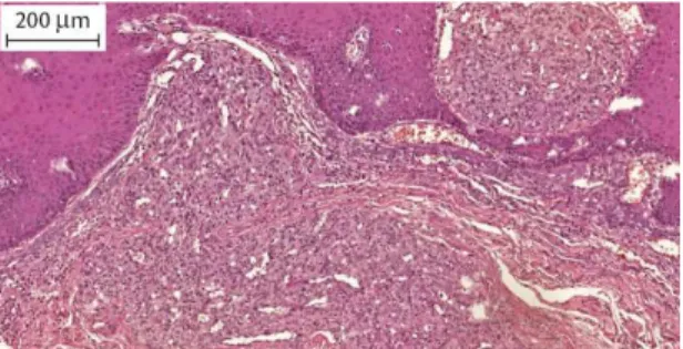
Histologically, clusters of proliferative endothelial cells form solid cords and masses, sometimes with formation of small vascular lumens (Figure 1). These cells tend to group together, forming lobes separated
by ine bundles of connective tissue. None of the lobes are encapsulated or ibrotic and many contain normal tissue and have a feeder artery. Additional indings
can include thrombosis and hemosiderin deposits,
which are limited to areas of acute inlammation or ulceration. Pericytes, ibroblasts and, particularly, mast
cells are numerous in the later stage of proliferation.7
In this phase, the tumor grows rapidly and can attain dimensions that are considerable in proportion to the size of the child. Depending on location, there may be functional, esthetic and psychological compromise.
Neoplastic growth can cause necrosis of the lesion due to vascular insuficiency, primarily in the central
portion, causing recurrent ulceration, bleeding and localized infectious processes, although these factors do not exhibit any relationship with the lesion’s prospects for regression. The proliferative phase is
most pronounced during the irst 3 to 6 months of life
and, in the majority of cases, the tumor will reach its maximum dimensions between nine and 12 months, although this can extend into the second year of life.5,23
The involution phase is characterized by differentiation of mesenchymal cells into adipocytes and the
Figure 1. Photomicrograph demonstrating the histopathological
endothelial cells surrounding the small vascular lumens undergo apoptosis, resulting in color changes (from bright red to pale or grey) and consequent resolution of the lesion.24 It has been estimated that
the pace of involution is at 10% per year and that 70% will already be involuted by 7 years of age. Once this
phase has stabilized, the hemangioma is deined as
involuted.23 However, reaching this phase does not
necessarily imply return to normality, since sequelae may remain at the site of the lesion, including tumor remnants, cutaneous atrophy, areas of scarring, telangiectasias, hypochromic or hyperchromic skin, alopecia and irregular borders.13
Although HIs are classically diagnosed on the basis of patient history, physical examination and predictable clinical course, these are the only vascular lesions that exhibit positive marking for GLUT1 when biopsy specimens are analyzed and they also exhibit greater endothelial cell volumes.25 It has been estimated that
just 10 to 20% of HIs truly require treatment.13 In the
majority of cases no intervention whatsoever is needed and lesions regress spontaneously. The skin or mucosa covering the lesion may exhibit mild atrophy, pallor
and, inally, resolution of the vascular telangiectasia.5
However, in some cases, the natural history of HIs leads to complications that make it impossible to restrict management to monitoring without interventions. Ulceration is the most common complication during the proliferative phase. Anatomic site is important and many complications are related to this factor, such as an expanding tumor causing compression of important structures, such as in the parotid region, involvement of the airways, and tumors located in the orbital area and the eyelids.5 Choice of treatment
should take into account patient age, size, number and location of lesions, stage of progression, and presence of other associated symptoms.22
Hemangiomas can also present as congenital lesions that, in contrast with HI, are completely formed at
birth. Congenital hemangiomas are classiied as
rapidly involuting congenital hemangiomas or as non-involuting congenital hemangiomas.21
These entities were considered variants of HI for a long time, but a number of studies have demonstrated histopathological and immunophenotypical differences between the two groups, indicating that it is unlikely that they are part of a single spectrum of lesions.8,21
North et al.26 demonstrated positive GLUT1 expression
in HI, whereas congenital hemangiomas do not exhibit this positivity, thereby classifying them as distinct lesions.
Isoform 1 of the human glucose transport protein (GLUT1)
Glucose is both the primary source of energy and an important substrate for synthesis of proteins and lipids in the cells of mammals. It provides energy in the form of ATP through glycolysis and the citric acid
cycle (Krebs cycle), and in the form of NADPH via
the pentose phosphate pathway. It is also utilized in synthesis of glycerol and for triglyceride production and provides intermediates for synthesis of nonessential amino acids.27
Since blood glucose levels in mammals are
maintained within a narrow range by a number of homeostasis mechanisms, the majority of cells receive
glucose from the interstitial luid through a process
of passive transport known as facilitated diffusion, pushed through the plasmatic membrane by the concentration gradient into the cell interior.28 Only
the epithelial cells of the small intestine brush border and the proximal tubules of the kidneys absorb or reabsorb glucose against its electrochemical gradient through a secondary mechanism of active transport, the sodium-potassium pump.27
Glucose transport processes are mediated by two distinct families of glucose transporters that are related structurally. The facilitated passive transport system is mediated by the facilitated glucose transporter family of proteins (GLUTs), while active transport is mediated by the sodium-dependent glucose cotransporter family
of proteins (SGLTs).29-31
The GLUT proteins have molecular weights from 50 to 60 kDa and are labeled GLUT1 to 12, HMIT-H+ – binds to myo-inositol – and GLUT14, according to the chronological order of their
identiication and description.31-33 Human GLUT1
has 492 amino acids and a molecular weight of approximately 54 kDa.27
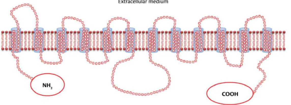
An in-depth review of the genome on GenBank indicated that GLUTs 1 through 12 and HMIT may represent all of the members of the facilitated glucose transport proteins in humans.29 Analysis of these
proteins indicates that this transporter family has a tertiary structure of 12 hydrophobic transmembrane domains. These domains are connected by intracellular
and extracellular hydrophilic segments and have NH2
and COOH cytoplasmic terminals (Figure 2).31,34,35
Studies comparing sequences of all members of
data suggest that the conserved amino acids play important roles in bonding to the substrate and/or in changing conformations during the transport process. Additionally, the presence of areas in common suggests this may have an origin in a shared ancestral gene.27
The most divergent regions are loops 1 and 9, and the two terminal regions, suggesting that these
domains may be responsible for speciic characteristics of each isoform, such as cell location, kinetic proile,
hormone regulation and immunogenicity.27,36
GLUT1 in diferential diagnosis of VAs
GLUT1 is highly expressed in the microvascular endothelium of barrier tissues in which selective
glucose low from the blood to tissues is extremely
important, such as in the central nervous system, retina, iris, ciliary muscle, endoneurium – a delicate web of loose connective tissues that surround each nerve
iber in peripheral nerves – and in the placenta,8,37-39
and may play a vital role in the entry of glucose into these strongly protected tissues.37 Expression is also
observed in erythrocytes, accounting for around 5% of the membrane proteins in this cell type, in germinal centers of lymphoid tissues and renal tubules, and in adipose tissue and certain hepatic cells.27,37-39
The protein is not expressed in the vasculature of any other normal tissue or in other vascular tumors. It has no relationship with mitotic activity
and is considered a sensitive and speciic marker for
diagnosis of HI, since it is present during all phases of progression2,4 (Figure 3).
North et al.8 studied immunohistochemical markers
of GLUT1 in a number of vascular anomalies. They observed intense immunoreactivity for GLUT1 in HIs, demonstrating greater than 50% marking in endothelial cells from lesion microvessels in 97% of cases. Additionally, no immunoreactivity for GLUT1
was observed in any other type of VA. These indings established GLUT1 as a highly sensitive and speciic marker for identiication of HIs in any organ.
Mo et al.40 concluded that there was a need for
appropriate terminology to describe hepatic vascular anomalies, which prompted them to conduct a study of 19 cases using several different vascular markers, including GLUT1. Their results indicate that there are two distinct groups of hepatic vascular lesions
in neonates and children: hepatic HI, which is
positive for GLUT1, and the hepatic VMs, which are GLUT1 negative. The authors therefore concluded that GLUT1 is an effective tool for differentiating between hepatic HIs and VMs.
Johann et al.,41 tested for GLUT1 immunoexpression
in oral “hemangiomas”, oral VMs and oral pyogenic granulomas (PG) in order to investigate the accuracy of histological diagnosis of these anomalies. They observed that none of the cases of oral benign vascular lesions exhibited immunopositivity for GLUT1. Cases initially diagnosed as oral hemangiomas also proved to be
negative for GLUT1 and were therefore reclassiied
after more detailed analysis of the cases. Additionally,
Figure 3. Photomicrograph showing immunoexpression of
GLUT-1 in HI - Panoramic Viewer 1.15.2 (3DHISTECH® Kft. 29-33, Konkoly-hege M. str. Budapest, Hungary, H-1121).
all cases initially classiied as GPs and VMs were
immunonegative for the protein, demonstrating the effectiveness of histological analysis for these lesions.
Osaki et al.42 conducted a study of orbital vascular
anomalies. They analyzed ten cases of HI and ten cases of orbital encapsulated cavernous venous lesions or orbital VMs. They found that all cases of HI were positive for GLUT1, whereas none of the VM cases were positive. According to the authors, the immunopositivity for GLUT1, observed only in the HIs, highlights an essential difference and as a marker makes the greatest contribution to improving diagnostic precision during later stages, when the solid areas of HIs undergo transformation into ectasic vascular channels, but remain GLUT1 positive.
Oliveira et al.43 analyzed immunohistochemical
expression of 30 cases initially diagnosed as “hemangiomas” and 30 cases diagnosed as oral PG. The immunohistochemical test showed that only seven of the “hemangioma” cases were true
HIs. Specimens that were GLUT1 negative were reclassiied as oral PG (ten) or VM (13). None of
the PG cases were positive for GLUT1 and so their initial histological diagnoses were maintained. In
view of these indings, the authors concluded that
histopathological characteristics are not alone enough to guarantee correct diagnosis of oral HIs.
DISCUSSION
VAs are disorders of vasculogenesis, angiogenesis or lymphangiogenesis, and are among the most common pediatric abnormalities.44 HIs have the
greatest relative frequency of the VAs.1,21 Despite
their elevated frequency, there is still a need to adopt a standardized terminology common to both clinicians and pathologists. Currently, the same term
can be used for different entities while, on the lip
side, the same lesion can be assigned to different categories by different health professionals. The term “hemangioma” has often been used to denote a wide range of VAs.7,12,18 This terminological confusion can
contribute both to incoherence that affects clinical management of patients and to introduction of biases into studies of the subject.12,45
Although almost 20 years have passed since the
ISSVA classiication was established, nomenclature
errors are still a common occurrence.12 Correct
identiication of the type of vascular anomaly is not
only essential because of their different clinical, radiographic and pathological characteristics and their morbidity, but also because different types require different treatments.9
Application of the term “hemangioma” for a varied range of VAs is not the only problem related to
naming these lesions. A variety of subclassiications
and neologisms that are not parametrized can still
be observed in current work, making it dificult
to standardize terminology and, as a consequence, diagnosis and treatment.
Correct treatment starts with correct diagnosis,
since a signiicant number of patients with vascular
lesions receive ineffective and potentially harmful treatment because of erroneous diagnoses.46
Many patients do receive the correct treatment
even when the lesion is misdiagnosed. Nevertheless,
incorrect designations are associated with a great risk of the patient receiving inappropriate treatment.12
It can be dificult to achieve differential diagnosis
between different VAs on the basis of histology, particularly between HIs, GPs and VMs, because HI in
the proliferative phase and with inlammation appears
very similar to PG, while histological similarities to VMs can be observed in the involution phase. As such, it is very important to compile well-described clinical information and patient history. Additionally, when necessary, biomarkers should be used to aid in
differential diagnosis and lesions should be reclassiied
as appropriate.8,41,47-51
According to Younes et al.,38 the observation that
lesions continue to exhibit GLUT1 immunoexpression in the proliferative phase could be explained by the increase in proliferative activity and in energy requirements, because the protein is capable of transporting glucose and also other molecules. However, the fact that this protein remains in the cells of these lesions during the involuted phase implies that its expression is not related to a temporary adaptation caused by a need for glucose to support a high rate of
proliferation, and one result is the marker’s speciicity for identiication of HIs.8
When VAs previously thought to be HIs exhibit negative immunomarcation for GLUT1, the solution is to reclassify them as other entities, on the basis of clinical and histopathological characteristics described in the literature.2,8,20,25,26,52 This is extremely important,
primarily with relation to prescribing treatment, since HIs generally exhibit spontaneous involution, which cannot be expected in cases of GPs and VMs.12,41
Many studies that analyzed GLUT1 expression in samples diagnosed as HIs on the basis of histopathology20,26,40,42,53-57 demonstrated that all of
the cases analyzed were immunopositive for the protein, providing support for the hypothesis advanced
by North et al.,8 when GLUT1 was conirmed as a
On the other hand, several studies41,43,58-60 demonstrated
that not all specimens diagnosed by histopathology as “hemangiomas” present immunopositivity for the GLUT1 protein and all of these studies considered that only those samples that were GLUT1 positive came from true HIs, showing that over the years this marker has been widely used for the purposes of
diagnosis, in addition to conirming its eficacy and
reliability as a diagnostic marker.
CONCLUSIONS
Patients with VAs continue to be misdiagnosed because of the complexity involved in differentiating these lesions. Many studies are still being conducted without correct parametrization, introducing biases that can compromise the results and return incorrect information. It is therefore important that centers where teaching, research or diagnosis take place
should adopt the ISSVA classiication when deining
new cases, using tools such as GLUT1. Additionally, older cases should be revisited and, if necessary,
reclassiied, in order to achieve better communication
between professionals and to ensure that research is more trustworthy and better parametrized.
REFERENCES
1. Mulliken JB, Glowacki J. Hemangiomas and vascular malformations in infants and children: a classification based on endothelial characteristics. Plast Reconstr Surg. 1982;69(3):412-22.http:// dx.doi.org/10.1097/00006534-198203000-00002. PMid:7063565. 2. Enjolras O, Mulliken JB. Vascular tumors and vascular malformations
(new issues). Adv Dermatol. 1997;13:375-423. PMid:9551150. 3. Metry DW, Hebert AA. Benign cutaneous vascular tumors of infancy:
when to worry, what to do. Arch Dermatol. 2000;136(7):905-14. http://dx.doi.org/10.1001/archderm.136.7.905. PMid:10890993. 4. Enjolras O, Wassef M, Chapot R. Introduction: ISSVA classification.
New York: Cambridge University Press; 1997. [citado 2014 abr 23]. http://assets.cambridge.org/97805218/48510/excerpt/9780521848510_ excerpt.pdf.
5. Tucci FM, De Vincentiis GC, Sitzia E, Giuzio L, Trozzi M, Bottero S. Head and neck vascular anomalies in children. Int J Pediatr Otorhinolaryngol. 2009;73(Supl 1):S71-6.http://dx.doi.org/10.1016/ S0165-5876(09)70014-X. PMid:20114160.
6. Drolet BA, Esterly NB, Frieden IJ. Hemangiomas in children. N Engl J Med. 1999;341(3):173-81.http://dx.doi.org/10.1056/ NEJM199907153410307. PMid:10403856.
7. Mulliken JB, Fishman SJ, Burrows PE. Vascular anomalies. Curr Probl Surg. 2000;37(8):517-84.http://dx.doi.org/10.1016/S0011-3840(00)80013-1. PMid:10955029.
8. North PE, Waner M, Mizeracki A, Mihm MC Jr. GLUT1: a newly discovered immunohistochemical marker for juvenile hemangiomas. Hum Pathol. 2000;31(1):11-22.http://dx.doi.org/10.1016/S0046-8177(00)80192-6. PMid:10665907.
9. Hiraki PY, Goldenberg DC. Diagnosis and treatment of infantile hemangioma. Rev. Bras. Cir. Plást. 2010;25(2):388-97.
10. Mulliken JB, Fishman SJ, Burrows PE. Vascular anomalies. Curr Probl Surg. 2000;37(8):518-84.
11. Gontijo B, Silva CMR, Pereira LB. Hemangiomaofinfancy. An Bras Dermatol. 2003;78:651-73.
12. Hassanein AH, Mulliken JB, Fishman SJ, Greene AK. Evaluation of terminology for vascular anomalies in current literature. Plast Reconstr Surg. 2011;127(1):347-51.http://dx.doi.org/10.1097/ PRS.0b013e3181f95b83. PMid:21200229.
13. Lowe LH, Marchant TC, Rivard DC, Scherbel AJ. Vascular malformations: classification and terminology the radiologist
needs to know. Semin Roentgenol. 2012;47(2):106-17.http://
dx.doi.org/10.1053/j.ro.2011.11.002. PMid:22370189.
14. Spiteri Cornish K, Reddy AR. The use of propranolol in the management of periocular capillary haemangioma--a systematic review. Eye (Lond). 2011;25(10):1277-83..http://dx.doi.org/10.1038/ eye.2011.164. PMid:21738233.
15. Gampper TJ, Morgan RF. Vascular anomalies: hemangiomas. Plast Reconstr Surg. 2002;110(2):572-85, quiz 585, discussion 587-8.http:// dx.doi.org/10.1097/00006534-200208000-00032. PMid:12142679. 16. Frieden IJ, Haggstrom AN, Drolet BA, et al. Infantile hemangiomas: current knowledge, future directions. Proceedings of a research workshop on infantile hemangiomas, april 7-9, 2005, Bethesda, Maryland, USA. Pediatr Dermato. 2005;22(5):383-406. 17. Chiller KG, Passaro D, Frieden IJ. Hemangiomas of infancy: clinical
characteristics, morphologic subtypes, and their relationship to race, ethnicity, and sex. Arch Dermatol. 2002;138(12):1567-76. http://dx.doi.org/10.1001/archderm.138.12.1567. PMid:12472344. 18. Werner JA, Dünne AA, Folz BJ, et al. Current concepts in the
classification, diagnosis and treatment of hemangiomas and vascular malformations of the head and neck. Eur Arch Otorhinolaryngol. 2001;258(3):141-9.http://dx.doi.org/10.1007/s004050100318. PMid:11374256.
19. Fevurly, RD, Fishman, SJ. Vascular anomalies in pediatrics. Surg Clin North Am. 2012;92(3):769-800.
20. North PE, Waner M, Mizeracki A, et al. A unique microvascular phenotype shared by juvenile hemangiomas and human placenta. Arch Dermatol. 2001;137(5):559-70. PMid:11346333.
21. Ballah D, Cahill AM, Fontalvo L, et al. Vascular anomalies: what they are, how to diagnose them, and how to treat them. Curr Probl Diagn Radiol. 2011;40(6):233-47. PMid:21939817. 22. Frieden IJ, Eichenfield LF, Esterly NB, Geronemus R, Mallory SB,
The Guidelines/Outcomes Committee. Guidelines of care for
hemangiomas of infancy. J Am Acad Dermatol. 1997;37(4):631-7.
http://dx.doi.org/10.1016/S0190-9622(97)70183-X. PMid:9344205. 23. Boye E, Jinnin M, Olsen BR. Infantile hemangioma: challenges, new insights, and therapeutic promise. J Craniofac Surg. 2009;20(Supl 1):678-84.http://dx.doi.org/10.1097/SCS.0b013e318193d6c1. PMid:19190505.
24. Yu Y, Fuhr J, Boye E, et al. Mesenchymal stem cells and adipogenesis in hemangioma involution. Stem Cells. 2006;24(6):1605-12.http:// dx.doi.org/10.1634/stemcells.2005-0298. PMid:16456130. 25. Mulliken JB, Enjolras O. Congenital hemangiomas and infantile
hemangioma: missing links. J Am Acad Dermatol. 2004;50(6):875-82.http://dx.doi.org/10.1016/j.jaad.2003.10.670. PMid:15153887. 26. North PE, Waner M, James CA, Mizeracki A, Frieden IJ, Mihm
27. Zhao FQ, Keating AF. Functional properties and genomics of glucose transporters. Curr Genomics. 2007;8(2):113-28.http:// dx.doi.org/10.2174/138920207780368187. PMid:18660845. 28. Widdas WF. Old and new concepts of the membrane transport
for glucose in cells. Biochim Biophys Acta. 1988;947(3):385-404. http://dx.doi.org/10.1016/0304-4157(88)90001-9. PMid:3048400. 29. Joost HG, Bell GI, Best JD, et al. Nomenclature of the GLUT/ SLC2A family of sugar/polyol transport facilitators. Am J Physiol Endocrinol Metab. 2002;282(4):E974-6.http://dx.doi.org/10.1152/ ajpendo.00407.2001. PMid:11882521.
30. Wright EM, Turk E. The sodium/glucose cotransport family SLC5. Pflugers Arch. 2004;447(5):513-5.http://dx.doi.org/10.1007/s00424-003-1202-0. PMid:12748858.
31. Machado UF, Schaan BD, Seraphim PM. Glucose transporters in the metabolic syndrome. Arq Bras Endocrinol Metabol. 2006;50(2):177-89.http://dx.doi.org/10.1590/S0004-27302006000200004. PMid:16767284.
32. Wu X, Freeze HH. GLUT14, a duplicon of GLUT3, is specifically expressed in testis as alternative splice forms. Genomics. 2002;80(6):553-7.http://dx.doi.org/10.1006/geno.2002.7010. PMid:12504846.
33. Scheepers A, Joost HG, Schürmann A. The glucose transporter families SGLT and GLUT: molecular basis of normal and aberrant function. JPEN J Parenter Enteral Nutr. 2004;28(5):364-71.http:// dx.doi.org/10.1177/0148607104028005364. PMid:15449578. 34. Joost HG, Thorens B. The extended GLUT-family of sugar/polyol
transport facilitators: nomenclature, sequence characteristics, and potential function of its novel members (review). Mol Membr Biol. 2001;18(4):247-56.http://dx.doi.org/10.1080/09687680110090456. PMid:11780753.
35. Thorens B, Charron MJ, Lodish HF. Molecular physiology of glucose transporters. Diabetes Care. 1990;13(3):209-18.http:// dx.doi.org/10.2337/diacare.13.3.209. PMid:2407476.
36. Kasanicki MA, Pilch PF. Regulation of glucose-transporter function. Diabetes Care. 1990;13(3):219-27.http://dx.doi.org/10.2337/ diacare.13.3.219. PMid:2407477.
37. Takata K, Kasahara T, Kasahara M, Ezaki O, Hirano H. Erythrocyte/ HepG2-type glucose transporter is concentrated in cells of blood-tissue barriers. Biochem Biophys Res Commun. 1990;173(1):67-73. http://dx.doi.org/10.1016/S0006-291X(05)81022-8. PMid:2256938. 38. Younes M, Brown RW, Stephenson M, Gondo M, Cagle PT.
Overexpression of Glut1 and Glut3 in stage I nonsmall cell lung carcinoma is associated with poor survival. Cancer. 1997;80(6):1046-51. http://dx.doi.org/10.1002/(SICI)1097-0142(19970915)80:6<1046::AID-CNCR6>3.0.CO;2-7 PMid:9305704.
39. Wood IS, Trayhurn P. Glucose transporters (GLUT and SGLT): expanded families of sugar transport proteins. Br J Nutr. 2003;89(1):3-9.http://dx.doi.org/10.1079/BJN2002763. PMid:12568659. 40. Mo JQ, Dimashkieh HH, Bove KE. GLUT1 endothelial reactivity
distinguishes hepatic infantile hemangioma from congenital hepatic vascular malformation with associated capillary proliferation. Hum Pathol. 2004;35(2):200-9.http://dx.doi.org/10.1016/j. humpath.2003.09.017. PMid:14991538.
41. Johann AC, Salla JT, Gomez RS, Aguiar MC, Gontijo B, Mesquita RA. GLUT-1 in oral benign vascular lesions. Oral Dis. 2007;13(1):51-5. http://dx.doi.org/10.1111/j.1601-0825.2006.01246.x. PMid:17241430. 42. Osaki TH, Jakobiec FA, Mendoza PR, Lee Y, Fay AM. Immunohistochemical
investigations of orbital infantile hemangiomas and adult encapsulated cavernous venous lesions (malformation versus hemangioma). Ophthal Plast Reconstr Surg. 2013;29(3):183-95. http://dx.doi.org/10.1097/IOP.0b013e31828b0f1f. PMid:23584448.
43. Oliveira DH, Silveira EJ, Medeiros AM, Alves PM, Queiroz LM. Study of the etiopathogenesis and differential diagnosis of oral
vascular lesions by immunoexpression of GLUT-1 and HIF-1α.
J Oral Pathol Med. 2014;43(1):76-80.http://dx.doi.org/10.1111/ jop.12092. PMid:23734967.
44. Tasnádi G. Epidemiology and etiology of congenital vascular malformations. Semin Vasc Surg. 1993;6(4):200-3. PMid:8305974. 45. Al-Adnani M, Williams S, Rampling D, Ashworth M, Malone M,
Sebire NJ. Histopathological reporting of paediatric cutaneous vascular anomalies in relation to proposed multidisciplinary classification system. J Clin Pathol. 2006;59(12):1278-82.http:// dx.doi.org/10.1136/jcp.2006.038240. PMid:16751300.
46. Burrows PE, Laor T, Paltiel H, Robertson RL. Diagnostic imaging in the evaluation of vascular birthmarks. Dermatol Clin. 1998;16(3):455-88. http://dx.doi.org/10.1016/S0733-8635(05)70246-1. PMid:9704205. 47. Freitas TM, Miguel MC, Silveira EJ, Freitas RA, Galvão HC.
Assessment of angiogenic markers in oral hemangiomas and
pyogenic granulomas. Exp Mol Pathol. 2005;79(1):79-85.http://
dx.doi.org/10.1016/j.yexmp.2005.02.006. PMid:16005715. 48. Leon-Villapalos J, Wolfe K, Kangesu L. GLUT-1: an extra diagnostic
tool to differentiate between haemangiomas and vascular malformations. Br J Plast Surg. 2005;58(3):348-52.http://dx.doi. org/10.1016/j.bjps.2004.05.029. PMid:15780229.
49. Seo J, Utumi ER, Zambon CE, et al. Escleroterapia de hemangioma labial. Rev Odonto. 2009;17(34):106-112.
50. Buckmiller LM, Richter GT, Suen JY. Diagnosis and management of hemangiomas and vascular malformations of the head and neck. Oral Dis. 2010;16(5):405-18.http://dx.doi.org/10.1111/j.1601-0825.2010.01661.x. PMid:20233314.
51. Vasconcelos MG, Alves PM, Vasconcelos RG, Silveira ÉJ, Medeiros AM, Queiroz LM. Expression of CD34 and CD105 as markers for angiogenesis in oral vascular malformations and pyogenic granulomas. Eur Arch Otorhinolaryngol. 2011;268(8):1213-7. http://dx.doi.org/10.1007/s00405-010-1472-z. PMid:21221622. 52. Berenguer B, Mulliken JB, Enjolras O, et al. Rapidly involuting
congenital hemangioma: clinical and histopathologic features. Pediatr Dev Pathol. 2003;6(6):495-510.http://dx.doi.org/10.1007/ s10024-003-2134-6. PMid:15018449.
53. Li Q, Yu Y, Bischoff J, Mulliken JB, Olsen BR. Differential expression of CD146 in tissues and endothelial cells derived from infantile
haemangioma and normal human skin. J Pathol.
2003;201(2):296-302.http://dx.doi.org/10.1002/path.1443. PMid:14517847. 54. Lyons LL, North PE, Mac-Moune Lai F, Stoler MH, Folpe AL, Weiss SW.
Kaposiform hemangioendothelioma: a study of 33 cases emphasizing its pathologic, immunophenotypic, and biologic uniqueness from juvenile hemangioma. Am J Surg Pathol. 2004;28(5):559-68.http:// dx.doi.org/10.1097/00000478-200405000-00001. PMid:15105642. 55. Drut RM, Drut R. Extracutaneous infantile haemangioma is also
Glut1 positive. J Clin Pathol. 2004;57(11):1197-200.http://dx.doi. org/10.1136/jcp.2003.012682. PMid:15509684.
56. Nguyen VA, Fürhapter C, Romani N, Weber F, Sepp N. Infantile hemangioma is a proliferation of beta 4-negative endothelial cells adjacent to HLA-DR-positive cells with dendritic cell morphology. Hum Pathol. 2004;35(6):739-44.http://dx.doi.org/10.1016/j. humpath.2004.02.005. PMid:15188141.
59. Srinivasan B, Ethunandan M, Van der Horst C, Markus AF. Intraosseous ‘haemangioma’ of the zygoma: more appropriately termed a venous malformation. Int J Oral Maxillofac Surg. 2009;38(10):1066-70. http://dx.doi.org/10.1016/j.ijom.2009.05.010. PMid:19574025. 60. Patiño-Seijas B, Lorenzo-Franco F, Rey-Sanjurjo JL,
González-Cuesta M, López-Cedrún Cembranos JL. Vascular Lesions: GLUT-1 expression as a diagnostic tool to discriminate tumors from malformations. J Oral Maxillofac Surg. 2012;70(10):2333-42.http:// dx.doi.org/10.1016/j.joms.2011.11.013. PMid:22330334.
*
Correspondence
Tiago João da Silva Filho Universidade Federal do Rio Grande do Norte – UFRN Av. Sen. Salgado Filho, 1787 – Lagoa Nova CEP 59056-000, Natal (RN), Brazil Tel.: +55 (84) 32154138 / + 55 (84) 98200469 E-mail: [email protected]
Author information
TJSF – PhD candidate at the Graduate Program in Oral Pathology, Universidade Federal do Rio Grande do Norte (UFRN). DHIPO – PhD candidate at the Graduate Program in Oral Pathology, Universidade Federal do Rio Grande do Norte (UFRN). ISM – Dental student at Universidade Federal do Rio Grande do Norte (UFRN). LKSM – Dental student at Universidade Federal do Rio Grande do Norte (UFRN). AKGG – Master’s candidate at the Graduate Program in Oral Pathology, Universidade Federal do Rio Grande do Norte (UFRN). VLMB – PhD candidate at the Graduate Program in Dentistry, Universidade Federal de Pernambuco (UFPE). LMGQ – Associate professor (level IV) at the School of Dentistry, Universidade Federal do Rio Grande do Norte (UFRN).
Author contributions
Conception and design: TJSF Analysis and interpretation: TJSF, ISM, LKSM Data collection: TTJSF, DHIPO, AKGG, VLMB Writing the article: TJSF, VLMB, AKGG Critical revision of the article: AKGG, DHIPO, LMGQ Final approval of the article*: TJSF, DHIPO, ISM, LKSM, AKGG, VLMB, LMGQ Statistical analysis: TTJSF, DHIPO, AKGG, VLMB Overall responsibility: TJSF