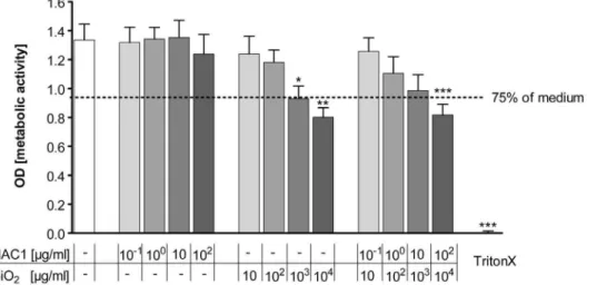
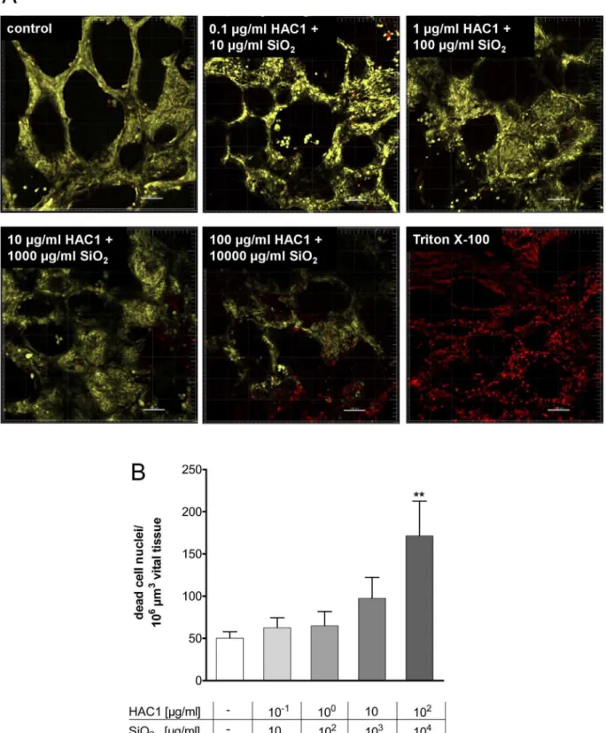
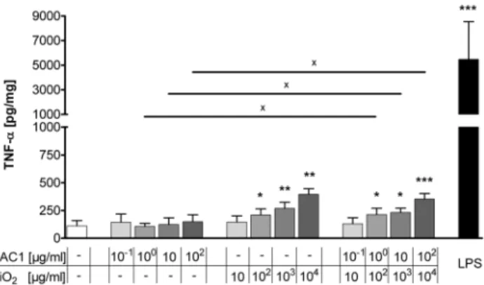
Functional testing of an inhalable nanoparticle based influenza vaccine using a human precision cut lung slice technique.
Texto
Imagem
Documentos relacionados
Abstract Faced with the historical role of or- ganized civil society in the social responses to AIDS and the global health governance, this paper analyzes the biography of
Recombinant adenoviruses as vaccine candidates against the erythrocytic stages of malaria - In principle, since human leukocyte antigen class I or II molecules
Expression of BCL11A protein significantly decreased in the K562 cell lines treated with BCL11A siRNA as compared with either the negative control siRNA group or
Thus, time is inconsistent (uniform and multiple) forming is in itself motionless and invariable and acts as numerical expression of occurring movements and changes, and time
The MRSA and MSSA pre-established biofilms were treated with different concentrations of eugenol or carvacrol alone or in combination, and the cell viability was determined by
For examining the C/EBP β protein stability in differentiating monocytic cells, THP-1 or MM-6 cells were treated with PMA for 24 h or with VitD3 for 72h and primary human monocytes
To determine if co-culturing leads to an up-regulation of KISS1R protein expression, MSCs were cultured either in conditioned media or directly with human (INA-6) or
time it is referred to as a drift, while a sudden genetic recombination of human and animal complete or major change in either or both influenza
