Glioma Cells: A Tale of Two Antibodies
Vaidehi Mahadev , Renate Starr , Sarah L. Wright , Catalina Martinez , Michael C. Jensen ,1 1 1 1 3 Michael E. Barish2, Stephen J. Forman1, Christine E. Brown1*
1Departments of Hematology and Hematopoietic Cell Transplantation, Cancer Immunotherapy & Tumor Immunology, Beckman Research Institute at the City of Hope National Medical Center, Duarte, California, United States of America,2Department of Neurosciences, Beckman Research Institute at the City of Hope National Medical Center, Duarte, California, United States of America,3Ben Towne Center for Childhood Cancer Research, Seattle Children’s Research Institute, Seattle, Washington, United States of America
Abstract
The interleukin-13 receptor alpha2 (IL13Ra2) is a cell surface receptor that is over-expressed by a subset of high-grade gliomas, but not expressed at significant levels by normal brain tissue. For both malignant and non-malignant cells, IL13Ra2 surface expression is reported to be induced by various cytokines such as IL-4 or IL-13 and tumor necrosis factor (TNF). Our group has developed a therapeutic platform to target IL13Ra2-positive brain tumors by engineering human cytotoxic T lymphocytes (CTLs) to express the IL13-zetakine chimeric antigen receptor. We therefore sought to investigate the potential of cytokine stimulation to induce IL13Ra2 cell surface expression, and thereby increase susceptibility to IL13Ra2-specific T cell killing. In the course of these experiments, we unexpectedly found that the commercially available putative IL13Ra2-specific monoclonal antibody B-D13 recognizes cytokine-induced VCAM-1 on glioblastoma. We provide evidence that the induced receptor is not IL13Ra2, because its expression does not consistently correlate with IL13Ra2 mRNA levels, it does not bind IL-13, and it is not recognized by IL13-zetakine CTL. Instead we demonstrate by immunoprecipitation experiments and mass spectrometry that the antigen recognized by the B-D13 antibody following cytokine stimulation is VCAM-1, and that VCAM-1, but not IL13Ra2, is induced on glioma cells by TNF alone or in combination with IL-13 or IL-4. Further evaluation of several commercial B-D13 antibodies revealed that B-D13 is bi-specific, recognizing both IL13Ra2 and VCAM-1. This binding is non-overlapping based on soluble receptor competition experiments, and mass spectrometry identifies two distinct heavy and light chain species, providing evidence that the B-D13 reagent is di-clonal. PE-conjugation of the B-D13 antibody appears to disrupt IL13Ra2 recognition, while maintaining VCAM-1 specificity. While this work calls into question previous studies that have used the B-D13 antibody to assess IL13Ra2 expression, it also suggests that TNF may have significant effects on glioma biology by up-regulating VCAM-1.
Citation:Mahadev V, Starr R, Wright SL, Martinez C, Jensen MC, et al. (2014) Cytokine Induction of VCAM-1 but Not IL13Ra2 on Glioma Cells: A Tale of Two Antibodies. PLoS ONE 9(5): e95123. doi:10.1371/journal.pone.0095123
Editor:Maria G. Castro, University of Michigan School of Medicine, United States of America
ReceivedAugust 21, 2013;AcceptedMarch 24, 2014;PublishedMay 2, 2014
Copyright:ß2014 Mahadev et al. This is an open-access article distributed under the terms of the Creative Commons Attribution License, which permits unrestricted use, distribution, and reproduction in any medium, provided the original author and source are credited.
Funding:This research was supported by NCI (P30 CA33572), California Institute for Regenerative Medicine (CIRM TR3-05641), and generous donations from Joan and Larry Flax and the Chubb Charity Golf Tournament. The funders had no role in study design, data collection and analysis, decision to publish, or preparation of the manuscript. The authors have no conflicting financial interests.
Competing Interests:The authors have declared that no competing interests exist.
* E-mail: cbrown@coh.org
Introduction
Malignant gliomas are highly aggressive and uniformly lethal human brain cancers for which tumor recurrence following conventional therapies remains a major challenge for successful treatment [1,2]. Immunotherapy is emerging as a promising therapeutic approach due to its potential to specifically seek-out and attack malignant cells, particularly the infiltrated cells often responsible for disease recurrence, while sparing cells of the normal brain parenchyma. For this reason, significant efforts are dedicated towards identifying targets amenable for immunother-apy of brain tumors.
One attractive immunotherapy target is IL13Ra2, a 42-kDa monomeric high affinity IL-13 receptor distinct from the more ubiquitously expressed IL-13Ra1/IL-4Ra receptor complex [3]. IL13Ra2 is expressed by a high percentage of gliomas, but not at significant levels on normal brain tissue [4–7], and in IL13Ra 2-expressing tumors has been identified on both stem-like malignant
cells and their more differentiated counterparts [8]. Targeting IL13Ra2 is currently the focus of ongoing clinical development for the treatment of brain tumors [8–12]. In one such effort, our group has constructed an IL13 (E13Y)-zetakine CAR for targeting IL13Ra2. Expanded ex vivo, IL13(E13Y)-zetakine+ CTL retain MHC-independent IL13Ra2-specific anti-glioma cytolytic activi-ty, maintain CAR-regulated Tc1 cytokine secretion and prolifer-ation, and mediate regression of established human glioblastoma xenograftsin vivo[12]. These pre-clinical studies have culminated in a FDA-authorized feasibility/safety clinical trial of intracranial adoptive therapy with autologous IL13-zetakine+ CD8+ CTL clones targeting recurrent/progressive malignant glioma.
multiple IL13Ra2-targeting treatment modalities including IL13(E13Y)-zetakine+
CTLs.
However, in the course of these studies we obtained divergent results with two IL13Ra2-directed antibodies: a goat polyclonal antibody from R&D Systems (cat#AF146) and a PE-conjugated mouse monoclonal antibody clone B-D13 from Cell Sciences. In reconciling these observations, we determined that the putative IL13Ra2-specific antibody B-D13 recognizes VCAM-1, and that cytokine induction is not a viable approach to increase cell surface expression of IL13Ra2 for therapeutic targeting of gliomas. Instead, we find that cytokine stimulation induces VCAM-1 expression by glioma cells, an observation of potential significance for understanding cytokine influences on glioma progression and dissemination.
Methods
Cell lines and culture conditions
The human monocytes line THP-1, glioblastoma line T98, medullablastoma line D283, and SV40 T antigen transformed human embryonic kidney line 293T were obtained from ATCC. The glioma line U251 originated from ATCC, and was a gift from Dr. Waldemar Debinsky (Wake Forest School of Medicine), and after being verified as tumorigenic designated U251T. D283 cells were engineered to express full length, human IL13Ra2 using lentiviral transduction. 293T cells were transiently tranfected using lipofectamine 2000 reagent (Invitrogen) to express either full length VCAM-1 (OriGene) or IL13Ra2 (Geneart). Primary glioma lines were derived from patients undergoing tumor resections at City of Hope. In some cases tumor explants were expanded by heterotopic subcutaneous (s.c.) passaging in mice prior to growth and characterization in culture; in such cases the s.c. passage number is reported after the PBT number (e.g., PBT003-4). Primary brain tumor lines were cultured in neural stem cell medium [DMEM:F12 (Irvine Scientific), 1:50 B27 (Invitrogen), 5 ug/mL heparin (Abraxis Pharmaceutical Products), and 2 mmol/L L-glutamine (Irvine Scientific)]. U251T and T98 cells were cultured in DMEM (high glucose; Gibco) supplemented with 10% heat inactivated fetal bovine serum (Omega Scientific), and 1% L-glutamine (Gibco).
For induction of the B-D13 antigen, cells were induced overnight (16–20 hours) with either 20 ng/mL of TNF-a (Peprotech) alone or in combination with 20 ng/mL 4 or IL-13 (Peprotech). To block transcription, cells were incubated with varying concentrations of Actinomycin D for 1.5 hours followed by incubation with 20 ng/mL of TNF/IL-4 for 5 hours. To block translation, cells were incubated with varying concentrations of Cyclohexamide for 3 hours followed by incubation with 20 ng/ mL of TNF for 5 hours.
Flow cytometry and Western analysis
Cell surface expression was evaluated by staining with phycoerythrin (PE)-conjugated anti-VCAM-1 (eBioscience, 12-1069-41), goat polyclonal anti-IL13Ra2 AF146 (R&D Systems) followed by detection with PE-conjugated donkey anti-goat (Novus Biologicals), PE-conjugated mouse monoclonal anti-IL13Ra2 B-D13 (Cell Sciences; lots 1107403, 1107404 and 1107405), or unconjugated B-D13 antibodies (Cell Sciences, Abcam and Santa Cruz) followed by detection with PE-conjugated goat anti-mouse (Jackson ImmunoResearch). In some cases B-D13 reagent was first pre-incubated for thirty minutes with soluble recombinant human IL13Ra2-Fc chimera or VCAM-1/CD106-Fc chimera reagents (R&D Systems). Percentages of positively staining cells were calculated using the subtraction method with FCS Express version
3 software (De Novo Software, Los Angeles, CA). Relative fluorescence intensity was calculated by dividing the MFI experimental by the MFI of the control. IL-13 biotin binding assays were done by biotinylating IL-13 (Thermoscientific) at a 2.5 biotin to recombinant IL-13 ratio using the EZ-Link Sulfo-NHS-Biotin kit (Thermoscientific) as per the manufacturer’s instructions. The biotinylated IL-13 was then incubated with various glioma lines, followed by staining with streptavidin-PE.
For immunoprecipitation assays, PBT008 and THP-1 cells were stimulated with cytokines as indicated and then incubated with or without unconjugated B-D13 antibody (Cell Sciences; 5mg antibody per 86106cells), washed 3 time in PBS (Irvine Scientific) to remove excess antibody. Cells were then lysed in RIPA assay buffer (PBS, 0.5% sodium deoxycholate, 0.1% SDS, 0.5% Igepal) and antibody/protein complexes were precipitated with Protein G beads. The bound antigen was gently eluted from the beads in 0.3 M glycine pH 2.8, in a series of elutions (E1, E2, E3).
For Western analysis, protein extracts were diluted in 1 mM BME-containing (Bio-Rad) Laemmli loading buffer (Bio-Rad) and separated on 4%–12% gradient SDS-PAGE gels (Bio-Rad) nitrocellulose membrane and probed with the indicated antibod-ies.
qPCR
To quantify mRNA levels of various genes, RNA samples were isolated from cells using the RNeasy Kit (Qiagen), and cDNA were prepared using the Superscript First Strand Synthesis System for reverse transcription PCR (RT-PCR) (Invitrogen). Real time PCR analysis was done using a custom plate from RT2Custom Profiler PCRArray CAPA9696-24 (SABiosciences) and analyzed using the Web-based PCR Array Data Analysis. Fold changes in mRNA levels were calculated after normalization to housekeeping genes [b-Actin (ACTB), glyceraldehyde-3-phosphate dehydrogenase (GAPDH), ubiquitin C (UBC), 60 S acidic ribosomal protein P0 (RPLP0)].
Chromium Release Assay
The cytolytic activity of T-cell effectors was determined by 4-hour chromium release assay [16]. Briefly, 56103 51Cr- labeled
target cells (Na251CrO4; (5 mCi/mL); Amersham Parmacia, Piscataway, NJ) seeded into triplicate wells containing the effector cells at various E:T cell ratios in 200mL of DMEM supplemented
with 10% FCS were seeded into V-bottom 96-well micro-plated and incubated for 4 hours at 5% CO2, 37uC. Plates were centrifuged, and 100ml of supernatant was removed from each well to assess chromium release using ac-counter (Packard Cobra II, Downer’s Grove, IL). The percentage of specific lysis (%) was calculated as follows: 1006(experimental release – spontaneous release)/(maximum release – spontaneous release). Maximal release represents the radioactivity released after total lysis of target cells with 1% SDS. Spontaneous release represents the radioactivity released by target cells present in the medium without effector cells.
Mass Spectrometry
Protein identification. Protein digestion: Proteins were dena-tured in 1:1 v/v trifluoroethanol (TFE), reduced, alkylated and digested with ChymoTrypsin (Promega) according to published protocols [17].
acid in acetonitrile), then eluted at 0.3 uL/min from 1% to 85% buffer B. The QTOF was operated in Auto MS/MS mode with the standard parameters for chip cube interface. MS spectra were acquired from 300 to 2500 m/z and MS/MS was on the six most intense ions in each MS scan, in the range 300–3000 m/z.
Data analysis: Data was searched using X-Tandem! (version: TORNADO 2009.04.01.1). The following parameters were used: Fragment Tolerance: 50 PPM (Monoisotopic), Parent Tolerance: 25 PPM (Monoisotopic), Fixed Modifications: +57 on C (Carba-midomethyl), Variable Modifications:+1 on NQ (Deamidation),+ 16 on MW (Oxidation),+32 on M (Sulphone),+42 on n (Acetyl), Database: the Human Ensembl database and crap.fasta.pro database (unknown version, 76704 entries), Digestion Enzyme: Chymotrypsin, Max Missed Cleavages: 3
Antibody characterization. Antibodies were reduced by mixing the antibody solution 1:1 with 100 mM dithiothreitol (Sigma) in 1 M ammonium bicarbonate and incubating for 1 hour at 57uC. Reduced antibodies were analyzed by LC/MS using the same mass spectrometer, HPLC, and source, but with an Agilent intact protein chip (part# G4240-63001 SPQ105). Antibodies were loaded onto the trapping column at 6ml/min in 1% Buffer B, then eluted through the analytical column with a linear gradient from 1% to 99% Buffer B. Multiply-charged protein spectra were deconvoluted using Agilent Bioconfirm software to give the uncharged protein masses.
Results
Differential recognition of cytokine-induced epitopes by two IL13Ra2-directed antibodies
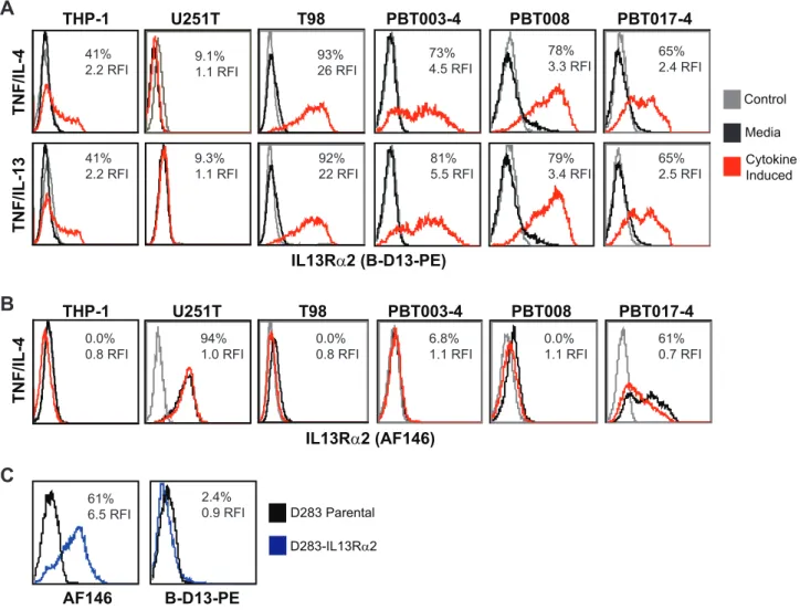
We investigated the possibility of up-regulating cell surface IL13Ra2 levels on glioma utilizing cytokine stimulation regimens previously reported to induce IL13Ra2 on other cell types [13– 15], including the monocytic cell line THP-1 [13]. Indeed, using the commercially available PE-conjugated B-D13 mouse mono-clonal antibody (B-D13-PE; Cell Sciences) reported to target IL13Ra2, we find that cell surface antigen expression on negative or low expressing glioma cell lines (T98, PBT003-4, PBT008 and PBT017-4) is up-regulated following incubation with either TNF and IL-4, or TNF and IL-13 (Figure 1A). Induction of the B-D13-PE target antigen was observed on all the glioma cell lines except U251T, to levels equivalent or greater than observed with the THP-1 line used as a positive control [13]. Further, B-D13-PE target antigen was up-regulated on glioma cell lines under a variety of cytokine conditions including TNF alone (Figure S1).
We have previously demonstrated that the goat polyclonal anti-IL13Ra2 antibody AF146 (R&D Systems) specifically recognizes human IL13Ra2 as shown by FACs, Western and qPCR [8]. However, inconsistencies were noted using B-D13-PE antibody that led us to consider the possibility of multiple IL13Ra2 isoforms or epitopes, or, alternatively, the induction of a different molecule recognized by B-D13-PE. First, the U251T glioma cell line does not express the B-D13-PE recognized antigen (Figure 1A), even though this glioma cell line is known to express high levels of IL13Ra2 [8]. Secondly, AF146 antibody did not recognize the cytokine induced putative IL13Ra2 antigen detected by B-D13-PE antibody on any of the cell lines evaluated (T98, PBT003-4, PBT008, PBT017-4 and THP-1) (compare Figure 1A to 1B). Interestingly, while most of the cell lines displayed mutually exclusive expression of the B-D13-PE and AF146 antigens, PBT017-4 expressed IL13Ra2 as detected by AF146 [8], and, following cytokine stimulation, also showed independent induction of the B-D13-PE antigen. Finally, the AF146 antibody recognizes cells engineered to express IL13Ra2 (D283- IL13Ra2), whereas
B-D13-PE does not (Figure 1C). These findings clearly demonstrate the differential recognition of constitutive IL13Ra2 (expressed by U251T and D283-IL13Ra2) and the cytokine-induced antigen (up-regulated on THP-1, PBT003-4, PBT008 and PBT017) by B-D13-PE as compared to the IL13Ra2-validated antibody AF146.
The cytokine induced antigen recognized by the B-D13 antibody is not IL13Ra2
Our observations led us to evaluate this discrepancy in IL13Ra2 recognition in more detail. Several lines of evidence, including evaluation of IL13Ra2 mRNA levels and IL-13 binding studies, suggest that the cytokine-induced antigen recognized by the B-D13-PE antibody is a molecule distinct from IL13Ra2.
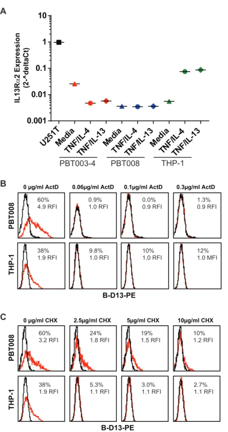
Cytokine-induced B-D13 antigen expression does not correlate with changes in IL13Ra2 mRNA levels. We reasoned that if the cytokine-induced B-D13-PE target antigen was an isoform of IL13Ra2, then IL13Ra2 mRNA levels would also increase upon cytokine stimulation when analyzed by qPCR. We focused on THP-1, PBT003 and PBT008 because these cell lines exhibited high B-D13-PE target antigen up-regulation following cytokine stimulation. Under basal conditions, all three cell lines expressed significantly lower levels of IL13Ra2 mRNA as compared to IL13Ra2-expressing U251T cells: 0.5% for THP-1, 0.3% for PBT003 and 2% for PBT008 cells (Figure 2A). Following cytokine stimulation, IL13Ra2 mRNA levels went down for PBT003 and did not increase in PBT008 cells, despite the significant increase in cell surface B-D13-PE target antigen expression detected by flow cytometry (PBT003: MFI increased 19-fold (RFI); PBT008: MFI increased 28-fold (RFI)) (Figure 1A). We did detect an increase in IL13Ra2 mRNA levels for the THP-1 monocyte line (THP-13–THP-15 fold increase; Figure 2A) consistent with previous reports [13]. However, IL13Ra2 mRNA levels for THP-1 cells after cytokine stimulation were more than THP-13–THP-15 fold lower than for steady state U251T cells (Figure 2A), and induced IL13Ra2 cell surface expression was below the limit of antibody AF146 detection (Figure 1B). Thus, by qPCR, induction of B-D13-PE target antigen does not consistently correlate with IL13Ra2 mRNA levels.
To rule out the possibility that discordance of cytokine induction of the B-D13-PE target antigen with mRNA levels was due to pre-existing mRNA or intracellular stores that could be mobilized to the cell surface following cytokine stimulation independently of transcription or translation, we evaluated the sensitivity to inhibitors of mRNA or protein synthesis [14]. To test a potential requirement for mRNA synthesis, we treated THP-1 and PBT008 cells for 1.5 hours with increasing concentrations of the transcription blocker actinomycin D, followed by 5 hours cytokine stimulation with TNF plus IL-4. Induction of the B-D13-PE target antigen was strongly inhibited by 0.06 ug/ml actino-mycin D treatment (Figure 2B). Similar inhibition was observed by blocking protein synthesis with cycloheximide (completely blocked at 10mg/ml; Figure 2C). These observations suggest that cytokine stimulation did not mobilize pre-existing B-D13-PE target antigen, and that new transcription was required for the observed B-D13-PE antigen induction.
not IL-4 [14]. To confirm authentic IL13Ra2 expression, we demonstrated that biotinylated IL-13 bound to IL13Ra 2-express-ing U251T cells, and is competed off by pre-incubation with 10-fold molar excess of unconjugated IL-13, but not by preincubation with the same molar excess of IL-4 (Figure 3B). In contrast, cytokine-stimulated THP-1 and PBT008 cells do not bind biotinylated IL-13 despite high cell surface induction of the B-D13 target antigen (Figure 3C). These experiments indicate that cells expressing IL13Ra2 which are reactive with AF146 antibody bind biotinylated IL-13, while cytokine-induced cells expressing the B-D13-PE target antigen fail to show biotinylated IL-13 binding.
In an independent approach to evaluate IL-13 binding, we used a highly sensitive cytotoxicity assay to determine whether IL13Ra2-specific redirected CTLs recognize the B-D13-PE target antigen. We have previously shown that IL-13-zetakine T cell lines specifically lyse IL13Ra2-positive target cells in an MHC-independent manner [8,12]. As expected, the IL13Ra2-positive U251T line is efficiently killed by IL13-zetakine+ T cells (Figure 3D). In contrast, IL-13-zetakine+T cells did not recognize
and kill PBT003 cell lines above background following cytokine induction of the B-D13-PE target antigen.
Taken together, the discordance between cytokine induction of B-D13-PE target antigen and IL13Ra2 mRNA levels, along with the demonstration that the cytokine-induced B-D13-PE target antigen does not bind IL-13 and is insensitive to IL13Ra2-directed killing, suggest that the inducible molecule is not IL13Ra2.
B-D13 antibody recognizes VCAM-1
To identify the cytokine-induced B-D13 target antigen we performed protein immunoprecipitation followed by tandem mass spectrometry. We focused on PBT008 glioma cells because this cell line displayed strong cell surface induction of the B-D13-PE target antigen. Following overnight cytokine stimulation with TNF, intact cells were incubated with B-D13 antibody (unconju-gated antibody; Cell Sciences), lysed and antigen-antibody complexes immunoprecipitated using Protein G beads. Immuno-precipitated proteins were eluted in low-pH buffer in three fractions (E1, E2 and E3), and a silver stained gel of E2 detected three distinct polypeptides between 100 to 150 kDa (Figure 4A). Figure 1. Differential recognition of constitutively-expressed versus cytokine-induced IL13Ra2 by commercially available
anti-IL13Ra2 antibodies.Flow cytometric analysis of monocytic line THP-1 and various glioma lines with (A) B-D13-PE (Cell Sciences) or (B) AF146 (R&D
Figure 2. Cytokine induction of B-D13 antigen is dependent on mRNA transcription and translation.(A) IL13Ra2 mRNA levels quantified by qPCR for U251T, PBT003-4, PBT008, and THP-1 cells after overnight culture in media, TNF/IL-4, or TNF/IL-13. mRNA levels were normalized to housekeeping genes (ACTB, UBC, GAPDH and RPLP0). (B, C) B-D13-PE immunoreactivity of PBT008 and THP-1 cells treated with increasing concentrations of (B) transcription blocker Actinomycin D (ActD) (0, 0.06, 0.1, 0.3mg/ml) or (C) translation blocker Cycloheximide (CHX) (0, 2.5, 5, 10mg/ml), then either cultured in media alone (black histograms) or stimulated with TNF/IL-4 (red histograms) for 5 hours. All data are representative of 2 separate experiments.
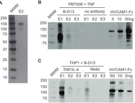
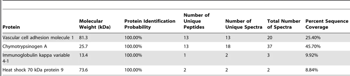
To identify the immunoprecipitated polypeptides, protein eluates were subjected to LC/MS/MS and in two independent experi-ments mass spectrometry identified the B-D13 immunoprecipitat-ed protein as VCAM-1 (IP#1: 3 peptides; IP#2: 13 peptides). The only other proteins detected in the eluates were either present in the control Protein G IP performed without B-D13 antibody or expected contaminants such as keratin and chymotrypsin (Table 1). To confirm that the protein product immunoprecipitated by B-D13 antibody is VCAM-1, Western analysis was performed on the immunoprecipitated eluates. For cytokine-induced PBT008 cells, the B-D13 antibody pulled-down polypeptides ranging from 100 to 150 kD protein which were immunoreactive for VCAM-1 (Figure 4B). Immunoprecipitation of VCAM-1 was specific, as it was not immunoprecipitated from cytokine induced (TNF) PBT008 cells in the absence of B-D13 antibody (Figure 4B; see no antibody E1-E3). For THP-1 cells, the B-D13 antibody also immunoprecipitated the VCAM-1 immunoreactive antigen fol-lowing cytokine induction (TNF/IL-4), and VCAM-1 was immunoprecipitated at significantly lower levels in non-cytokine exposed cells (media) (Figure 4C). Although THP-1 cells did show
an increase in IL13Ra2 mRNA levels following cytokine induction (13–15 fold, Figure 2A), the B-D13-immunoprecipitated protein was not recognized by the IL13Ra2-specific antibody AF146 (not shown).
Further, authentic VCAM-1 immunoreactivity correlated with B-D13-PE staining on all lines exhibiting cytokine-induced antigen expression (Figure 5A, see THP-1, T98, PBT003-4, and PBT008), while cytokine-stimulated U251T cells showed neither induction of VCAM-1 nor B-D13-PE immunoreactivity. In contrast, and consistent with our previous findings (Figure 1), no induction of IL13Ra2, as detected by the AF146 antibody, was seen on any of the lines responding to TNF/IL-4, and U251T cells expressed constitutively high levels of IL13Ra2.
The VCAM-1 specificity of B-D13-PE was confirmed by ligand competition, in which we compared the potential of either soluble recombinant human VCAM-1-Fc (sVCAM-1-Fc) or IL13Ra2-Fc (sIL13Ra2-Fc) to compete for B-D13-PE immunoreactivity on TNF/IL-4-stimulated PBT008 cells (Figure 5B). Pre-incubation of sVCAM-1-Fc, but not sIL13Ra2-Fc, inhibited B-D13-PE anti-body binding on cytokine induced PBT008 cells, indicating that Figure 3. The cytokine-induced cell surface antigen recognized by B-D13-PE does not bind IL-13.(A) D283 cells engineered to express IL13Ra2 (D283- IL13Ra2) (blue histograms) and D283 parental (black histograms) cells were evaluated by flow cytometry for expression of constitutive IL13Ra2 using the IL13Ra2-specific antibody AF146, and biotinylated recombinant human IL-13 (IL13-bio) followed by PE-conjugated strepavidin (SA-PE). Data are representative of 2 separate experiments. (B) U251T (grown in the absence of cytokines) were evaluated by flow cytometry for constitutive IL13Ra2 expression using AF146, and for binding to IL13-bio/SA-PE in the presence and absence of 10-fold molar excess of recombinant human IL-4 or IL-13. Black histograms represent staining with istoype control antibody or SA-PE alone. Data are representative of 2 separate experiments. (C) THP-1 and PBT008 grown in media alone (black histograms) or induced overnight with TNF and IL-4 (red histograms) were analyzed by flow cytometry for expression of constitutive IL13Ra2 (AF146), for expression of the induced antigen (B-D13-PE), and for binding to IL13-bio/SA-PE. Data are representative of 3 separate experiments. (D) IL13-zetakine+CD8+ CTL recognize and kill U251T glioma targets expressing constitutive IL13Ra2 (AF146-positive), but not cytokine-induced PBT003 cells (B-D13-positive). Percentage specific lysis (mean6S.D.) of triplicate wells is depicted. *, p#0.0002 using an unpaired Student’s t-test to compare U251T vs. PBT003-4 targets.#, p.0.05 using an unpaired Student’s t-test to compare PBT003-4 targets with and without overnight cytokine stimulation. Data are representative of at least 2 separate experiments. doi:10.1371/journal.pone.0095123.g003
Figure 4. The B-D13 antibody recognizes VCAM-1, not IL13Ra2. (A) Silver stain gel of the second elution (E2) from the B-D13
immunoprecipitation of PBT008 stimulated overnight with TNF. (B) Western blot detecting B-D13 pull-down of VCAM-1 in immunoprecipitation eluates (E1, E2, E3) for PBT008 cells stimulated overnight with TNF. VCAM-1 was not immunoprecipitated in the absence of B-D13 antibody (no antibody: E1, E2, E3). Titrated soluble recombinant human VCAM-1-Fc shows specificity of the VCAM-1 antibody. (C) Western blot detecting B-D13 pull-down of VCAM-1 in immunoprecipitation eluates (E1, E2, E3) for THP-1 cells in both media and cytokine overnight-stimulated conditions. All data are representative of 2 separate experiments.
soluble VCAM-1 blocks the binding site for the epitope recognized by antibody B-D13-PE.
Thus by four independent measures — mass spectrometry, Western analysis, direct comparison of antibody immunoreactiv-ity, and soluble ligand competition — the protein induced by cytokine (TNF, TNF/IL-4 or TNF/IL-13) and recognized by antibody B-D13-PE was identified as VCAM-1.
Characterization of additional commercially available B-D13 antibodies
To provide further insight into the specificity discrepancies revealed by our studies for the B-D13 antibody, we compared the behaviors of two lots of PE-conjugated B-D13 (B-D13-PE; Cell
Sciences lots 1107404 and 1107405), as well as unconjugated B-D13 antibodies (B-B-D13-unc) from Cell Sciences, Santa Cruz and Abcam. We confirmed by flow cytometric analysis that each commercially available B-D13 reagent — two B-D13-PE lots and three B-D13-unc antibodies — stained cytokine-stimulated PBT008 cells in a manner similar to that of the VCAM-1 specific antibody (Figure 6A). These B-D13 reagents also recognized 293T cells engineered to express VCAM-1 by transient transfection, but not parental 293T cells (Figure 6B, top and middle panels). Thus recognition of VCAM-1 by the B-D13 antibodies is independent of antibody lot or commercial source.
Interestingly, however, the unconjugated B-D13 antibodies (B-D13-unc) also recognized 293T cells transiently transfected to Table 1.Peptides identified by tandem mass spectrometry.
Protein
Molecular Weight (kDa)
Protein Identification Probability
Number of Unique Peptides
Number of Unique Spectra
Total Number of Spectra
Percent Sequence Coverage
Vascular cell adhesion molecule 1 81.3 100.00% 13 13 20 25.40%
Chymotrypsinogen A 25.7 100.00% 13 18 37 45.70%
Immunoglobulin kappa variable 4-1
13.4 100.00% 1 2 3 9.92%
Heat shock 70 kDa protein 9 73.6 100.00% 2 2 2 8.84%
doi:10.1371/journal.pone.0095123.t001
Figure 5. Antibody immunoreactivity, and soluble ligand competition confirms B-D13-PE specificity for VCAM-1.(A) Flow cytometric analysis of monocytic line THP-1 and various glioma lines with B-D13-PE (top), or PE-conjugated anti-VCAM-1 (VCAM-1-PE, bottom), after overnight culture in media alone (black histogram) versus cytokine (TNF/IL-4; red histogram) conditions. Data are representative of at least 2 separate experiments. (B) PBT008 cells cultured overnight in media alone (black histogram) versus cytokine (TNF/IL-4; red histogram) conditions were stained with B-D13-PE antibody that was pre-incubated with soluble recombinant human IL13Ra-Fc or VCAM-1-Fc. As a control, cells were stained with B-D13-PE alone. Data are representative of 2 separate experiments.
express IL13Ra2, as well as constitutively IL13Ra2-expressing U251T cells (Figure 6B lower panels, and 6C). This is in contrast to the two lots of PE-conjugated B-D13 from Cell Sciences that did not strongly recognize IL13Ra2 on either U251T or IL13Ra 2-engineered 293T cells. These findings suggest that PE-conjugation impairs IL13Ra2 recognition, and that unconjugated B-D13 antibody, independent of commercial source, recognizes both IL13Ra2 and VCAM-1.
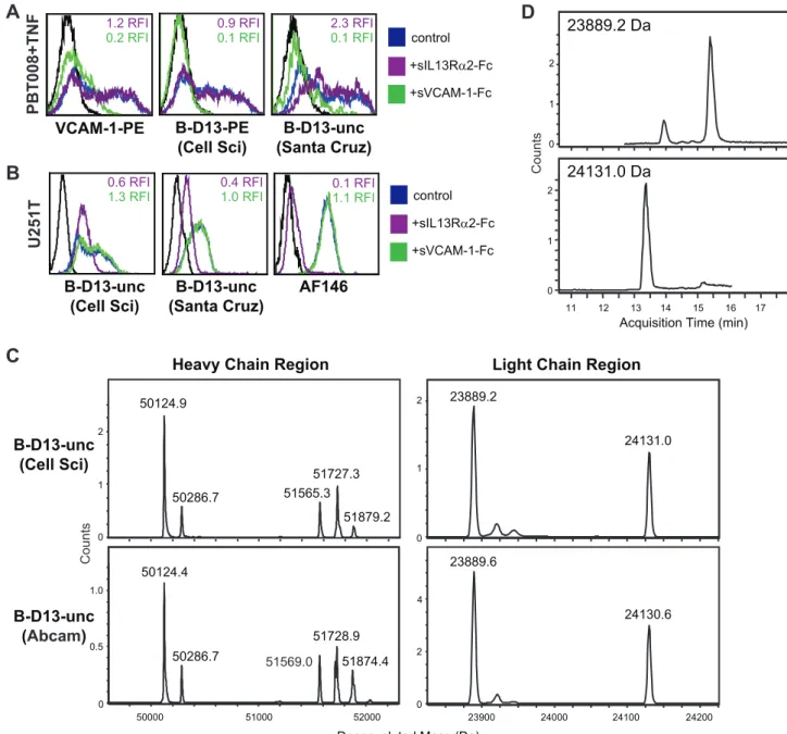
We next set out to understand the bi-specificity of the B-D13-unc antibodies, and the results of several experiments suggest the presence of two distinct monoclonal antibodies in the B-D13
preparation, which recognize either IL13Ra2 or VCAM-1. First, in competition experiments, only soluble VCAM-1-Fc, but not soluble IL13Ra2-Fc, could compete for recognition of VCAM-1 on cytokine-induced PBT008 cells (Figure 7A). Reciprocally, only soluble IL13Ra2-Fc, but not soluble VCAM1-Fc, could compete for recognition of IL13Ra2 on U251T cells (Figure 7B). These results demonstrate that the IL13Ra2 and VCAM-1 antigen binding sites of the B-D13-unc antibody are non-overlapping. Second, mass spectrometry analysis identified two distinct heavy and light chains in the B-D13-unc antibody preparations, that were identical between those purchased from either Cell Sciences Figure 6. Evaluation of additional commercially available B-D13 antibodies.(A) PBT008 cells that had been cultured overnight in media alone (black histogram) versus cytokine (TNF; red histogram) conditions, (B) Parental 293T cells or 293T cells engineered to express either VCAM-1 or IL13Ra2, and (C) U251T cells were stained with VCAM-1-PE, AF146 or various B-D13 reagents – two lots of PE-conjugated B-D13 antibody (B-D13-PE; Cell Sciences) and two to three unconjugated B-D13 antibodies (B-D13-unc) purchased from either Cell Sciences (Cell Sci), Abcam or Santa Cruz as indicated. (B, C) Black histograms represent staining with istoype control antibody or SA-PE alone.
or Abcam (Figure 7C) (B-D13-unc from Santa Cruz was not evaluated due to the presence of 0.1% gelatin in the preparation that would interfere with this analysis). The deconvoluted mass of the two heavy chains differed by more than 1,500 Da (e.g., 50124.9 vs. 51565.3 for the Cell Sciences B-D13-unc reagent), and the multiple mass species for each (i.e., the clusters of peaks) reflect differences in glycosylation states (Figure 7C). The deconvoluted mass of the two light chains differed by more than 240 Da (e.g., 23889.2 vs. 24131.0 for the Cell Sciences B-D13-unc reagent; Figure 7C), and further were chromatographically separated by HPLC with a retention time difference of approximately 2
minutes, which is indicative of differential amino acid composition and/or hydrophobicity (Figure 7D). Taken together, these data are most consistent with the B-D13 antibody reagent being di-clonal due to the presence of two distinct monodi-clonal antibodies in the preparation.
Discussion
These experiments were prompted by efforts to use cytokine-stimulation paradigms to increase IL13Ra2 expression on glioma cells and thereby increase the efficacy of IL13Ra2 targeted
Figure 7. B-D13 reagent appears to contain two distinct monoclonal antibodies.Soluble receptor competition study evaluating the specificity of IL13Ra2 and VCAM-1 recognition by the B-D13-unc antibodies (Cell Sciences and Santa Cruz) using (A) PBT008 cells cultured overnight in media alone (black histogram) versus cytokine (TNF; blue histogram); or (B) IL13Ra2-expressing U251T cells (blue histogram). Cells were stained with the indicated antibody that was pre-incubated with soluble recombinant human IL13Ra-Fc (purple histograms) or VCAM-1-Fc (green histograms). Relative fluorescence index (RFI) compared to staining without the soluble competitors (i.e., the control/blue histograms) are indicated in each histogram. (C) Unconjugated B-D13 antibodies from Cell Sciences (top) and Abcam (bottom) were reduced and analyzed by LC/MS. Shown is the spectra of the deconvoluted protein masses depicting two distinct mass species for both the heavy and light chains. (D) Extracted ion chromatograms (EIC) for the two light chain species, of the Cell Sciences B-D13-unc reagent from (C).
therapies for brain tumors [8–12]. Based on the many studies which reported induction of IL13Ra2 on a variety of cell types following cytokine stimulation [13–15], we envisioned that this strategy for IL13Ra2 induction may be conserved for glioblastoma as well. Indeed, following cytokine stimulation, we did observe induction of a cell surface antigen on both primary glioblastoma cell lines and the monocyctic cell line THP-1, which was strongly detected by the commercially available putative IL13Ra2-targeted monoclonal antibody B-D13-PE (Cell Sciences). However, during the course of these studies, we encountered a series of discrepan-cies in the behavior of the B-D13-PE antibody that led us to question its binding specificity, and whether the induced antigen following cytokine stimulation was really IL13Ra2. In particular, following cytokine stimulation, B-D13-PE immunoreactivity did not correlate with either the immunoreactivity of the highly-characterized IL13Ra2-specific goat polyclonal antibody AF146 (R&D Systems) [18] or IL13Ra2 mRNA levels. Further, the cytokine induced B-D13-PE antigen did not bind IL-13 or elicit lysis by IL13Ra2-redirected CAR T cells, and B-D13-PE binding on cytokine stimulated cells could not be blocked by soluble IL13Ra2-Fc. Therefore, our data indicate that neither TNF/IL-4, TNF/IL-13, nor TNF alone induce cell surface IL13Ra2 up-regulation on glioma cells (or at significant levels on monocytes), and therefore that these cytokine treatments are not a viable strategy for expanding the targetability of IL13Ra2 by immuno-therapy.
Instead, our data definitively demonstrate that the cytokine induced antigen recognized by B-D13-PE is VCAM-1, as demon-strated by B-D13 immunoprecipitation/mass spectrometry, as well as soluble receptor competition studies. Many studies have reported induction of IL13Ra2 on a variety of cell types following cytokine stimulation [13–15], and induction of IL13Ra2 has been reported to be involved in TGF-b1production [13,15,19]. However, all of these studies used the B-D13 antibody to evaluate protein induction, and thus may have inadvertently mis-identified the induction of IL13Ra2 protein following cytokine stimulation. It should be noted, however, that qPCR and knockdown studies do support IL13Ra2 induction in some cell types [13]. In fact, consistent with previous reports [13], we find that THP-1 cells show induction of IL13Ra2 mRNA 13 to 15-fold after overnight treatment with TNF and IL-13 or IL-4, although the level of IL13Ra2 expression was more than 13.6-fold lower than that expressed by the U251T glioma cell line and not at sufficient levels to be detected by flow cytometry using the IL13Ra2-specfic AF146 antibody. We do not, however, find any evidence for induction of IL13Ra2 through 4 or TNF/IL-13 cytokine regimens on glioma cell lines, either established (T98 or U251T) and/or low-passage primary lines (PBT003-4, PBT008, PBT017-4).
The B-D13 antibody has also been used to evaluate constitutive IL13Ra2 expression on a variety of cell types, including a new model cell line for oral cavity squamous cell carincoma [20], tumor initiating cells in adult human gliomas [20,21], and
IL13Ra2 expression in human pediatric brain tumors [7]. Our findings possibly explain discrepancies between publications evaluating IL13Ra2 expression. For example, when evaluating cancer stem cells, our group found that IL13Ra2 is expressed by approximately 50% of glioma stem cell lines when using the AF146 antibody [8]. However, in a separate study of glioma stem cells isolated from glioblastomas, IL13Ra2 expression was not reported when using the B-D13-PE antibody (Cell Sciences) (8 samples characterized) [22]. Our findings may also help explain inconsistencies noted in previous studies using BD-13 to evaluate IL13Ra2 expression by immunohistochemistry and flow cytom-etry, as compared to qPCR to measure mRNA levels [8,23]. Our results thus highlight the necessity of re-evaluating reports using the B-D13 antibody to characterize cell surface expression of IL13Ra2.
One of the principle observations resulting from these studies is that cytokine treatment of primary low-passage glioma cell lines induces the expression of VCAM-1, expanding the previously observed cytokine induction of VCAM-1 on astrocytoma cell lines [24], as well as on other transformed and non-transformed cell types [25,26]. As an adhesion molecule, induction of VCAM-1 on glioma cells by inflammatory cytokines is of potential clinical significance. VCAM-1 expression emerges late in tumorigenesis and is positively correlated with malignancy grades in various tumors including glioma [27]. In breast and colon cancers, VCAM-1 mediates prometastatic tumor stromal interactions, proliferation, apoptosis and invasion, all of which ultimately contribute to malignancy and tumor progression [28]. That similar processes may influence glioma progression will be the subject of future investigations.
Supporting Information
Figure S1 B-D13 is induced on glioma cell lines in a various cytokine conditions.THP-1, T98, and PBT003 cells were incubated overnight with 20 ng/ml of the indicated cytokines and analyzed for cell surface expression of the B-D13 target antigen (grey histogram) vs. isotype control staining (black line). (PDF)
Acknowledgments
We thank Dr. Gabriel Gugiu, Roger Moore and Helen Ge from the Mass Spectrometry and Proteomics core at City of Hope for technical guidance and assistance in generating data. We also thank Dr. Patrick Perrigue for thoughtful scientific discussions, and Dr. Julie Ostberg for assisting in manuscript revisions.
Author Contributions
Conceived and designed the experiments: CB VM. Performed the experiments: VM RS CM SW. Analyzed the data: VM RS CB CM SW. Contributed reagents/materials/analysis tools: CB MJ SF. Wrote the paper: CB MB VM.
References
1. Han SJ, Zygourakis C, Lim M, Parsa AT (2012) Immunotherapy for glioma: promises and challenges. Neurosurg Clin N Am 23: 357–370.
2. Ruzevick J, Jackson C, Phallen J, Lim M (2012) Clinical trials with immunotherapy for high-grade glioma. Neurosurg Clin N Am 23: 459–470. 3. Lupardus PJ, Birnbaum ME, Garcia KC (2010) Molecular basis for shared
cytokine recognition revealed in the structure of an unusually high affinity complex between IL-13 and IL-13Ralpha2. Structure 18: 332–342. 4. Debinski W, Gibo DM, Slagle B, Powers SK, Gillespie GY (1999) Receptor for
interleukin 13 is abundantly and specifically over-expressed in patients with glioblastoma multiforme. Int J Oncol 15: 481–486.
5. Jarboe JS, Johnson KR, Choi Y, Lonser RR, Park JK (2007) Expression of interleukin-13 receptor alpha2 in glioblastoma multiforme: implications for targeted therapies. Cancer Res 67: 7983–7986.
6. Joshi BH, Puri RA, Leland P, Varricchio F, Gupta G, et al. (2008) Identification of interleukin-13 receptor alpha2 chain overexpression in situ in high-grade diffusely infiltrative pediatric brainstem glioma. Neuro Oncol 10: 265–274. 7. Kawakami M, Kawakami K, Takahashi S, Abe M, Puri RK (2004) Analysis of
interleukin-13 receptor alpha2 expression in human pediatric brain tumors. Cancer 101: 1036–1042.
Targeted and Killed by IL13-Zetakine-Redirected T Cells. Clin Cancer Res 18: 2199–2209.
9. Kunwar S, Chang S, Westphal M, Vogelbaum M, Sampson J, et al. (2010) Phase III randomized trial of CED of IL13-PE38QQR vs Gliadel wafers for recurrent glioblastoma. Neuro Oncol 12: 871–881.
10. Saka M, Amano T, Kajiwara K, Yoshikawa K, Ideguchi M, et al. (2009) Vaccine therapy with dendritic cells transfected with Il13ra2 mRNA for glioma in mice. J Neurosurg 113: 270–279.
11. Yaghoubi SS, Jensen MC, Satyamurthy N, Budhiraja S, Paik D, et al. (2009) Noninvasive detection of therapeutic cytolytic T cells with 18F-FHBG PET in a patient with glioma. Nat Clin Pract Oncol 6: 53–58.
12. Kahlon KS, Brown C, Cooper LJ, Raubitschek A, Forman SJ, et al. (2004) Specific recognition and killing of glioblastoma multiforme by interleukin 13-zetakine redirected cytolytic T cells. Cancer Res 64: 9160–9166.
13. Fichtner-Feigl S, Strober W, Kawakami K, Puri RK, Kitani A (2006) IL-13 signaling through the IL-13alpha2 receptor is involved in induction of TGF-beta1 production and fibrosis. Nat Med 12: 99–106.
14. Daines MO, Hershey GK (2002) A novel mechanism by which interferon-gamma can regulate interleukin (IL)-13 responses. Evidence for intracellular stores of IL-13 receptor alpha -2 and their rapid mobilization by interferon-gamma. J Biol Chem 277: 10387–10393.
15. Strober W, Kitani A, Fichtner-Feigl S, Fuss IJ (2009) The signaling function of the IL-13Ralpha2 receptor in the development of gastrointestinal fibrosis and cancer surveillance. Curr Mol Med 9: 740–750.
16. Cooper LJ, Topp MS, Serrano LM, Gonzalez S, Chang WC, et al. (2003) T-cell clones can be rendered specific for CD19: toward the selective augmentation of the graft-versus-B-lineage leukemia effect. Blood 101: 1637–1644.
17. Meza JE, Miller CA, Fischer SM (2004) Improved tryptic digestion of proteins using 2,2,2-Trifluoroethanol (TFE). In: Agilent Technologies SC, CA, editor. The Association of Biomolecular Resource Facilities (ABRF) Annual Meeting. 18. Jonnalagadda M, Brown CE, Chang WC, Ostberg JR, Forman SJ, et al. (2013)
Efficient selection of genetically modified human T cells using methotrexate-resistant human dihydrofolate reductase. Gene Ther 20: 853–860.
19. Fichtner-Feigl S, Young CA, Kitani A, Geissler EK, Schlitt HJ, et al. (2008) IL-13 signaling via IL-IL-13R alpha2 induces major downstream fibrogenic factors mediating fibrosis in chronic TNBS colitis. Gastroenterology 135: 2003–2013, 2013 e2001–2007.
20. Russell SM, Lechner MG, Gong L, Megiel C, Liebertz DJ, et al. (2011) USC-HN2, a new model cell line for recurrent oral cavity squamous cell carcinoma with immunosuppressive characteristics. Oral Oncol 47: 810–817.
21. Ogden AT, Waziri AE, Lochhead RA, Fusco D, Lopez K, et al. (2008) Identification of A2B5+CD133- tumor-initiating cells in adult human gliomas. Neurosurgery 62: 505–514; discussion 514–505.
22. Di Tomaso T, Mazzoleni S, Wang E, Sovena G, Clavenna D, et al. (2010) Immunobiological characterization of cancer stem cells isolated from glioblas-toma patients. Clin Cancer Res 16: 800–813.
23. Saikali S, Avril T, Collet B, Hamlat A, Bansard JY, et al. (2007) Expression of nine tumour antigens in a series of human glioblastoma multiforme: interest of EGFRvIII, IL-13Ralpha2, gp100 and TRP-2 for immunotherapy. J Neurooncol 81: 139–148.
24. Rosenman SJ, Shrikant P, Dubb L, Benveniste EN, Ransohoff RM (1995) Cytokine-induced expression of vascular cell adhesion molecule-1 (VCAM-1) by astrocytes and astrocytoma cell lines. J Immunol 154: 1888–1899.
25. Osborn L, Hession C, Tizard R, Vassallo C, Luhowskyj S, et al. (1989) Direct expression cloning of vascular cell adhesion molecule 1, a cytokine-induced endothelial protein that binds to lymphocytes. Cell 59: 1203–1211.
26. Rice GE, Bevilacqua MP (1989) An inducible endothelial cell surface glycoprotein mediates melanoma adhesion. Science 246: 1303–1306. 27. Wang YB, Hu Y, Li Z, Wang P, Xue YX, et al. (2013) Artemether combined
with shRNA interference of vascular cell adhesion molecule-1 significantly inhibited the malignant biological behavior of human glioma cells. PLoS One 8: e60834.