Induced maturation of hepatic progenitor
cells
in vitro
Y. Bi
1, Y. He
1, J.Y. Huang
2, L. Xu
2, N. Tang
2, T.C. He
1,2and T. Feng
1,2 1Stem Cell Biology and Therapy Laboratory, Ministry of Education Key Laboratory of Child Development and Disorders, Department of Pediatric Surgery, Chongqing Stem Cell Therapy Engineering Technical Center, The Children’s Hospital, Chongqing Medical University, Chongqing, China 2Faculty of Basic Medical Sciences, Chongqing Medical University, Chongqing, China
Abstract
Hepatic progenitor cells (HPCs) are a potential cell source for liver cell transplantation but do not function like mature liver cells. We sought an effective and reliable method to induce HPC maturation. An immortalized HP14.5 albumin promoter-driven Gaussian luciferase (ALB-GLuc) cell line was established from HPCs isolated from fetal mouse liver of post coitus day 14.5 mice to investigate the effect of induction factors on ALB promoter. HP14.5 parental cells were cultured in DMEM with different combinations of 2% horse serum (HS), 0.1mM dexamethasone (DEX), 10 ng/mL hepatic growth factor (HGF), and/or 20 ng/mL fibroblast growth factor 4 (FGF4). Trypan blue and crystal violet staining were used to assess cell proliferation with different induction conditions. Expression of hepatic markers was measured by semi-quantitative RT-PCR, Western blot, and immunofluorescence. Glycogen storage and metabolism were detected by periodic acid-Schiff and indocyanine green (ICG) staining. GLuc activity indicated ALB expression. The combination of 2% HS++0.1mM Dex++10 ng/mL HGF++20 ng/mL FGF4 induced the highest ALB-GLuc activity. Cell proliferation decreased in 2% HS but increased by adding FGF4. Upon induction, and consistent with hepatocyte development, DLK, AFP, and CK19 expression decreased, while ALB, CK18, and UGT1A expression increased. The maturity markers tyrosine aminotransferase and apolipoprotein B were detected at days 3 and 6 post-induction, respectively. ICG uptake and glycogen synthesis were detectable at day 6 and increased over time. Therefore, we demonstrated that HPCs were induced to differentiate into functional mature hepatocytesin vitro, suggesting that factor-treated HPCs may be further explored as a means of liver cell transplantation.
Key words: Hepatic progenitor cells; Induction; Maturation; Dexamethasone; Hepatic growth factor; Fibroblast growth factor 4
Introduction
Hepatic stem cells (HSCs) are capable of self-renewal and multi-potential differentiation into hepatocytes, biliary epithelial cells, and other cells. HSCs may be involved in the repair and regeneration of liver, and may also serve as an important cell source for liver cell transplantation and generation of bioartificial livers. It has been found that HSCs transplantation for acute and chronic liver diseases has a promising therapeutic effect (1,2). HSCs may include extrahepatic and intrahepatic sources of stem cells, such as embryonic stem cells, hematopoietic cells, bone marrow mesenchymal stem cells, hepatic oval cells, and small hepatic cells (3). Liver stem cells from different sources have been shown to differentiate into functional hepatocytesin vitro and in vivo. However, the induction efficiency of hepatocyte maturation varies significantly. In vitrostudies have shown that lineage-specific hepatic differentiation from embryonic stem cells and bone
marrow mesenchymal stem cells into hepatic functional cells is difficult to achieve. The induced cells expressed surface markers with limited hepatocyte function, the differentiation efficiency was relatively low, and terminal differentiation into completely functional hepatocytes has not been realized (4,5).
Hepatic progenitor cells (HPCs) are the major compo-nent of the hepatic parenchyma in early liver development, exhibiting the bio-potential characteristics to directly differ-entiate into hepatocytes and cholangiocytes. This inter-mediate state is an essential process of hepatic maturation, not only in liver organogenesisin vivo, but also in hepatic differentiation from various stem cells into mature hepato-cytes in vitro (6,7). HPCs derived from embryonic liver retain the capability of self-renewal and differentiation potential, and have low immunogenicity, indicating potential significant value in clinical applications (8). Thus, HPCs are
Correspondence: T. Feng, Stem Cell Biology and Therapy Laboratory, The Children’s Hospital, Faculty of Basic Medical Sciences, Chongqing Medical University, Chongqing 400014, China. E-mail: [email protected]
very useful cell sources for studying the mechanisms behind liver development and for developing novel cell-based therapies for liver diseases. Nonetheless, HPCs have to undergo maturation to become functional liver cells. Most studies thus far have shown that the differentiation efficiency of HPCs is too low to generate sufficient numbers of functional mature hepatocytes (4,9-10).
In this study, we investigated the effect of different induction factors on maturation of HPCs in order to identify an effective and reliable method to induce maturation of
HPCs in vitro. We found that HPCs can be effectively
induced to differentiate into functional mature hepatocytes
in vitro by the combination of 2% horse serum
(HS)++0.1mM dexamethasone (Dex)++10 ng/mL
hepato-cyte growth factor (HGF)++20 ng/mL fibroblast growth factor 4 (FGF4). Thisin vitromodel is useful for elucidating the mechanism of liver development and the directed differentiation of liver stem cells into mature liver cells, which would improve the efficiency and biosafety profile of possible clinical applications for liver stem cell transplantation (11).
Material and Methods
Cell culture and chemicals
Primary HPCs, designated as HP14.5, were isolated from embryonic liver of post coitus day 14.5 mice as previously described (12). Reversibly immortalized HP14.5 containing a simian virus 40 large T (SV40T) antigen flanked by Cre/loxP sites were established by infecting HP14.5 with the retroviral vector SSR#69 and selecting the cells in hygromycin B at a concentration of 0.3 mg/mL (Invitrogen, USA) for 7-10 days. Two-week hepatocytes, designated as LC14d, were isolated from the liver of 14-day old mice in a similar fashion. Cells were cultured in Dulbecco’s modified Eagle’s medium (DMEM) supplemented with 10% (v/v) fetal bovine serum (FBS, Hyclone, USA), 100 U/mL penicillin, and 100mg/mL streptomycin at 376C in 5% CO2. Cells at a confluency of 90% were passaged every
3-4 days. Unless otherwise indicated, all chemicals were purchased from Sigma-Aldrich (USA).
An HP14.5 albumin promoter-driven Gaussian (ALB-GLuc) cell line was established as follows. A 2.5-kb genomic fragment containing mouse ALB promoter was amplified by PCR and cloned into the luciferase reporter plasmid pSEB-GLuc to construct a pSEB-ALB-pSEB-GLuc plasmid in which the expression of GLuc is driven by the ALB promoter. ALB-GLuc retrovirus was packaged by co-transfecting pSEB-ALB-GLuc and a pCL-Ampho plasmid into HEK293 cells, and then infecting HP14.5 cells to establish a stable cell line, designated as HP14.5 ALB-GLuc.
Gaussia luciferase reporter assay
HP14.5 ALB-GLuc cells were seeded in 24-well culture plates at an initial confluence of 20% and then treated with different induction factors including 0.1mM
Dex, HGF or FGF4 at concentrations of 0, 5, 10, 20, 40, and 80 ng/mL, 10% FBS or 2% HS (Hyclone). Relative ALB promoter-driven GLuc activity can indirectly measure the ALB expression and maturation of hepatocytes. Therefore, the effects of single factors and different combinations of culture conditions on induced maturation of HP14.5in vitrowere detected by GLuc assay. Culture medium was collected from HP14.5 ALB-GLuc cells exposed to different treatments at each of the indicated times. GLuc activity was assayed by using the Gaussian Luciferase Assay Kit (New England Biolabs, USA). All measurements were performed in triplicate.
Cell proliferation assessed by trypan blue staining and crystal violet staining
Trypan blue staining was carried out at D3, D6, D9, D12, and D15 after treatments. Both adherent and floating cells were collected in 2X trypan blue buffer (Beyotime, China) to make suspensions of approximately 106cells/
mL. A 10mL volume of cell suspension was placed in a
hemocytometer counting chamber and the cells in each large square of the grid were counted by light microscopy (TS100, Nikon, Japan). Blue-stained cells were recorded as dead cells. Crystal violet staining was performed at D12 after treatments. Briefly, 20,000 HP14.5 cells per well were seeded in 24-well culture plates and treated with different induction factors. After 12 days, cells were fixed in 4% paraformaldehyde for 10 min and stained with 0.05% crystal violet for 30 min. The plates were washed twice with tap water, drained upside down on paper towels, and photographed. Five hundred microliters of 100% methanol was added to each well to dissolve the dye, which was measured for absorbance at 540 nm. Three independent experiments were performed in duplicate, and representative results are shown.
RT-PCR analysis
Immunofluorescence staining
As previously described (13), methanol was used to fix the treated cells at -206C for 15 min, 5% goat serum was used to block cells at room temperature (RT) for 1 h. Then, cells were incubated with primary antibodies against delta-like protein (DLK), ALB, a-fetoprotein
(AFP), or glucuronosyltransferase 1 A (UGT1A) (Santa Cruz Biotechnology, USA) at RT for 1 h, followed by incubation with DyLight 488-labeled secondary antibodies (Jackson ImmunoResearch Laboratories, USA) at RT for 30 min. The presence of those proteins was recorded under a fluorescence microscope (TE2000-S, Nikon). Untreated cells stained with nonspecific IgG (Santa Cruz Biotechnology, USA) were used as negative controls.
Periodic acid-Schiff (PAS) staining
Cells were seeded in 24-well plates, treated for 12 days and fixed with 4% paraformaldehyde for 10 min, followed by washing with water. Cells were oxidized by staining with 0.5% periodic acid solution for 5 min, and then incubated with Schiff’s reagent for 15 min with tap water rinses between treatments. The stained cells were counterstained with hematoxylin solution for 2 min, and thoroughly washed in tap water. In each group, more than 10 non-overlapping fields with positive purple-red cells were recorded under a microscope.
Indocyanine green (ICG) uptake and release
Cells were treated as described previously for 12 days (14). ICG was prepared in DMSO (25 mg/mL stock) and freshly diluted in complete DMEM medium at a final concentration of 1 mg/mL. Cells were washed with PBS and incubated in ICG working solution at 376C for 1 h. Positive stained cells (green color in the nucleus) were photographed under a microscope after careful washing with several changes of PBS. Complete medium was added to the same cells, which were cultured at 376C for an additional 6 h, and then observed again to document ICG release. At least 10 non-overlapping vision fields were recorded.
Statistical analysis
Data are reported as means±SD and analyzed using the SPSS 15.0 software (USA). Significant differences among more than three groups were evaluated by analysis of variance, while differences between two groups were evaluated by two-tailed Studentt-tests. A P value,0.05 was considered to be statistically significant.
Results
Relative ALB-GLuc activity with treatment by different factors
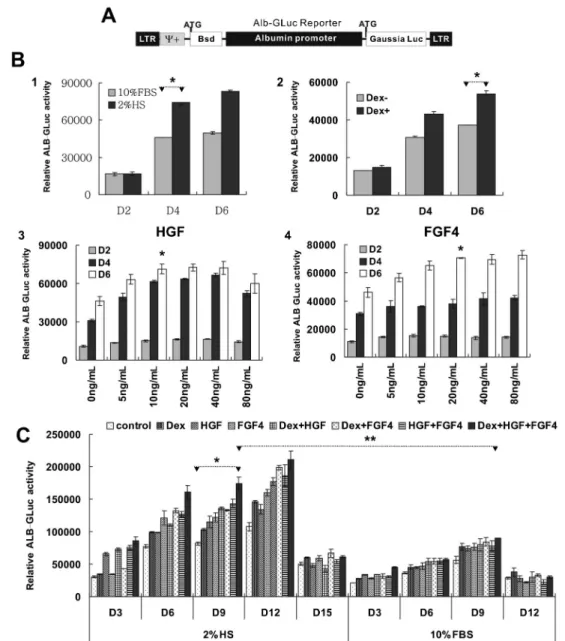
First, we detected an effect of different induction factors on maturation of HP14.5. ALB is the most abundant protein produced by the liver and its expression is correlated with
the maturation of hepatocytes. In HP14.5 cells, an ALB promoter was used to drive the expression of GLuc, which is an indirect indicator of the level of ALB in cells (Figure 1A). Treatment with 2% HS induced higher ALB-GLuc activity than 10% FBS. The activity of ALB-GLuc in 0.1mM
Dex-treated HP14.5 was higher at 4 days than in controls (P,0.05). Both HGF and FGF4 induced ALB-GLuc activity at the beginning of induction day 4 and day 6, respectively. The ALB-GLuc activity plateaued with HGF at concentra-tions ranging from 10 to 40 ng/mL (P.0.05 among HGF groups, P,0.05vscontrol), but decreased with 80 ng/mL. The effect of FGF4 was dose dependent at concentrations between 5 and 20 ng/mL (P,0.05 among FGF 4 groups, P,0.05 vs control; Figure 1B). Thus, the concentrations of HGF and FGF4 chosen to test the optimal induction condition in the following experiment were 10 and 20 ng/mL, respectively.
Next, we sought to determine the effect of combina-tions of different induction factors on maturation of HP14.5. While single factors or combinations of two factors could improve the expression of ALB-GLuc, a combination of 0.1mM Dex++10 ng/mL HGF++20 ng/mL
FGF4 was the best induction combination in both 10% FBS and 2% HS (P,0.05vscontrol). Meanwhile, HP14.5 cells were more sensitive to induction factors in 2% HS than in 10% FBS (P,0.05, 2% HS vs 10% FBS in same treatment of 0.1mM Dex++10 ng/mL HGF++20 ng/mL
FGF4). When the initial cell density was 20-30% in culture medium with 2% HS, the expression of ALB-GLuc increased with induction time, peaked at day 12, and sharply decreased at day 15. However, in culture medium with 10% FBS, the ALB-GLuc activity increase was apparent and peaked at day 9 (Figure 1C). Therefore, our results indicated that the combination of 2% HS++0.1mM Dex++10 ng/mL HGF++20 ng/mL FGF4
may be the best induction condition for ALB activity. We thus chose this condition for the following experiments.
Proliferation of cells treated with different single factors
assays was correlated with the growth curve at day 12 (Figure 2C). Thus, our results indicated that cell prolifera-tion was inhibited in 2% HS but enhanced by FGF4.
In vitroinduced maturation of HPCs
We investigated whether the optimal induction condition for ALB-GLuc activity could also induce maturation of
RT-PCR was performed to detect the expression of liver cell markers over 12 days of induction. As shown in Figure 3B, DLK and cytokeratin-19 (CK19), two hepatic stem cell markers, began to decline at day 3 and continued to decline until day 12. The expression of AFP initially increased, followed by a decrease from D6 onward. The expression of the liver cell specific markers ALB and cytokeratin-18 (CK18) continuously increased during the whole induction period. Two other liver-specific proteins, tyrosine amino-transferase (TAT) and apolipoprotein B (ApoB) were detectable at day 3 and day 6, respectively, and continued to increase during induction. Immunofluorescence staining results of DLK, AFP, and ALB were consistent with those obtained using RT-PCR. DLK protein was localized on cell membranes while the other proteins were largely distributed in the cytoplasm. The liver microsomal marker UGT1A also appeared in the cytoplasm and its expression increased significantly during induction (Figure 3C). Real-time PCR results further indicated that the expression of AFP, ALB, CK18, and TAT in the treated cells at day 12 was significantly greater than in the untreated cells, and was nearly the same as the expression level seen in LC14d cells, which were used as positive liver cell controls (Figure 3D).
Glycogen storage and ICG uptake/release function of induced HP14.5 cellsin vitro
Mature hepatocytes are able to carry out the function
of glycogen storage and accumulation in granule form in the cytosol, which can be demonstrated by PAS staining (15). The untreated HP14.5 cells were mostly PAS-negative. After 6 days of induction, PAS-positive granules started to appear in the cytoplasm and significantly increased at day 9 and day 12 of induction. ICG is a cyanine dye used to determine hepatic function (16). No ICG-positive cells were observed in the untreated group. At day 3 of induction, fewer than 5% of the cells took up ICG (green nuclear stain) from the medium and excluded the absorbed ICG 6 h later. The ICG positivity gradually increased along the induction time, and more than 70% of the cells were positive at day 12. The above results demonstrated that the reported induction method could induce not only the expression of liver cell markers but also the function of HP14.5 cells, providing an effective means to induce the maturation of HPCsin vitro.
Discussion
transplantation is a simple, safe, and relatively less costly procedure that can take advantage of freshly discarded liver segments, and shows great promise for the treatment of many liver diseases (18). Embryonic stem cells, bone marrow hematopoietic cells, mesenchymal stem cells, and umbilical cord blood cells have all been shown to differentiate into a hepatic linage with hepato-cyte-like morphology and cell markers. But few hepatic functions have been demonstratedin vivo(4,19). HPCs are progenitor cells originated from liver and capable of differentiating into hepatocytes and biliary cells. HPCs also express the stem cell-related markers with self-renewal capacity, serving as a continual and readily available source of cells for liver cell transplantation (20). However, HPCs are not mature enough to recover liver functionin vivo. In this study, we tested various induction conditions of in vitro culture of HP14.5 cells in order to identify the best method to induce maturation of HPCs.
Dex, HGF, and/or FGF4 have been shown to induce bone marrow mesenchymal stem cells, hematopoietic cells, or mouse embryonic stem cell differentiation to
hepatic cell lines (21,22). However, there are still no corresponding reports on the optimal concentration and combinations of induction factors in the differentiation of HPCs to mature hepatocytes. ALB is a marker of mature liver cells and is widely used to detect the maturation of hepatic cells (23). Here, we constructed a stable cell line expressing an ALB-promoter-driven GLuc reporter gene. During the liver cell differentiation process, many transcrip-tional factors regulate the expression levels of ALB by activating or inhibiting the ALB promoter. Moreover, GLuc is a natural secretary luciferase isolated from the marine copepodGaussia princeps that can be released into the culture medium (24). Here, we observed that the combina-tion of 2% HS++0.1mM Dex++10 ng/mL HGF++20 ng/mL
cells are more committed fetal HPCs and hence more responsive to the exogenous induction factors.
ALB-GLuc activity was only an indicator. We further confirmed the effect of this combination induction method. Compared with untreated cells, induced cells became neatly arranged and exhibited the typical liver cell morphology of polyhedron-shape. As previously reported (10), HP14.5 highly expressed the hepatic stem/progenitor cell markers DLK, AFP, and CK19. Upon induction, the expression of DLK and CK19 gradually decreased. CK19 is not a unique marker for liver stem cells, as it is also a marker of biliary differentiation (27), its marked reduction reflects HP14.5 cell differentiation into mature hepatic cells, but not bile duct cells. AFP is a major plasma protein produced by the liver during fetal development and is thought to be the fetal form of serum albumin. The highest AFP levels are present in the fetus and decrease at the end of the first trimester (28). HP14.5 cells were isolated from post-coitus day 14.5 embryonic liver, which may not represent the high point of the APF level. Thus, APF expression firstly increased and then decreased by day 6 during the induction process. Furthermore, other hepatic-specific proteins ALB, CK18, UGT1A produced by mature liver cells continued to rise. TAT became detectable by day 3, and ApoB, by day 6 after induction. On day 12, the induced cells exhibited expression of liver markers comparable to adult mouse liver cells. Therefore, our result revealed that HP14.5 cells could be effectively differentiated into mature hepatocytes.
To become a reliable cell source for liver cell transplantation, the induced HPCs should have good liver
function. PAS staining is primarily used to identify glycogen in tissues. Functional hepatocytes are capable of glycogen synthesis and accumulation (15). ICG is a nontoxic cyanine dye used in hepatic function diagnostics. ICG is metabolized microsomally in liver cells and removed from the liver exclusively in bile juice (16). Microsomes were present in the nucleus, thus the green nuclear staining reflected the function of ICG metabolism. With the induction conditions used here, ICG uptake/ release and PAS-positive cells were observed at day 6, and gradually increased to more than 70% by day 12, suggesting the induced HP14.5 cells had hepatic function. Taken together, the results demonstrate that the combination of 2% HS++0.1mM Dex++10 ng/mL
HGF++20 ng/mL FGF4 effectively induced the maturation and function of HPCs. It is conceivable that more effective methods need to be further explored because fewer than 100% of the induced cells had hepatic functions. Nevertheless, we report a relatively effective method to induce hepatic differentiation and maturation of HPCs.
Supplementary Material
Click here to view [pdf]
Acknowledgments
References
1. Feldmann G. Liver transplantation of hepatic stem cells: potential use for treating liver diseases. Cell Biol Toxicol 2001; 17: 77-85, doi: 10.1023/A:1010954020488.
2. Kakinuma S, Nakauchi H, Watanabe M. Hepatic stem/ progenitor cells and stem-cell transplantation for the treatment of liver disease.J Gastroenterol2009; 44: 167-172, doi: 10.1007/s00535-008-2297-z.
3. Navarro-Alvarez N, Soto-Gutierrez A, Kobayashi N. Hepatic stem cells and liver development.Methods Mol Biol2010; 640: 181-236, doi: 10.1007/978-1-60761-688-7_10. 4. Sharma AD, Cantz T, Vogel A, Schambach A, Haridass D,
Iken M, et al. Murine embryonic stem cell-derived hepatic progenitor cells engraft in recipient livers with limited capacity of liver tissue formation. Cell Transplant 2008; 17: 313-323, doi: 10.3727/096368908784153896.
5. Tomiyama K, Miyazaki M, Nukui M, Takaishi M, Nakao A, Shimizu N, et al. Limited contribution of cells of intact extrahepatic tissue origin to hepatocyte regeneration in transplanted rat liver.Transplantation 2007; 83: 624-630, doi: 10.1097/01.tp.0000253942.16061.d9.
6. Sangan CB, Tosh D. Hepatic progenitor cells.Cell Tissue Res2010; 342: 131-137, doi: 10.1007/s00441-010-1055-8. 7. Brolen G, Sivertsson L, Bjorquist P, Eriksson G, Ek M, Semb H, et al. Hepatocyte-like cells derived from human embryonic stem cells specifically via definitive endoderm and a progenitor stage.J Biotechnol 2010; 145: 284-294, doi: 10.1016/j.jbiotec.2009.11.007.
8. Zhang W, Li W, Liu B, Wang P, Li W, Zhang H. Efficient generation of functional hepatocyte-like cells from human fetal hepatic progenitor cellsin vitro. J Cell Physiol2012; 227: 2051-2058, doi: 10.1002/jcp.22934.
9. Ichinohe N, Tanimizu N, Ooe H, Nakamura Y, Mizuguchi T, Kon J, et al. Differentiation capacity of hepatic stem/progenitor cells isolated from D-galactosamine-treated rat livers. Hepatology2013; 57: 1192-1202, doi: 10.1002/hep.26084. 10. Ichinohe N, Kon J, Sasaki K, Nakamura Y, Ooe H, Tanimizu
N, et al. Growth ability and repopulation efficiency of transplanted hepatic stem cells, progenitor cells, and mature hepatocytes in retrorsine-treated rat livers. Cell Transplant2012; 21: 11-22.
11. Gridelli B, Vizzini G, Pietrosi G, Luca A, Spada M, Gruttadauria S, et al. Efficient human fetal liver cell isolation protocol based on vascular perfusion for liver cell-based therapy and case report on cell transplantation. Liver Transpl2012; 18: 226-237, doi: 10.1002/lt.22322. 12. Bi Y, Huang J, He Y, Zhu GH, Su Y, He BC, et al. Wnt
antagonist SFRP3 inhibits the differentiation of mouse hepatic progenitor cells. J Cell Biochem2009; 108: 295-303, doi: 10.1002/jcb.22254.
13. Bi Y, Gong M, Zhang X, Zhang X, Jiang W, Zhang Y, et al. Pre-activation of retinoid signaling facilitates neuronal differ-entiation of mesenchymal stem cells. Dev Growth Differ 2010; 52: 419-431, doi: 10.1111/j.1440-169X.2010.01182.x. 14. He Y, Zhang WY, Gong M, Huang JY, Tang N, Feng T, et al. Low serum concentration facilitates the differentiation of hepatic progenitor cells.Saudi Med J2011; 32: 128-134. 15. Liu YN, Zhang J, He QH, Dai X, Shen L. Isolation and
characterization of epithelial progenitor cells from human fetal liver. Hepatol Res2008; 38: 103-113, doi: 10.1111/ j.1872-034X.2007.00163.x.
16. Yamada T, Yoshikawa M, Kanda S, Kato Y, Nakajima Y, Ishizaka S, et al.In vitrodifferentiation of embryonic stem cells into hepatocyte-like cells identified by cellular uptake of indocyanine green. Stem Cells 2002; 20: 146-154, doi: 10.1634/stemcells.20-2-146.
17. Clavien PA, Lesurtel M, Bossuyt PM, Gores GJ, Langer B, Perrier A. Recommendations for liver transplantation for hepatocellular carcinoma: an international consensus con-ference report. Lancet Oncol 2012; 13: e11-e22, doi: 10.1016/S1470-2045(11)70175-9.
18. Yu Y, Fisher JE, Lillegard JB, Rodysill B, Amiot B, Nyberg SL. Cell therapies for liver diseases.Liver Transpl2012; 18: 9-21, doi: 10.1002/lt.22467.
19. Passier R, Mummery C. Origin and use of embryonic and adult stem cells in differentiation and tissue repair. Cardiovasc Res2003; 58: 324-335, doi: 10.1016/S0008-6363(02)00770-8.
20. Parveen N, Aleem AK, Habeeb MA, Habibullah CM. An update on hepatic stem cells: bench to bedside.Curr Pharm Biotechnol 2011; 12: 226-230, doi: 10.2174/ 138920111794295765.
21. Banas A, Yamamoto Y, Teratani T, Ochiya T. Stem cell plasticity: learning from hepatogenic differentiation strategies. Dev Dyn2007; 236: 3228-3241, doi: 10.1002/dvdy.21330. 22. Kang XQ, Zang WJ, Bao LJ, Li DL, Song TS, Xu XL, et al.
Fibroblast growth factor-4 and hepatocyte growth factor induce differentiation of human umbilical cord blood-derived mesenchymal stem cells into hepatocytes. World J Gastroenterol2005; 11: 7461-7465.
23. Heo J, Factor VM, Uren T, Takahama Y, Lee JS, Major M, et al. Hepatic precursors derived from murine embryonic stem cells contribute to regeneration of injured liver. Hepatology2006; 44: 1478-1486, doi: 10.1002/hep.21441. 24. Wiles S, Ferguson K, Stefanidou M, Young DB, Robertson BD. Alternative luciferase for monitoring bacterial cells under adverse conditions. Appl Environ Microbiol 2005; 71: 3427-3432, doi: 10.1128/AEM.71.7.3427-3432.2005. 25. Oh SH, Witek RP, Bae SH, Zheng D, Jung Y, Piscaglia AC,
et al. Bone marrow-derived hepatic oval cells differentiate into hepatocytes in 2-acetylaminofluorene/partial hepatect-omy-induced liver regeneration. Gastroenterology 2007; 132: 1077-1087, doi: 10.1053/j.gastro.2007.01.001. 26. Song Z, Cai J, Liu Y, Zhao D, Yong J, Duo S, et al. Efficient
generation of hepatocyte-like cells from human induced pluripotent stem cells.Cell Res2009; 19: 1233-1242, doi: 10.1038/cr.2009.107.
27. Zong Y, Panikkar A, Xu J, Antoniou A, Raynaud P, Lemaigre F, et al. Notch signaling controls liver develop-ment by regulating biliary differentiation.Development2009; 136: 1727-1739, doi: 10.1242/dev.029140.