Antigen Recognition to Toll-Like Receptor 9 Activation in
Late Endosomes
Margaret Veselits1, Azusa Tanaka1, Stanley Lipkowitz2, Shannon O’Neill3, Roger Sciammas1, Alison Finnegan4, Jian Zhang5, Marcus R. Clark1*
1Section of Rheumatology, Department of Medicine and Knapp Center for Lupus and Immunological Research, University of Chicago, Chicago, Illinois, United States of America,2Laboratory of Cellular and Molecular Biology, Center for Cancer Research, National Cancer Institute, National Institutes of Health, Bethesda, Maryland, United States of America,3Integrated Department of Immunology, National Jewish Medical and Research Center and University of Colorado and Health Sciences Center, Denver, Colorado, United States of America,4Department of Immunology and Microbiology, and Department of Internal Medicine, Section of Rheumatology, Rush University Medical Center, Chicago, Illinois, United States of America,5Section of Nephrology, Department of Medicine, University of Chicago, Chicago, Illinois, United States of America
Abstract
Casitas B-lineage lymphoma-b (Cbl-b) is a ubiquitin ligase (E3) that modulates signaling by tagging molecules for degradation. It is a complex protein with multiple domains and binding partners that are not involved in ubiquitinating substrates. Herein, we demonstrate that Cbl-b, but not c-Cbl, is recruited to the clustered B cell antigen receptor (BCR) and that Cbl-b is required for entry of endocytosed BCRs into late endosomes. The E3 activity of Cbl-b is not necessary for BCR endocytic trafficking. Rather, the ubiquitin associated (UBA) domain is required. Furthermore, the Cbl-b UBA domain is sufficient to confer the receptor trafficking functions of Cbl-b on c-Cbl. Cbl-b is also required for entry of the Toll-like receptor 9 (TLR9) into late endosomes and for thein vitroactivation of TLR9 by BCR-captured ligands. These data indicate that Cbl-b acts as a scaffolding molecule to coordinate the delivery of the BCR and TLR9 into subcellular compartments required for productively delivering BCR-captured ligands to TLR9.
Citation:Veselits M, Tanaka A, Lipkowitz S, O’Neill S, Sciammas R, et al. (2014) Recruitment of Cbl-b to B Cell Antigen Receptor Couples Antigen Recognition to Toll-Like Receptor 9 Activation in Late Endosomes. PLoS ONE 9(3): e89792. doi:10.1371/journal.pone.0089792
Editor:Wasif N. Khan, University of Miami, United States of America
ReceivedJuly 23, 2013;AcceptedJanuary 27, 2014;PublishedMarch 20, 2014
Copyright:ß2014 Veselits et al. This is an open-access article distributed under the terms of the Creative Commons Attribution License, which permits unrestricted use, distribution, and reproduction in any medium, provided the original author and source are credited.
Funding:This study was supported by National Institutes of Health (MRC, GM088847 and GM101090 and JZ, AI090901). This research was supported in part by the Intramural Research Program of the NIH, National Cancer Institute, Center for Cancer Research.
Competing Interests:The authors have declared that no competing interests exist.
* E-mail: [email protected]
Introduction
Antigen presentation by B lymphocytes is required to mount high affinity humoral immune responses, for coordinating antigen specific cytotoxicity, and for propagating some T cell responses [1]. B lymphocytes differ from other antigen presenting cells in several fundamental ways. The most important difference is that B cells are clonotypic, and they usually only efficiently capture and process antigens recognized by the B cell antigen receptor (BCR) [2]. The primacy of the BCR as the portal for entry of antigen ensures coordination of B and T cell responses.
In B cells, most antigens are processed in specialized MHC class II containing late endosomes (MIIC) [3] which are Lamp-1+
, acidic and contain cathepsins, thiol reductases, and other molecules required for efficient antigen processing [4]. MIIC vesicles consist of a limiting membrane studded with Lamp-1 and a lumen containing multivesicular bodies [5]. These intraluminal vesicles are derived from BCR-laden transport vesicles that have gained access to the MIIC compartment [6].
BCR trafficking to late endosomes is also required for coupling antigen recognition to the activation of the toll-like receptors (TLRs) 7 and 9 [7,8]. This is because these receptors only productively bind ligands in late endosomes. The mechanisms
underlying this requirement have been best defined for TLR9. In resting B cells, TLR9 resides outside the MIIC. Upon BCR ligation, TLR9 rapidly transits into the MIIC [9,10] where the receptor can bind DNA containing complexes captured by the endocytosed BCR [11–13]. Analysis of BCR and TLR9 endocytic trafficking in anergic B cells, in which the trafficking of both receptors is aberrant, indicates that entry of the BCR and TLR9 into late endosomes is coordinated and that both receptors enter on common transport vesicles [10]. Presumably, this facilitates the transfer of BCR captured ligands to the TLRs.
the core machinery for the delivery of receptors to late endosomes, several other molecular complexes are involved in facilitating and regulating ESCRT-mediated endocytic transit [15].
Previously, we have demonstrated that the BCR subunit Igbis ubiquitinated and that this is required for sorting to late endosomes [16]. Normal receptor ubiquitination required Itch, a member of the Nedd4 family of E3s. This is in apparent contrast to the T cell receptor (TCR) [17], and other receptors [15], where recruitment of the Casitas B-lineage Lymphoma (Cbl) E3s to the tyrosine phosphorylated receptor induce ubiquitination. We now report that Cbl-b is also required for BCR endocytic trafficking, and that it contributes to receptor ubiquitination following receptor stimulation. However, Cbl-b ligase activity is dispensible for BCR endocytic trafficking. Rather, Cbl-b provides a necessary scaffolding function that is dependent upon the carboxyterminal tail. Surprisingly, transit of TLR9 into late endosomes was also dependent upon Cbl-b. These and other findings demonstrate a unique, unexpected, and functionally important role for Cbl-b in directing the delivery of both the BCR and TLR9 to late endosomes.
Materials and Methods
Mice
Wild-type, Cblb2/2 (Balb/c), and CblbC373A
(C57BL/6J) [18] mice were housed in the animal facilities of the University of Chicago. Mice were used at 8 to 12 weeks of age, and experiments were in accordance with the guidelines of the Institutional Animal Care and Use Committee of the University of Chicago (Protocol Number: 71577, approval date 4/6/13). All animal studies were carried out in strict accordance with the recommendations in the Guide for the Care and Use of Laboratory Animals of the National Institutes of Health. All mice were sacrificed by CO2inhalation
followed by cervical dislocation.
Cells and culture conditions
Splenic B cells were isolated by negative selection using biotinylated CD11b (M1/70), CD11c (HL3), anti-NK1.1 (PK136), anti-Ter-119, anti-CD3e1452C11, anti-CD4 (RM4-5), anti-CD8a(53-6.7), anti-Ly-6G, and Ly-6C (RB6-8C5, all BD Bioscience) followed by streptavidin magnetic beads (MACS; Miltenyi Biotec) [16]. Cells were cultured for 20 hours in complete DMEM supplemented with 10% FCS, 50 ng/ml rIL-4 (R & D Systems), and 7.5mg/ml anti-CD-40 (BD Biosciences).
Receptor internalization and ubiquitination assays
BCR internalization assays on wild-type andCblb2/2splenic B cells were performed as previously described [19]. Receptor ubiquitination assays were performed as described [16].
Retroviral gene transduction
The cDNA encoding human Cbl-b and truncation mutants as well as human c-Cbl [20] were subcloned into the plasmid MIGR1. Calcium Phosphate transfection of PlatE cells with MIGR1 was carried out as previously described [21,22]. Aliquots of cell lysates were immunoblotted to confirm the size of the expressed proteins. Cbl-b (H-454 and H-121) and c-Cbl (C-15) antibodies were from Santa Cruz Biotechnology. Wt andCblb2/2
B cells were transduced by resuspending in viral supernatant with 8 mg/ml polybrene in the presence of rIL-4 (50 ng/ml) and anti-CD40 (7.5mg/ml) and centrifuging at 10006g for 1.5 hr at room temperature. The cells were then washed and placed into fresh culture medium and allowed to grow for 48 hrs.
Confocal microscopy
Confocal Microscopy was performed as previously described [10]. Images were collected by using a Leica TCS SP2 AOBS confocal microscope. Antibodies used for visualization were Lamp-1 (1D4B) (BD Biosciences), TLR9 (26C593.2) (Imgenex), Cathepsin L (CPLH-3G10) and Cbl-b (H-121 and H-454) (Santa Cruz Biotechnology). All secondary antibodies were Alexa Fluor conjugated IgG (H & L) from Invitrogen. To quantitate the extent of colocalization between 2, 3 or 4 fluorescent markers in individual cells, we used the JACoP plug-in of ImageJ. The specific algorithm used was based on the Mander’s coefficient with a threshold of 40 [23]. Cells that had a Mander’s coefficient of 0.25 or higher were scored as positive for co-localization. For each experiment, at least 30 randomly selected cells were scored.
T-bet assay and quantitative real-time PCR
Biotinylated ODN 1826 and biotinylated control ODN were from Invivogen. Purified splenic B cells from WT and Cblb2/2
mice were incubated with 10mg/ml of streptavidin-conjugated F(ab)2Ig (H & L) followed by either the biotinylated ODN 1826 or
control ODN (Invivogen). Cells were then incubated for 6 hrs at 37uC. RNA was isolated and quantitative PCR performed as previously described [10].
Western blotting and immunoprecipitations
Splenic B cells were purified by negative selection as described above. Cells were stimulated with 20mg/ml F(ab)2 goat anti–
mouse Ig (H+L) (Jackson ImmunoResearch Laboratories) at 37uC for indicated times. Cell aliquots were lysed in 1% NP40 buffer (150 mM NaCl, 10 mM Tris HCl pH 7.7, 5 mM EDTA, 0.4 mM sodium orthovanadate, and 10 mM sodium pyrophosphate) containing mini EDTA-free protease inhibitor cocktail tablets (Roche), phenylmethylsulfonyl fluoride (PMSF)(Sigma-Aldrich), 20 mM N-Ethylmaleimide (NEM) (Sigma-Aldrich), 10 mM 1,10-phenatholine monohydrate (OPD) (Sigma-Aldrich), and 50mM PR-619 (LifeSensors). Lysates were clarified by centrifugation at 4uC. Lysates were precleared with protein A–Sepharose (Pierce), incubated with primary antibodies specific for Igb (Hm79b, BD Biosciences) and captured with protein G–Sepharose (Pierce). Lysates or immunoprecipitates were resolved on a 4–15% Mini-Protean TGX gel (Bio-Rad) and transferred onto Immun-Blot PVDF membrane (Bio-Rad). Membranes were probed with antibodies specific for ubiquitin (P4D1, Santa Cruz), Igb[24] or Cbl-b.
Statistical Analyses
Statistical comparisons were made using the Student’s t-test.
Results
Cbl-b is required for BCR endocytic trafficking to late endosomes
To explore if Cbl-b played a role in BCR endocytic trafficking, splenic B lymphocytes [10] from wild type (WT) orCblb2/2Balb/
c mice were isolated and then stimulatedin vitrothrough the BCR with FITC-conjugated anti-Ig F(ab)2antibodies for 30 minutes at
37uC. Aliquots were then fixed, stained with anti-Lamp-1 antibodies (Alexa Fluor 647, Invitrogen), and visualized by confocal microscopy [10]. Representative images are provided in
Figure 1A, and a quantitative analysis of the fraction of cells demonstrating significant overlap (.25%) between the BCR and Lamp-1 is provided inFigure 1B [10]. In WT cells, the BCR rapidly targeted Lamp-1+
late endosomes with almost all cells
demonstrating strong co-localization between the BCR and Lamp-1. In contrast, in Cblb2/2 cells there was little co-localization
between the BCR and Lamp-1. Rather, the endocytosed BCR was in close proximity with Lamp-1+
late endosomes. Similar results were obtained if cells were stimulated for 60 minutes (data not shown).
We hypothesized that in the absence of Cbl-b, endocytosed BCRs might target terminal lysosomes which in B lymphocytes, do not contain Lamp-1 [25]. As demonstrated inFigures 1A and
1B, the Lamp-12subcellular compartment targeted by the BCR
in Cblb2/2 splenocytes contained cathespin L. Interestingly,
Cathepsin L was much more apparent in the Lamp-1+
compart-ments of WT than Cblb2/2 splenocytes. This latter observation
suggests that Cbl-b lies in the signaling pathway necessary for BCR-mediated MIIC maturation [10,26,27].
To determine if Cbl-b contributed to BCR endocytic trafficking by ubiquitinylating Igb, splenic B lymphocytes from WT or
Cblb2/2 mice were stimulated with anti-Ig F(ab)2antibodies for
Figure 1. Cbl-b is required for normal trafficking to late endosomes.(A–B) Trafficking to Lamp-1+
and Cathepsin L+
endocytic compartments by the ligated BCR in WT andCblb2/2
splenic B cells. (A) Representative confocal images of BCR stimulated cells (30 min) and (B) quantitative analysis of three independent experiments (30 cells/exp) (*p = 0.0003 and **p = 0.0004). Average Mander’s coefficient of BCR vs Lamp-1 for WT cells was 0.55660.12 vs 0.14660.07 forCblb2/2
cells (p,0.0001). Average Mander’s coefficient of Cathepsin-L vs Lamp-1 for WT cells was 0.41560.05 vs 0.17960.03 forCbl-b2/2
cells (p,0.002). (C) Ubiquitination of IgbinCblb2/2
and WT splenocytes. Unstimulated or BCR stimulated splenocytes were lysed, immunoprecipitated with anti-Igbantibodies, and then sequentially immunoblotted with anti-Ub (upper panel) or anti-Igb(lower panel) antibodies. Results are representative of three experiments. (D) Cells were stimulated as in ‘‘A’’ for two minutes, fixed, and visualized by confocal microscopy. Shown are the percentages of each cell population that formed a receptor cap on the cell surface containing more than 50% of visualized BCRs (n = 3 experiments, p = 0.005). (E) Internalization of BCR inCblb2/2(blue, diamonds) and WT (purple, squares) splenocytes in response
to anti-IgM F(ab)2antibodies. Results are representative of three experiments.
15 minutes at 37uC. Aliquots were then lysed in a buffer containing 1% NP40, and lysates were precipitated with anti-Igbantibodies. Precipitations were resolved by SDS-PAGE, transferred to PVDF membranes, and probed with anti-ubiquitin (Ub) antibodies. As can be seen in Figure 1C, there was approximately a three-fold decrease in the amount of ubiquiti-nated Igb immunoprecipitated from Cblb2/2 B cells following
BCR stimulation. These results indicate that Cbl-b, along with the E3 ligase Itch [16], contributes to inductive Igbubiquitination.
Recently, in chicken B cell lines, Cbl has been demonstrated to be required for ligand-induced BCR clustering on the cell surface [28]. Therefore, cells were stimulated as above for two minutes and then visualized by confocal microscopy. As demonstrated in
Figure 1D, while more than 80% of WT B cells formed a cap following BCR stimulation, only approximately 35% ofCbl-b2/2
B cells formed caps (p = 0.005). These data indicate that Cbl-b is required for BCR clustering in primary murine splenocytes.
Despite the difference in capping, subsequent internalization of the ligated BCR on both WT andCblb2/2splenic B cells were
similar (Figure 1E). Furthermore, transit of the ligated BCR to EEA1+
early endosomes was similar in WT andCblb2/2splenic B
cells (Figure S1) [10]. However, Cblb2/2 BCRs left this
compartment much more rapidly that WT BCRs. These data indicate that Cbl-b is specifically required for two discrete processes, BCR clustering and trafficking between early and late endosomes.
Rapid recruitment of Cbl-b following BCR stimulation
To examine if Cbl-b directly participated in BCR endocytic trafficking, we first determined if Cbl-b was recruited to the BCR following receptor engagement. Splenic WT Balb/c B lympho-cytes were stimulated as above with Texas Red-conjugated anti-Ig F(ab)2 antibodies for various times up to 60 minutes. Cells were
then fixed, stained with Lamp-1 (Alexa Fluor 647) and anti-Cbl-b antibodies (Alexa Fluor 488, Invitrogen), and visualized by confocal microscopy. Representative images are provided in
Figure 2A with the corresponding quantitations from three independent experiments provided inFigure 2B. As can be seen, Cbl-b was rapidly and almost completely recruited to the BCR within two minutes of ligation. The BCR and Cbl-b remained co-localized as the receptor was internalized and targeted to late endosomes. However, the association between the BCR and Cbl-b diminished as the BCR started to enter Lamp-1+
late endosomes. These data indicate that Cbl-b is rapidly and robustly recruited to the BCR and then traffics with the receptor through the endocytic pathway. However, the majority of Cbl-b does not enter Lamp-1+ late endosomes with the BCR.
The Cbl-b UBA domain, but not the RING finger domain, are required for BCR endocytic trafficking
We next examined which functional domains of Cbl-b were required for BCR endocytic trafficking. For these experiments, cDNAs encoding WT Cbl-b and a series of Cbl-b mutants (Figure 3A) [20,29] were cloned into the retroviral vector MIGR1. These vectors were then packaged in PlatE cells [21]. Aliquots of cell lysates from packaging cells were immunoblotted to confirm expression of Cbl-b proteins of the expected relative molecular weights (Figure 3B). Splenic B cells were harvested fromCblb2/2mice and cultured for 24 hours in IL-4 (50 ng/ml)
and anti-CD40 antibodies (7.5mg/ml). Aliquots of proliferating
Cblb2/2 splenic B cells were then infected with viruses encoding the indicated Cbl-b molecules. After an additional 48 hours, cells were stimulated with Texas Red conjugated anti-BCR F(ab2)
antibody fragments for 30 minutes, then fixed and stained with
anti-Lamp-1 and anti-Cbl-b antibodies. Finally, cells expressing each construct (typically 30–40% of cells) were visualized by confocal microscopy (Figure 3C). A quantitative assessment of the degree of colocalization between the BCR and Lamp-1 in MIGR1 infectedCblb2/2 cells is provided in Figure 3D(n = 3
experiments). As can be seen, retroviral mediated expression of WT Cbl-b reconstituted BCR trafficking to late endosomes, while there was no reconstitution mediated by vector alone. Examina-tion of the various mutants indicated that the C-terminal domain, which lacks the tyrosine kinase-binding (TKB) domain, could reconstitute BCR trafficking as could a Cbl-b molecule lacking E3 activity (Cbl-bC373A). In contrast, a mutant lacking the C-terminal UBA domain could not. Interestingly, expression of Cbl-bGYDAQ, in which the Ub binding motif within the UBA domain [20,29] was mutated, reconstituted BCR trafficking. These data indicate that the Cbl-b UBA domain, but not the ability of the UBA domain to bind ubiquitin, is necessary for BCR endocytic trafficking.
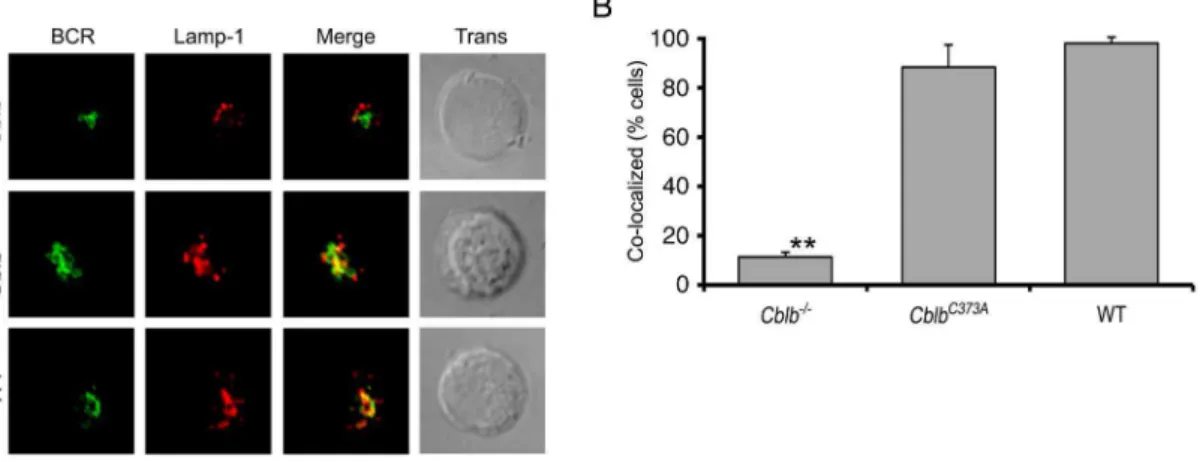
To confirm that the E3 activity of Cbl-b was dispensable for BCR endocytic trafficking, we obtained mice in which gene targeting had been used to derive mice expressing Cbl-b bearing the C373A mutation [18]. Spleens from these mice or WT controls were harvested and splenic B cells isolated and stimulated as described above. Aliquots were then fixed, stained, and visualized by confocal microscopy. Representative images are provided inFigure 4A, and quantitative assessments from three independent experiments are depicted inFigure 4B. These data indicate that BCR endocytic trafficking was similar in WT and
CblbC373AB splenocytes. These results indicate that, although Cbl-b contriCbl-butes to the uCbl-biquitination of Igb, this function is dispensible or redundant for BCR endocytic trafficking.
Our data indicated that the carboxyterminal domain of Cbl-b, which contains the UBA domain, was required for BCR endocytic trafficking. This finding was potentially revealing as the amino acid sequences of c-Cbl and Cbl-b are divergent in the UBA domain. Therefore, we next examined if ectopic expression of c-Cbl in Cblb2/2 splenocytes could restore BCR endocytic
trafficking.
A cDNA encoding WT c-Cbl (Figure 5A) was cloned into MIGRI and packaged in PlatE cells (anti-c-Cbl immunoblots provided in Figure 5B). Packaged virus was used to infect
Cblb2/2splenocytes which were assayed by confocal microscopy
(Figure 5C) and scored for reconstitution of BCR endocytic trafficking (Figure 5D) as described above. As can be seen, ectopic expression of c-Cbl did not reconstitute BCR endocytic trafficking inCblb2/2B splenocytes. In keeping with these results,
confocal microscopic analysis of WT splenocytes revealed that, in contrast to Cbl-b, c-Cbl was not recruited to the ligated BCR complex (Figure S2).
We next examined if the UBA domain of Cbl-b, in the context of c-Cbl, was sufficient to reconstitute BCR endocytic trafficking. To this end, we constructed a cDNA encoding a chimeric c-Cbl molecule in which the UBA domain of c-Cbl was replaced with that of Cbl-b (c-CblDUBA-Cbl-b)(Figure 5A–D). This chimeric molecule was then expressed inCblb2/2splenocytes and cells were analyzed by confocal microscopy. Expression of c-CblDUBA-Cbl-b restored normal BCR endocytic trafficking. These observations indicate that the unique ability of Cbl-b to mediate endocytic trafficking is fully encoded within the carboxyterminal region containing the UBA domain.
Cbl-b is required for TLR9 endocytic trafficking and for TLR9-dependent B cell responses in vivo
In addition to inducing rapid endocytic transit of the BCR, receptor stimulation induces the translocation of TLR9 into late endosomes [9,10]. Therefore, we next examined if Cbl-b was required for BCR ligation-induced TLR9 transit. B splenocytes from WT, Cblb2/2, and CblbC373A
mice were stimulated as in Figure 1 for 30 minutes and then analyzed by confocal microscopy for the co-localization of the BCR, Lamp-1 and TLR9. As can be seen inFigures 6A–6C, inCblb2/2mice, stimulation of the BCR
induced the co-localization of the endocytosed BCR with TLR9. However, both receptors were excluded from Lamp-1+ late endosomes. As expected, this compartment was Cathepsin L+ consistent with terminal lysosomes (Figure S3). In contrast, both the BCR and TLR9 entered late endosomes in B splenocytes from
CblbC373Amice. No staining of TLR9 was observed in splenic B cells fromTLR92/2mice [10]. These data indicate that TLR9 has
similar Cbl-b requirements as the BCR for entry into late endosomes.
As in Cblb2/2 splenocytes TLR9 was sequestered within
terminal lysosomes, we examined if these cells were unresponsive to TLR9 ligands delivered through the BCR. A unique downstream target of TLR9 is the transcription factor T-box expressed in T cells (T-bet), which can be directly activated by CpG via TLR9 in B cells through an IFNcR/STAT1-independent pathway [30]. Therefore, WT andCblb2/2 splenic B cells were
incubated with streptavidin-conjugated F(ab)2anti-IgG antibodies
followed by biotinylated ODN 1826 or control ODN (Invivogen). After six hours, RNA was isolated from stimulated B cells, and T-bet mRNA expression was assayed by quantitative PCR. IFNc stimulated cells were used as a positive control [30]. As can be seen in Figure 6D, BCR targeted ODN 1826 stimulated T-bet expression in WT cells. The control ODN did not induce T-bet expression. However, inCblb2/2splenocytes ODN 1826 failed to
induce T-bet. These data reveal a defect in T-bet responses to TLR9 ligands inCblb2/2 splenocytes. T-bet induction was also
defective through IFNcsuggesting the presence of more global activation defects.
Discussion
Herein, we demonstrate that recruitment of Cbl-b to the BCR is necessary for both clustering the BCR on the cell surface and for guiding internalized BCRs into late endosomes. This activity of Cbl-b was unique and could not be supplanted by c-Cbl. Furthermore, Cbl-b was required for TLR9 entry into late endosomes and for activation of TLR9 by BCR captured ligands. In the absence of Cbl-b, the BCR and TLR9 became sequestered together within terminal lysosomes. These data indicate that Cbl-b plays an important role in coupling adaptive and innate immune signaling responses in B lymphocytes.
Our findings both confirm and extend recent observations in chicken cell lines on the importance of Cbl in determining BCR endocytic fate. As observed in chicken cell lines [28], Cbl-b was required for ligand-induced clustering on the cell surface. In these cell line studies, Cbl played a role in coupling the BCR to the motor protein dynein. Dynein has also been implicated in endocytic trafficking [31] indicating that the observed defects in clustering and endocytic trafficking could reflect a common underlying mechanism.
The unique ability of Cbl-b to facilitate receptor endocytic trafficking mapped to the Cbl-b carboxyl-terminal tail that contains the UBA domain. One well-described difference between Cbl-b and c-Cbl is that the Cbl-b UBA domain can bind Ub while the c-Cbl UBA cannot. Ubiquitin binding mediates Cbl-b dimerization and increases E3 ligase activity [29]. However, the ability of Cbl-b to enable BCR endocytic trafficking to late endosomes was not dependent on the Cbl-b ubiquitin-binding motif. In chicken B cells, c-Cbl is recruited to the BCR and is required for BCR surface clustering [28]. Chicken c-Cbl is also predicted to not bind Ub, and therefore, there is likely another unidentified functional domain, conserved between chicken c-Cbl Figure 2. Cbl-b is recruited to the BCR and co-traffics to late endosomes.WT splenic B cells were stimulated through the BCR for the indicated times. (A) Representative confocal images demonstrating relative locations of BCR (green), Lamp-1 (red), and Cbl-b (blue) over time. (B) Quantitative analysis (n = 3, 30 cells/exp for each condition and time point) of co-localization over time of BCR and Cbl-b (diamond, red), BCR and Lamp-1 (triangle, light blue), and Cbl-b and Lamp-1 (circle, dark blue).
Figure 3. BCR endocytic trafficking is dependent on the UBA domain of Cbl-b and independent of its ligase activity.(A) Schematic representation of different Cbl-b mutants that were used to reconstitute Cblb2/2
splenocytes (TKB, tyrosine kinase binding domain; RF, ring finger domain; PRO, proline rich regions; UBA, ubiquitin binding domain). Numbers above schematic refer to amino acid positions. (B) Immunoblots of packaging cell lysates with anti-Cbl-b antibodies demonstrating the relative molecular weights of the indicated expressed Cbl-b mutants. (C) Representative confocal images demonstrating the ability of each indicated Cbl-b mutant to reconstitute BCR endocytic trafficking in Cblb2/2
splenocytes. For these experiments, cells were stained with anti-BCR (green), anti-Lamp-1 (red), and anti-Cbl-b antibodies (blue). (D) Quantitative analysis (n = 3, 30 cells/exp for each condition) for co-localization of BCR with Lamp-1 (* statistically similar, p,0.001 for Cbl-b WT vs. N1/2,DUBA). doi:10.1371/journal.pone.0089792.g003
Figure 4. BCR endocytic trafficking is normal in splenocytes expressing an E3 ligase dead mutant of Cbl-b (CblbC373A).Indicated
splenocytes were stimulated through the BCR and imaged after 30 min by confocal microcopy. (A) Representative images. (B) Quantitative analysis of three independent experiments (p,0.001 forCblb2/2
vsCblbC373A
or WT). doi:10.1371/journal.pone.0089792.g004
and human and murine Cbl-b, that enables BCR endocytic trafficking.
Previous publications have focused on Cbl-b as a negative regulator of lymphocytes [32]. Mice deficient in Cbl-b develop an autoimmune syndrome associated with lymphocytic infiltrates in multiple organs [33], and they are highly susceptible to experimental autoimmune encephalomyelitis [34] and collagen induced arthritis [35]. Cblb2/2 T cells do not need CD28
costimulation for activation and cannot be tolerized [34]. These phenotypic changes have been related to Cbl-b-mediated regula-tion of PI3K [36] and PLCc1 [35], respectively. In B cells, Cbl-b negatively regulates signaling through CD40 [37] and has been demonstrated to bind and ubiquitinylate multiple proximal BCR signaling components [33,38–41]. Our observation that Cbl-b is recruited to the aggregated BCR complex provides a mechanism by which Cbl-b could gain access to many of these signaling substrates.
However, given the multiple negative signaling functions ascribed to Cbl-b, the phenotype of Cblb2/2 mice is relatively
mild. Autoimmunity does not develop until after six months of age, and potentially pathogenic immune complex deposits in glomeruli are only observed in some aged mice (10 months old) in which bothCblandCblbhad been targeted in B cells [42]. By ELISA, anti-dsDNA antibodies are detected inCblb2/2 mice. However,
such ELISAs can be falsely positive and are not as relevant as
Crithidia luciliaeimmunofluorescence (IF) assays. In fact, inMRL/ Mplpr/lpr mice, TLR9 deficiency greatly diminishes anti-dsDNA responses as measured by IF but not as measured by ELISA [43]. Thus, the available evidence indicates that the intrinsic B cell defect in Cblb2/2 mice is not severe. We propose that this is
because Cbl-b is a complex molecule with both negative and positive effects on peripheral B cell activation.
While the Cbl-b UBA domain is uniquely required for BCR endocytic trafficking, it is likely that one or more additional Cbl-b domains, conserved in c-Cbl, contribute by linking the receptor to downstream effector mechanisms. When tyrosine phosphorylated, Cbl proteins bind to CD2AP (CD2 adaptor protein) and the
homolog CIN85 [44–46]. CIN85 has mostly been studied in the context of epidermal growth factor receptor (EGFR) trafficking, while CD2AP appears to play a prominent role in TCR function [47]. CIN85 can localize to late endosomes [48], and has been implicated in the endocytic degradation of the EGFR [46]. T cells from CD2AP2/2 mice have a defect in ligand-induced TCR degradation that appears to be due to a block in trafficking to lysosomes [47]. The exact function of CD2AP and CIN85 is not known, although the latter has been demonstrated to function as a scaffold for multiple molecules implicated in endocytic trafficking [49].
Cbl-b and c-Cbl likely share another function important for BCR endocytic trafficking. The region immediately carboxyterm-inal to the RF domain (the RF tail) mediates phosphorylation and degradation of Hrs [50]. Mutation of this domain both abrogates Hrs phosphorylation and prevents fusion of EGFR-containing early endosomes with acidic late endosomes. This phenotype is similar to that observed for BCR trafficking in Cblb2/2
splenocytes.
Surprisingly, Cbl-b was also required for entry of TLR9 into late endosomes. It is possible that Cbl-b regulates one or more signaling pathways mediating TLR9 transit into late endosomes. This possibility is consistent with our findings in anergic B cells that defective JNK activation prevents the transit of both the BCR and TLR9 into late endosomes [10]. It is also consistent with the apparent dependency of MIIC maturation on Cbl-b and with the general known interplay between BCR endocytic trafficking and intracellular signaling [9]. Alternatively, Cbl-b could provide a necessary linker or scaffolding function for TLR9. As Cbl-b is recruited to the BCR, such a model would require that the BCR and TLR9 come into close proximity before entry into late endosomes. The strong co-localization observed between the BCR and TLR9 in BCR-stimulatedCblb2/2splenocytes indicates that
this is the case.
By demonstrating a unique, non-E3 dependent function for Cbl-b, our data provide insights into how the family of Cbl ligases may mediate different cellular functions. Deletion of Cbl-b results Figure 5. The UBA domain of Cbl-b, in the context of c-Cbl, is sufficient for BCR endocytic trafficking.(A) Schematic representation of Cbl-b and c-Cbl encoding constructs used to reconstituteCblb2/2
splenocytes. Percentages represent homology between c-Cbl and Cbl-b in indicated domains. (B) Immunoblot of PlatE cell lysates expressing indicated constructs with anti-c-Cbl antibodies. (C) Representative confocal images of Cblb2/2
splenocytes reconstituted with virus encoding c-Cbl or c-CblDUBA-Cbl-b
in immune hyperactivity and some features of autoimmunity. In this regard, Cbl-b is broadly similar to c-Cbl. However, specific mechanistic dissection indicates that the Cblb2/2 phenotype
reflects the net effect of deleting a molecule that can both enable and suppress specific immune mechanisms. Our data also demonstrate that Cbl-b is more than a biochemical component Figure 6. Cbl-b is required for the endocytic transit of TLR9.(A) Representative confocal microscopic images of splenocytes from mice with indicated genotypes. For experiments, cells were stimulated through the BCR (green) for 30 minutes then fixed and stained for TLR9 (blue) and Lamp-1 (red)(n = 3). (B) Quantitation of experiments shown in (A) for fraction of cells demonstrating significant co-localization of TLR9 with Lamp-Lamp-1 (n = 3, 30 cells/exp) (Cbl-b2/2vs. CblbC373Aor WT, p
,0.001). (C) Quantitation of fractions of cells in (A) demonstrating significant co-localization between BCR and TLR9 (n = 3, 30 cells/exp). (D)In vitroassay of T-bet induction in response to ODN 1826 or control ODN targeted through the BCR (n = 3, p,0.01).
doi:10.1371/journal.pone.0089792.g006
of several signaling pathways. Rather, it performs an important cell biological function by coordinating the intracellular movement of immune recognition receptors. Therefore, Cbl-b functions in two or more fundamental cellular processes that determine immune responses to both self and non-self antigens.
Supporting Information
Figure S1 Normal trafficking of the ligated BCR to early endosomes in Cblb2/2
splenocytes. (A) Cblb2/2 or WT
splenocytes were stimulated as in Figure 1 for 15 or 45 minutes and then fixed and stained with antibodies to the early endosomal marker EEA1 (C45B10, Cell Signaling). Shown are representative images of results obtained from three independent experiments. In (B) is a quantitation of the co-localization between the BCR and EEA1 in Cblb2/2 (light grey) and WT (dark grey) (n = 3
experiments, *p = 0.002). (TIF)
Figure S2 C-Cbl is not recruited to the aggregated BCR complex.Cells were stimulated for 0 to 20 minutes through the BCR, fixed, stained with either anti-Cbl-b or anti-c-Cbl antibodies
and analyzed as in Figure 2. In (A) is provided representative images obtained two minutes after stimulation (n = 3). In (B) is a quantitation of the co-localization between the BCR and either c-Cbl (red, squares) or c-Cbl-b (blue, diamonds)(n = 3).
(TIF)
Figure S3 Both the BCR and TLR9 target the same Lamp-12Cathepsin L+
compartment. (A) Representative four color confocal micrographs of WT andCblb2/2splenocytes stimulated with TexasRed-conjugated anti-BCR antibodies for 30 minutes. Cells were then fixed, stained with antibodies specific for TLR9, Cathepsin L and Lamp-1 and visualized by confocal microscopy. (B) Quantification of co-localization between different markers in WT (dark grey) andCblb2/2(light grey) splenocytes.
(n = 3, *p = 8.6561026, **p = 0.0015 and ***p = 0.0005). (TIF)
Author Contributions
Conceived and designed the experiments: MRC. Performed the experi-ments: MV AT SO. Analyzed the data: MV AF MRC. Contributed reagents/materials/analysis tools: SL RS JZ. Wrote the paper: MRC.
References
1. Cascalho M, Platt JL (2007) Novel functions of B cells. Crit Rev Immunol 27: 141–151.
2. Clark MR, Massenburg D, Siemasko K, Hou P, Zhang M (2004) B-cell antigen receptor signaling requirements for targeting antigen to the MHC class II presentation pathway. Cur Opin Immunol 16: 382–387.
3. Ferrari G, Knight AM, Watts C, Pieters J (1997) Distinct intracellular compartments involved in invariant chain degradation and antigenic peptide loading of major histocompatibility complex (MHC) class II molecules. J Cell Biol 139: 1433–1446.
4. Watts C (2001) Antigen processing in the endocytic compartment. Cur Opin Immunol 13: 26–31.
5. Siemasko K, Clark MR (2001) The control and facilitation of MHC class II antigen processing by the BCR. Curr Opin Immunol 13: 32–36.
6. Riberdy JM, Avva RR, Geuze HJ, Cresswell P (1994) Transport and intracellular distribution of MHC class II molecules associated with invariant chain in normal and antigen processing mutant cell lines. J Cell Biol 125: 1215– 1237.
7. Viglianti GA, Lau CM, Hanley TM, Miko BA, Shlomchik MJ, et al. (2003) Activation of autoreactive B cells by CpG dsDNA. Immunity 19: 837–847. 8. Lau CM, Broughton C, Tabor AS, Akira S, Flavell RA, et al. (2005)
RNA-associated autoantigens activate B cells by combined B cell antigen receptor/ Toll-like receptor 7 engagement. J Exp Med 202: 1171–1177.
9. Chaturvedi A, Dorward D, Pierce SK (2008) The B cell receptor governs the subcellular location of Toll-like receptor 9 leading to hyperresponses to DNA-containing antigens. Immunity 28: 799–809.
10. O’Neill SK, Veselits ML, Zhang M, Labno C, Cao Y, et al. (2009) Endocytic sequestration of the B cell antigen receptor and toll-like receptor 9 in anergic B cells. Proc Nat Acad Sci, USA 106: 6262–6267.
11. Park B, Brinkmann MM, Spooner E, Lee CC, Kim YM, et al. (2008) Proteolytic cleavage in an endolysosomal compartment is required for activation of Toll-like receptor 9. Nat Immunol 12: 1407–1414.
12. Barton GM, Kagan JC (2009) A cell biological view of Toll-like receptor function: regulation through compartmentalization. Nat Rev Immunol 9: 535– 542.
13. Ewald SE, Engel A, Lee J, Wang M, Bogyo M, et al. (2011) Nucleic acid recognition by Toll-like receptors is coupled to stepwise processing by cathepsins and asparagine endopeptidase. J Exp Med 208: 643–651.
14. Railborg C, Stenmark H (2009) The ESCRT machinery in endosomal sorting of ubiquitylated membrane proteins. Nature 458: 445–452.
15. Mukherjee S, Tessema M, Wandinger-Ness A (2006) Vesicular trafficking of tyrosine kinase receptors and associated protiens in regulation of signaling and vascular function. Circ Res 98: 743–756.
16. Zhang M, Veselits M, O’Neill S, Hou P, Reddi AL, et al. (2007) Ubiquitinylation of Ig-beta dictates the endocytic fate of the B cell antigen receptor. J Immunol 179: 4435–4443.
17. Myers MD, Sosinowski T, Dragone LL, White C, Band H, et al. (2006) Src-like adaptor protein regulates TCR expression on thymocytes by linking the ubiquitin ligase c-Cbl to the TCR complex. Nat Immunol 7: 57–66. 18. Oksvold MP, Dagger SA, Thien CB, Langdon WY (2008) The Cbl-b RING
finger domain has a limited role in regulating inflammatory cytokine production by IgE-activated mast cells. Mol Immunol 45: 925–936.
19. Hou P, Araujo E, Zhao T, Massenburg D, Veselits M, et al. (2006) B cell antigen receptor signaling and internalization are mutually exclusive events. PLoS Biology e200.
20. Davies GC, Ettenberg SA, Coats AO, Mussante M, Ravichandran S, et al. (2004) Cbl-b interacts with ubiquitinated proteins; differential functions of the UBA domains of c-Cbl and Cbl-b. Oncogene 23: 7104–7115.
21. Mandal M, Powers SE, Ochiai K, Georgopoulos K, Kee B, et al. (2009) Ras orchestrates cell cycle exit and light chain recombination during early B cell development. Nat Immunol 10: 1110–1117.
22. Johnson K, Hashimshony T, Sawai CM, Pongubala JM, Skok JA, et al. (2008) Regulation of immunoglobulin light-chain recombination by the transcription factor IRF-4 and the attenuation of interleukin-7 signaling. Immunity 28: 335– 345.
23. Bolte S, Cordelieres FP (2006) A guided tour into subcellular colocalization analysis in light microscopy. J Microscopy 224: 213–232.
24. Luisiri P, Lee YJ, Eisfelder BJ, Clark MR (1996) Cooperativity and segregation of function within the Iga/b heterodimer of the B cell antigen receptor complex. J Biol Chem 271: 5158–5163.
25. Li C, Siemasko K, Clark MR, Song W (2002) Cooperative interaction of Ig-alpha and Ig-beta of the BCR regulates kinetics and specificity of antigen targeting. Int Immunol 14: 1179–1191.
26. Siemasko K, Eisfelder BJ, Williamson E, Kabak S, Clark MR (1998) Cutting Edge: Signals from the B lymphocyte antigen receptor regulate MHC class II containing late endosomes. J Immunol 160: 5203–5208.
27. Lankar D, Vincent-Schneider H, Briken V, Yokozeki T, Raposo G, et al. (2002) Dynamics of major histocompatibility complex class II compartments during B cell receptor-mediated cell activation. J Exp Med 195: 461–472.
28. Schnyder T, Castello A, Feest C, Harwood NE, Oellerich T, et al. (2011) BCR-mediated antigen gathering requires Grb2, Dok-3 and Cbl to recruit dynein to the signalling microcluster. Immunity 34: 905–918.
29. Peschard P, Kozlov G, Lin T, Mirza IA, Berghuis AM, et al. (2007) Structural basis for ubiquitin-mediated dimerization and activation of the ubiquitin protein ligase Cbl-b. Mol Cell 27: 474–485.
30. Liu N, Ohnishi N, Ni L, Akira S, Bacon KB (2003) CpG directly induces T-bet expression and inhibits IgG1 and IgE switching in B cells. Nat Immunol 4: 687– 693.
31. Hunt SD, Stephens DJ (2011) The role of motor proteins in endosomal sorting. Biochem Soc Trans 2011: 1179–1184.
32. Paolino M, Penninger JM (2010) Cbl-b in T-cell activation. Semin Immuno-pathol 32: 137–148.
33. Bachmaier K, Krawczyk C, Kozieradzki I, Kong Y-Y, Takehiko S, et al. (2000) Negative regulation of lymphocyte activation and autoimmunity by the molecular adaptor Cbl-b. Nature 403: 211–216.
34. Chiang YP, Kole HK, Brown K, Maramura M, Fukuhara S, et al. (2000) Cbl-b regulates the CD28 dependence of T-cell activation. Nature 403: 216–220. 35. Jeon MS, Atfield A, Venuprasad K, Krawczyk C, Sarao R, et al. (2004) Essential
role of the E3 ubiquitin ligase Cbl-b in T cell anergy induction. Immunity 21: 167–177.
36. Fang D, Liu YC (2001) Proteolysis-independent regulation of PI3K by Cbl-b-mediated ubiquitination in T cells. Nat Immunol 2: 870–875.
38. Sohn HW, Gu H, Pierce SK (2003) Cbl-b negatively regulated B cell antigen receptor signaling in mature B cells through ubiquitination of the tyrosine kinase syk. J Exp Med 197: 1511–1524.
39. Bustelo XR, Crespo P, Lopez-Barahona M, Gutkind JS, Barbacid M (1997) Cbl-b, a member of the Sli-1/c-Cbl protein family, inhibits Vav-mediated c-Jun N-terminal kinase activation. Oncogene 15: 2511–2520.
40. Rao N, Dodge I, Band H (2002) The Cbl family of ubiquitin ligases: critical negative regulators of tyrosine kinase signaling in the immune system. J Leukoc Biol 71: 753–763.
41. Yasuda T, Maeda A, Kurosaki M, Tezuka T, Hironaka K, et al. (2000) Cbl suppresses B cell receptor-mediated phospholipase C (PLC)-g2 activation by regulating B cell linker protein-PLC-g2 binding. J Exp Med 191: 641–650. 42. Kitaura YJ, Jang IK, Wang Y, Han Y-C, Inazu T, et al. (2007) Control of B
cell-intrinsic tolerance programs by ubiquitin ligasess Cbl and Cbl-b. Immunity 26: 567–578.
43. Christensen SR, Shupe J, Nickerson K, Kashgarian M, Flavell RA, et al. (2006) Toll-like receptor 7 and TLR9 dictate autoantibody specificity and have opposing inflammatory and regulatory roles in a murine model of lupus. Immunity 25: 417–428.
44. Hicke L, Schubert HL, Hill CP (2005) Ubiquitin-binding domains. Nat Rev Mol Cell Biol 6: 610–621.
45. Kirsch KH, Georgescu M-M, Shishido T, Langdon WY, Brige RB, et al. (2001) The adapter type protein CMS/CD2AP binds to the proto-oncogenic protein c-Cbl through a tyrosine phosphorylation-regulated Src homology 3 domain interaction. J Biol Chem 276: 4957–4963.
46. Haglund K, Shimokawa N, Szymkiewicz I, Dikic I (2002) Cbl-directed monoubiquitination of CIN85 is involved in regulation of ligand-induced degradation of EGF receptors. Proc Natl Acad Sci 99: 12191–12196. 47. Lee KH, Dinner AR, Tu C, Campi G, Raychaudhuri S, et al. (2003) The
immunological synapse balances T cell receptor signaling and degradation. Science 302: 1218–1222.
48. Zhang J, Zheng XX, Yang X, Liao K (2009) CIN85 associates with endosomal membrane and binds phosphatidic acid. Cell Res 19: 733–746.
49. Kowanetz K, Husnjak K, Holler D, Kowanetz K, Soubeyran P, et al. (2004) CIN85 associates with multiple effectors controlling intracellular trafficking of epidermal growth factor receptors. Mol Cell Biol 15: 3155–3166.
50. Visser Smit GD, Place TL, Cole SL, Clausen KA, Vemuganti S, et al. (2009) Cbl controls EGFR fate by regulating early endosome fusion. Sci Signal 2: ra86.