Submitted19 August 2016
Accepted 4 November 2016
Published7 December 2016
Corresponding author
Chia-Chi Wang,
chiachiwang@kmu.edu.tw
Academic editor
Howard Young
Additional Information and Declarations can be found on page 22
DOI10.7717/peerj.2758
Copyright
2016 Cheng et al.
Distributed under
Creative Commons CC-BY 4.0
OPEN ACCESS
Attenuation of antigen-specific T helper 1
immunity by
Neolitsea hiiranensis
and its
derived terpenoids
Yin-Hua Cheng1, Ih-Sheng Chen2, Ying-Chi Lin1,2, Chun-Wei Tung1,2, Hsun-Shuo Chang2and Chia-Chi Wang1,2
1Ph.D. Program in Toxicology, College of Pharmacy, Kaohsiung Medical University, Kaohsiung, Taiwan 2School of Pharmacy, Kaohsiung Medical University, Kaohsiung, Taiwan
ABSTRACT
Background. T cells play a pivotal role in the adaptive immunity that participates in a wide range of immune responses through a complicated cytokine network. Imbalance of T-cell responses is involved in several immune disorders. Neolitsea species, one of the biggest genera in the family Lauraceae, have been employed widely as folk medicines for a long time in Asia. Previous phytochemical investigations revealed the abundance of terpenes in the leaves of N. hiiranensis, an endemic Neolitsea in Taiwan, and demonstrated anti-inflammatory activities. However, the effect of N. hiiranensison the functionality of immune cells, especially T cells, is still unclear. In this study, we utilizein vitroandin vivoapproaches to characterize the effects of leaves of N. hiiranensisand its terpenoids on adaptive immune responses.
Methods. Dried leaves ofN. hiiranensiswere extracted three times with cold methanol to prepare crude extracts and to isolate its secondary metabolites. The ovalbumin (OVA)-sensitized BALB/c mice were administrated with N. hiiranensis extracts (5– 20 mg/kg). The serum and splenocytes of treated mice were collected to evaluate the immunomodulatory effects ofN. hiiranensis on the production of OVA-specific antibodies and cytokines. To further identify the N. hiiranensis-derived compounds with immunomodulatory potentials, OVA-primed splenocytes were treated with compounds isolated from N. hiiranensis by determining the cell viability, cytokine productions, and mRNA expression in the presence of OVAin vitro.
Results. Crude extracts of leaves ofN. hiiranensissignificantly inhibited IL-12, IFN-γ, and IL-2 cytokine productions as well as the serum levels of antigen-specific IgM and IgG2ain vivo. Two of fourteen selected terpenoids and one diterpenoid derived from the leaves of N. hiiranensis suppressed IFN-γ in vitro. In addition, β-caryophyllene oxide attenuated the expression of IFN-γ, T-bet, and IL-12Rβ2 in a dose-dependent manner. N. hiiranensis-derived β-caryophyllene oxide inhibited several aspects of adaptive immune responses, including T-cell differentiation, IFN-γ production, and Th1-assocaited genes.
Conclusion. As IFN-γ is the key cytokine secreted by T helper-1 cells and plays a pivotal role in Th1 immune responses, our results suggested that theN. hiiranensis and its terpenoids may possess potential therapeutic effects on Th1-mediated immune disorders.
INTRODUCTION
T helper (Th) cells play a pivotal role in our immune system against environmental stimulations. They participated in a wide range of immune responses via cell–cell interaction with other cells through a complicated cytokine network. Th1 cells, producing interferon-gamma (IFN-γ), interleukin-2 (IL-2) in the regulation of cellular immunity. On the other side, Th2 cells promoted humoral immunity via secreting IL-4, IL-5 and IL-13 (Lazarevic & Glimcher, 2011;Lin & Lin, 2011). Abnormal immunostimulation and the imbalance of Th1/Th2 responses may lead to a variety of immune diseases. For example, the dominant of Th1 cells is associated with multiple sclerosis, Crohn’s disease, rheumatoid arthritis, and delayed type hypersensitivity (DTH) (Agnello et al., 2003;Lazarevic & Glimcher, 2011).
The immunosuppressive drugs, such as glucocorticoids, cyclosporine A, infliximab, and etanercept, were developed to treat the over-reactive immune responses, inflammation or T-cell mediated immune disorders (Diluvio et al., 2010;Lee et al., 2002;Lin & Wang, 2016;
Rezzani, 2004;Rezzani, 2006;Srivastava, Alexander & Tuthill, 2005;Tan et al., 2007;Torres et al., 2011;Weinberg et al., 2005;Wolff, McKay & Brugarolas, 2014; Zeevi et al., 1987). However, severe adverse effects have been associated with the long-term usage of these immunosuppressants. To discover new botanicals with differential immunomodulatory effects on T-cell function may provide more therapeutics for different T-cell-mediated immune disorders. Many natural compounds isolated from medicinal plants have been shown to possess therapeutic potentials for Th1-associated diseases (Jeon et al., 2015;Wu et al., 2014).
Neolitseais one of the major genera in Lauraceae family. There are about 85 species in Asiatic and Malaysia, including six endemic species in Taiwan (Liao, 1996;Liou et al., 2011). These evergreen shrubs or trees have long been used as traditional folk medicines to treat carcinomatous swelling, abdominal pain, diarrhea, rheumatism, nausea and vomiting (Xie, 1996). These plants contain various bioactive components including sesquiterpenes which are known to have anti-inflammatory effects (Chang et al., 2002;Chen et al., 2005) and terpenoids which have been demonstrated to possess immunomodulatory effects on LPS-stimulated splenocytesin vitro(Ku & Lin, 2013).
N. hiiranensis is an endemic Neolitsea in Taiwan containing a rich amount of sesquiterpenoids which have been documented to possess anti-inflammation activity (Liou et al., 2011;Wu & Li, 1995). Hiiranlactone B and hiiranlactone D, the sesquiterpenoids isolated from the leaves of N. hiiranensis, suppressed theN -formyl-methionyl-leucyl-phenylalanine (fMLP)-induced generation of the superoxide anion by human neutrophils (Ho et al., 2011;Liou et al., 2011). Pseudoneolinderane and isolinderalactone isolated from the roots ofN. hiiranensishave been shown their anti-inflammatory activities (Wu & Li, 1995). These data indicated the potential immunomodulatory effects ofN. hiiranensison innate immune responses. However, the effect ofN. hiiranensis on T-cell functionality remains unclear.
immunomodulatory effects of the therapeutic botanicals for Th1 immune disorders. We report here that the administration ofN.hiiranensisdidn’t affect body weight, spleen index, and spleen cellularity in vivo. Several antigen-specific immune responses were attenuated byN. hiiranensisand its terpenoids.
MATERIALS AND METHODS
Extraction and isolation from the Taiwanese N. hiiranensis
The crude extracts and the secondary metabolites were prepared and isolated from the leaves ofN. hiiranensisaccording to the previous report (Liou et al., 2011). Briefly, TaiwaneseN. hiiranensiswere collected at Mudan (Pingtung County, Taiwan) and identified by Dr. Ih-Sheng Chen, one of the authors. The dried leaves were extracted with three times cold MeOH, and then the different partition of crude extracts was prepared with the differential proportion solvents system, including EtOAc:H2O,n-hexane: EtOAc, acetone: H2O, and
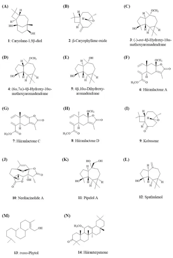
n-hexane: acetone for further isolating the secondary metabolites. Seven sesquiterpenoids, (e.g., (-)-ent-6α-methoxyeudesm-4(15)-en-1β-ol, hiiranlactones A–D, (+)-villosine, hiiranepoxide), one triterpenoid (hiiranterpenone), and 22 known compounds were identified and elucidated by spectroscopic analysis and single crystal X-ray diffraction (Liou et al., 2011). An established QSAR model for drug-induced liver injury (DILI) was utilized for prediction of non/less toxic pure compounds for further functionality tests (Huang et al., 2015).
Reagents and chemicals
All reagents were purchased from Sigma (St Louis, MO) unless otherwise stated. Fetal bovine serum (FBS) and cell culture medium RPMI 1640 were used from Hyclone (Logan, UT). Enzyme-linked immunosorbent assay (ELISA) sets for cytokine and antibody measurement were purchased from BD Biosciences (San Diego, CA). Isol-RNA lysis reagent were purchased from 5-Prime (Gaithersburg, MD). RevertAid RT kit was purchased from Thermo for Reverse transcription-polymerase chain reactions (RT-PCR).
Animals
Male BALB/c mice (5 weeks old) were purchased from BioLasco (Ilan, Taiwan). On arrival, mice were randomly transferred to plastic cages containing aspen bedding (five mice per cage) and acclimatized for at least one week before initiating experiments. Mice were housed in a temperature (22±2◦C), humidity (50±20%) and light (12-hour light/dark
cycle)-controlled environment. Food and water were suppliedad libitum.
Animal model for antigen-specific T-cell function
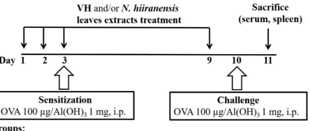
Figure 1 Protocol of administration ofN. hiiranensisand ovalbumin (OVA) sensitization and
challenge in BALB/c mice.Male BALB/c mice were randomly divided into the following groups: naïve (NA), vehicle (VH; Saline, and 4% of DMSO) andN. hiiranensis-treated (5 and 20 mg/kg) plus ovalbumin-sensitized and challenged groups. The mice were administered with VH and/or crude extracts by intraperitoneal injection for four doses. The dosing regimen for administration ofN. hiiranensisand immunization protocol were described in the materials and methods.
were administered to mice daily by intraperitoneal injection for three consecutive days (day 1–3). Except for the NA group, mice were sensitized with OVA 12 h after the third dose of VH orN. hiiranensison day 3 by an intraperitoneal injection with 0.1 mL per mouse of sensitization solution containing 100µg OVA and 1 mg alum (as adjuvant) in saline. The
mice and then challenged with OVA/alum at day 9. After OVA challenge, the mice were sacrificed at day 10 and their spleens were prepared and made into single-cell suspensions. The splenocytes were re-stimulated with OVA (100µg/mL) in culture for 72 h to induce
cell proliferation and cytokine production.
Cell proliferation assay
Splenocytes from the mice were aseptically cultured in RPMI 1640 medium supplemented with 5% heat-inactivated FBS, 100 µg/mL streptomycin, and 100 U/mL penicillin at
37◦C in 5% CO
2. Splenocytes (7×10⁶cells/mL) were seeded into 96-well plates. The cells were either left unstimulated or stimulated with OVA for 72 h. The viability of splenocytes was determined by the 3-(4, 5-dimethylthiazol-2-yl)-2, 5-diphenyl-tetrazolium bromide (methylthiazol tetrazolium) assay. A methylthiazol tetrazolium stock solution (5 mg/mL in phosphate buffered saline) was then added to each well (10 µL/well) and
Enzyme-linked immunosorbent assay (ELISA) for serum antibodies
ELISA plates were coated with 0.05% OVA in coating buffer (0.1 M NaHCO3) and blocked with 1% bovine serum albumin in phosphate-buffered saline containing 0.05% Tween 20 (PBST). After washing with PBST, the serum samples were added into wells (50
µL/well) and incubated for 1 h. After another washing, horseradish peroxidase-conjugated
anti-mouse IgG1, IgG2aor IgM was added (50µL/well) and incubated for 1 h. Finally, wells
were washed and a tetramethylbenzidine solution (50µL/well) was added for colorimetric
detection of bound peroxidase conjugate. The reaction was terminated by adding 150µL of
3 N H2SO4per well. The optical density (OD) was measured at 450 nm using a microplate reader (Dynatech Laboratories, Chantilly, VA, USA). Total IgE was measured according to manufacture’s instruction (BD Pharmingen).
Cytokine measurement by ELISA
To examine the effects ofN. hiiranensison specific subsets of T cells, splenocytes (7×10⁶ cells/mL) were cultured in 48-well plates (300µL/well) followed by OVA re-stimulated
(100µg/mL) for 72 h. The supernatants were harvested and quantified for IL-2, IL-4,
IL-12p70, IL-10, IL-13 and IFN-γ by ELISA kits according to manufacture’s instruction (BD Pharmingen).
In silicoprediction of hepatotoxicity and genotoxicity
Quantitative structure-activity relationship (QSAR) models are useful tools for in silico estimating toxicity properties of chemicals according to toxicity-related descriptors of physicochemical properties and fingerprints (Perkins et al., 2003). QSAR models have been extensively applied to prioritize chemicals for potential toxicity (Cronin et al., 2003;
Gramatica, Cassani & Sangion, 2016). In this study, toxicity properties of tested compounds were predicted by both the admetSAR server (Cheng et al., 2012) and our hepatotoxicity prediction model (Huang et al., 2015). The genotoxicity, carcinogenicity and acute oral toxicity of tested compounds are predicted by admetSAR with probabilities representing the confidence of prediction. The hepatotoxicity model is a special QSAR model utilizing toxicity information in human that no cross-species extrapolation is required. Similar to admetSAR, a confidence score is given by the hepatotoxicity prediction model. Generally, a score/probability close to 1 indicates a higher probability that a toxicity is associated with a given chemical. In contrast, a score closed to 0 indicates that a toxicity is unlikely associated with a given chemical.
RNA isolation and real-time reverse transcription-polymerase chain reactions (RT-PCR)
Total RNA from whole splenocytes (stimulated with OVA for 48 h) was isolated using an isol-RNA lysis reagent (5-Prime). The RNA samples (5 µg) were then treated
USA). One µg of total RNA of each sample was reverse-transcribed by RevertAid RT
Kit (Thermo) into cDNA products using oligo (dT) as primer. Real-time PCR was performed in a 96-well optic tray by an ABI PRISMR 7900HT Sequence Detection
System (Applied Biosystems, UK). During real-time RT-PCR process, we used Luminaris Color HiGreen High ROX qPCR Master Mix (Thermo Fisher Scientific, Waltham, MA, USA) which provides a highly specific and sensitive method to quantify mRNA expression. The HPRT gene was used as an endogenous control to normalize the expression of target genes. The primers are: 5′-GCCAGGGAACCGCTTATATG-3′ and 5′-GACGATCATCTGGGTCACATTCT-3′ for T-bet, 5′
-TACCCTCCGGCTT-CATCCT-3′ and 5′-TGCACCTGATACTTGAGGCAC-3′ for GATA-3, 5′ -GCC-AAGTTTGAGGTCAACAAC-3′ and 5′-CCGAATCAGCAGCGACTC-3′ for IFN-γ, 5′
-CCATATCCACGGATGCGACA-3′ and 5′-AAGCCCGAAAGAGTCTCTGC-3′ for IL-4,
5′-TCAGTCAACGGGGGACATAAA-3′and 5′-GGGGCTGTA- CTGCTTAACCAG-3′for HPRT (Shi et al., 2010), and 5′-CCTCAATGGTATAGCAGAAC and 5′ -TAGCCTTGG
AATCCTTGG for IL-12Rβ2 (Chognard et al., 2014).
In vitroscreening of cytokine production by antigen-specific T cells
OVA-primed splenocytes were generated according to previous protocol. Briefly, 6–8 week mice were sensitized by intraperitoneal injection of OVA (10 mg OVA absorbed to 100 mg alum as adjuvant) twice on day 1 and 14. On day 15, the mice were sacrificed and their spleens were harvested and made into single-cell suspensions. The OVA-primed splenocytes (7 ×10⁶ cells/mL) were either left untreated (control), 0.05% DMSO (VH) and/or secondary metabolites (1–10µM) followed by re-stimulation with OVA (100µg/mL) for
72 h. The cell proliferation activity, cytokine productions, and mRNA expression of target genes were measured as described above.
Flow cytometry analysis of intracellular cytokine and transcription factor staining in CD4+cells
OVA-primed splenocytes were cultured in a 12-well plate and stimulated with OVA and
β-caryophyllene oxide for 36 h. For analysis of intracellular cytokine production, the cells then treated with GolgiStop (0.6µL/mL; BD Biosciences) for 10 h prior to being harvested
for antibody staining. The cells were next stained with FITC-conjugated anti-mouse CD4 mAb (clone GK1.5; Biolegend, CA, USA) for 30 min on ice. The splenocytes then were fixed and permeabilized using Fixation and Perm/Wash buffers (BD Biosciences) before staining for intracellular IFN-γ by PE-conjugated anti-mouse IFN-γ mAb (clone XMG1.2; Biolegend) for 30 min on ice. Ten thousand CD4+ cells were acquired on a BD LSR II
flow cytometer (BD Biosciences). The mean fluorescence intensity (MFI) of IFN-γ in total CD4+ cells was quantified by gating CD4+ cells and then analyzed using FlowJo
GATA-3 mAb (clone 16E10A23; Biolegend) were applied to detect protein level of T-bet and GATA-3 in CD4+cells. Ten thousand CD4+cells were acquired on a BD LSR II flow
cytometer (BD Biosciences). The mean fluorescence intensity (MFI) of T-bet or GATA-3 in total CD4+ cells was quantified by gating CD4+ cells and then analyzed using FlowJo
software (Treestar, Inc., Ashland, OR, USA).
Statistical analysis
Homogeneous data were evaluated by a parametric analysis of variance (ANOVA) with Dunnett’s test to assess the statistical differences between the treatment groups and the VH control group by software Prism 5.0 (GraphPad Software Inc., San Diego, CA, USA). P<0.05 was defined as statistical significance. The mean±standard error was presented for individual experiments.
RESULTS
Crude extracts of leaves of N. hiiranensis did not affect body weight, spleen index, and cellularityin vivo
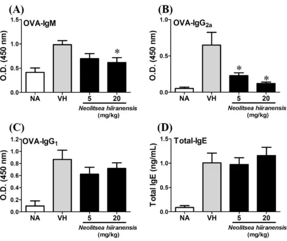
To investigate the potential of N. hiiranensis for in vivouse, we first investigated the direct immunotoxicity of N. hiiranensis in vivo. As shown inTable 1, intraperitoneal injection of N. hiiranensisextracts (5 and 20 mg/kg) didn’t affect the body weight, the spleen index, and the population of CD4+, CD8+, B220+, and CD11b+in spleens of the mice received treatment. We then examined the effect of theN. hiiranensis extracts on T-cell mediated humoral and cell-mediated immune responsesin vivo. TheN. hiiranensis extracts decreased the serum level of OVA-specific IgM and IgG2a. The serum level of OVA-specific IgG2a was dose-dependently suppressed byN. hiiranensisextracts. At the dose of 20 mg/kg, the level of OVA-specific IgM was significantly decreased. In contrast,N. hiiranensisdidn’t affect OVA-specific IgG1and total-IgE production. The result indicated that repeating administration ofN. hiiranensis significantly suppressed Th1-associated antibody production (Fig. 2)
Crude extracts of leaves of N. hiiranensisattenuated antigen-specific IL-2, IL-12, and IFN-γ cytokine productionin vivo
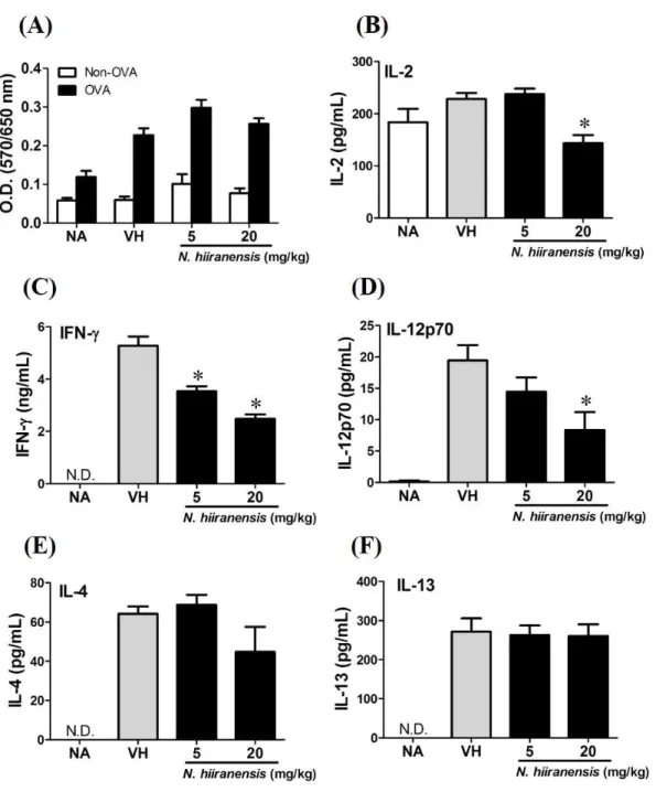
We then proceed to examine the effects of the crude extracts on the functionality of antigen-specific T cells by measuring the production of IL-2 (T-cell growth factor for clonal expansion), IFN-γ and IL-4 (the signature Th1 and Th2 differential cytokines), IL-12 (induction of IFN-γ production and Th1 differentiation), and IL-13 (Th2 cytokine closely related IL-4 to induce allergic Th2 responses). To check whether or not the induction of OVA was successful, the splenocytes were isolated and divided into non-stimulated (Non-OVA) and OVA 100µg/mL re-stimulated (OVA) groups and culture for 72 h to induce
Table 1 No effect ofN. hiiranensisleaves extracts on the body weight, spleen index and cellularity in
BALB/c mice.
NAa VH Leaves extracts ofN. hiiranensis
5 mg/kg 20 mg/kg
Body weight (g)
Day1 22.0±0.3 22.7±0.5 22.0±0.3 22.5±0.4
Day9 22.6±0.3 23.7±0.5 23.8±0.2 23.7±0.4
Spleen weight (mg) 91.4±3.1 103.5±4.2 97.1±4.5 91.8±3.4 Spleen indexb 4.1±0.2 4.4±0.2 4.2±0.2 3.9±0.2
Spleen cellularity (%)c
CD4+ 21.1±0.3 22.6±0.3 23.2±0.7 22.6±0.7
CD8+ 10.2±0.3 11.3±0.2 12.3±0.5 11.5±0.2
B220+ 45.9±1.0 45.4±1.0 46.2±1.1 46.1±0.2
CD11b+ 2.5±0.3 2.5±0.3 2.5±0.1 2.5±0.0
Notes.
aNA, untreated; VH, vehicle-treated and OVA-sensitized and challenged and leaves:N. hiiranensis-treated and OVA-sensitized
and challenged.
bSpleen index was calculated as the spleen weight (mg) per body weight (g). Data are expressed as mean±SE of eight (control
groups) and eleven (treatment groups) mice from three independent experiments.
cThe percentage of CD4+, CD8+, B220+, and CD11b+cells in spleen was determined by flow cytometry. Data are expressed as
mean±SE of four samples pooled from three independent experiments
group (Figs. 3Band3D). The production of IFN-γ by the cells was also attenuated by the treatment. IFN-γ was significantly lowered at both 5 mg/kg and 20 mg/kg treatment groups (P<0.05 for each group compared to the vehicle control group;Fig. 3C). By contrast, IL-4 and IL-13 were not affected (Figs. 3Eand3F). The results indicated that the extracts of the leaves ofN. hiiranensishave differential effects on Th1 responses.
In silicoselection of potential secondary metabolites of N. hiiranensis without undesired toxicity
Figure 2 N. hiiranensisattenuated OVA-specific IgM and IgG2aproduction.(A–D) The serum levels of
OVA-specific IgM, IgG2aIgG1, and total-IgE were determined by ELISA. Data are expressed as the mean ±standard error of eight samples per group. Results are pooled from three independent experiments. *p
<0.05 compared to the vehicle-treated group.
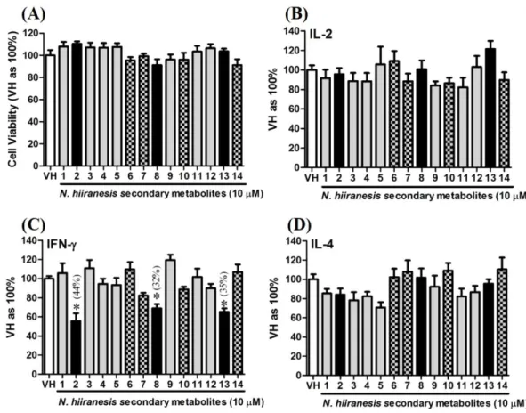
Secondary metabolites ofN. hiiranensisattenuated IFN-γ production in vitro
OVA-prime splenocytes were generated for screening the immunomodulatory effects of the compounds fromN.hiiranensis. The OVA-primed splenocytes were re-stimulated with OVA (100µg/mL) in the presence of vehicle and/or 10µM of selected compounds for 72
hin vitro. The selected pure compounds didn’t affect the proliferation activity nor induce the direct cytotoxicity (Fig. 5A). IL-2 and IL-4 were not significantly affected by the 14 selected compounds compared to VH. Interestingly, the antigen specific IFN-γ cytokine production were significantly suppressed byβ-caryophyllene oxide (2), hiiranlactone D (8), andtrans-phytol (13) with the inhibition rate of 44%, 32%, and 35%, respectively, comparing to VH (referred as 100%) (P<0.05). Spathulenol (12) slightly inhibited IFN-γ
and IL-4 productions without statistical significance (Fig. 5).
We further investigated the concentration-dependent effects of these compounds (2,8,
Figure 3 Suppression of IL-2, IFN-γ, and IL-12 production by leave extracts ofN. hiiranensis in vivo.
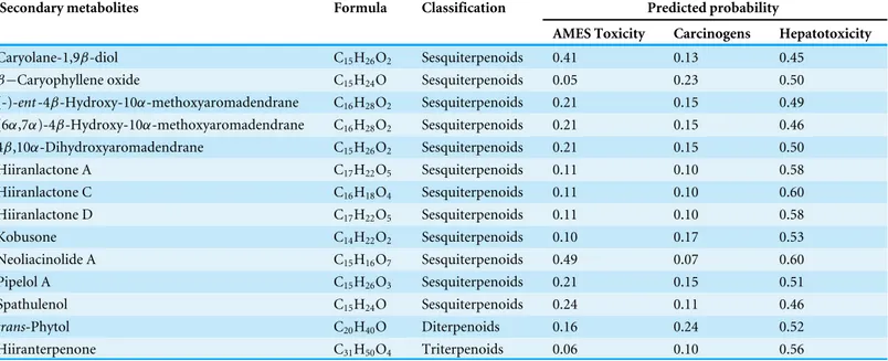
Table 2 The toxicity profile of 14 selected secondary metabolites fromN. hiiranensis. In order to select less toxic compounds for further study,
in silicoQSAR models were applied to filter out the compounds with potential toxicity concerns. A probability≦0.5 indicates no toxicity concern. A probability≦0.6 indicates a less hepatotoxicity concern.
Secondary metabolites Formula Classification Predicted probability
AMES Toxicity Carcinogens Hepatotoxicity
Caryolane-1,9β-diol C15H26O2 Sesquiterpenoids 0.41 0.13 0.45
β−Caryophyllene oxide C15H24O Sesquiterpenoids 0.05 0.23 0.50
(-)-ent-4β-Hydroxy-10α-methoxyaromadendrane C16H28O2 Sesquiterpenoids 0.21 0.15 0.49 (6α,7α)-4β-Hydroxy-10α-methoxyaromadendrane C16H28O2 Sesquiterpenoids 0.21 0.15 0.46
4β,10α-Dihydroxyaromadendrane C15H26O2 Sesquiterpenoids 0.21 0.15 0.50
Hiiranlactone A C17H22O5 Sesquiterpenoids 0.11 0.10 0.58
Hiiranlactone C C16H18O4 Sesquiterpenoids 0.11 0.10 0.60
Hiiranlactone D C17H22O5 Sesquiterpenoids 0.11 0.10 0.58
Kobusone C14H22O2 Sesquiterpenoids 0.10 0.17 0.53
Neoliacinolide A C15H16O7 Sesquiterpenoids 0.49 0.07 0.60
Pipelol A C15H26O3 Sesquiterpenoids 0.21 0.15 0.51
Spathulenol C15H24O Sesquiterpenoids 0.24 0.11 0.46
trans-Phytol C20H40O Diterpenoids 0.16 0.24 0.52
Hiiranterpenone C31H50O4 Triterpenoids 0.06 0.10 0.56
proliferation activity as well as IL-2 cytokine production of OVA-specific cells. By contrast,
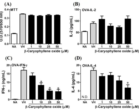
β-caryophyllene oxide inhibited IFN-γ production in a dependent manner and inhibited IL-4 production with an approximately 40% of inhibition rate at the concentrations higher than 25µM (Fig. 6). Hiiranlactone D (8) didn’t affect cell viability at the concentration
of 50µM. Hiiranlactone D attenuated IL-2 production of OVA-specific cells at 50 µM
and inhibited IFN-γ production in a concentration dependent manner starting from concentrations above 10µM (P<0.05). No effect on IL-4 was observed for hiiranlactone
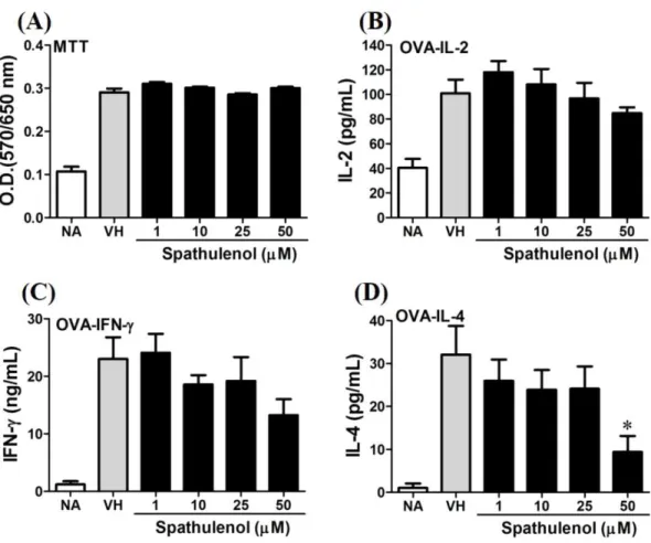
D (Fig. 7). Spathulenol also didn’t affect cell viability at the concentration of 50µM.
Spathulenol inhibited IL-4 production at 50 µM, while neither IL-2 nor IFN-γ were
significantly affected (Fig. 8).trans-Phytol didn’t affect cell viability at the concentration of 50µM. Interestingly,trans-phytol enhanced antigen-specific IL-2, and IL-4 production
in an concentration-dependent manner with significant inhibitions started from 25 and 50
µM for IL-2 and IL-4, respectively.trans-Phytol significantly inhibited IFN-γ production
in a concentration-dependent manner at the concentrations between 10 and 50µM (Fig.
9). The results demonstrated a differential immunomodulatory effects oftrans-phytol on the Th1/Th2 cytokine expression in antigen-specific T cells.
In the present data, β-caryophyllene oxide and trans-phytol are the most effective secondary metabolites from N. hiiranensis to suppress IFN-γ production. We next determined the effects of β-caryophyllene oxide and trans-phytol on the production of other Th1 and Th2 cytokines, including IL-12, IL-13, and IL-10. InFig. 10,trans-phytol significantly decreased IL-12 production, while both IL-13 and IL-10 were not altered (Figs. 10Eand10F). Interestingly, althoughβ-caryophyllene oxide significantly suppressed IFN-γ
Figure 5 The effects of secondary metabolites of leaves ofN. hiiranensison antigen-induced
produc-tion of cytokines and the metabolic activity in OVA-primed splenocytes.OVA-primed splenocytes (7 ×10⁶cells/mL) isolated from OVA-sensitized BALB/c mice were pretreated with secondary metabolites (10µM) and/or VH (0.05% DMSO) for 30 min followed by re-stimulated OVA (100µg/mL). After 72 h of culture, (A) the cell proliferation activity was determined using an MTT assay, the level of (B) IL-2, (C) IFN-γ, and (D) IL-4 in the supernatants was quantified by ELISA. The data were expressed as the mean ±SEM of quadruplicate cultured. Results were pooled from two or three independent experiments. *p< 0.05 was significant compared to the VH group.
IFN-γ production by CD4+cells, the intracellular cytokine staining approach was applied.
In the supplemental data (Fig. S1),β-caryophyllene oxide significantly suppressed the cellular level of IFN-γ in the CD4+cells.
β-caryophyllene oxide differentially modulated the development of Th1 and Th2 cells at transcription level
T-bet is a Th1-specific T-box transcription factor which controls the expression of Th1 cytokines and directs Th1 lineage commitment, while GATA-binding protein 3 (GATA-3) is a Th2-specific transcription factor which augments Th2-specific cytokines and Th2 differentiation to suppress Th1 immune responses. These transcription factors play crucial roles to regulate the homeostasis of Th cells (Agnello et al., 2003;Koch et al., 2009;
Figure 6 Antigen-specific IFN-γwas suppressed byβ-caryophyllene oxidein vitro.OVA-primed
splenocytes (7×10⁶cells/mL) were either left untreated (NA) or re-stimulated with OVA (100 µg/mL) in the absence or the presence ofβ-caryophyllene oxide (1–50µM) for 72 h. (A) The cell proliferation activity of viable cells was determined using the MTT assay. The levels of (B) IL-2, (C) IFN-γ, and (D) IL-4 in the supernatants were quantified by ELISA. Data were expressed as the mean±SE of quadruplicate cultures. Results were pooled from two independent experiments. *p<0.05 was significant compared to the VH group.
We next determined how β-caryophyllene oxide regulates Th1/Th2 associated gene expression. IFN-γ, IL-4, and Th1/Th2 differential transcription factors, T-bet and GATA-3 were determined. The mRNA expression of T-bet was significantly down-regulated by approximately 2-3-fold at 1 and 10µM (Fig. 11A), whereas GATA-3 wasn’t significantly
altered after treatment ofβ-caryophyllene oxide (Fig. 11D). IFN-γ was also down-regulated by approximately 1.5-2-fold (Fig. 11B); however, IL-4 was not changed (Fig. 11E). As T-bet response to IFN-γ may lead to the up-regulation of IL-12Rβ2 expression on Th1 cell surface for Th1 cell responsiveness to IL-12 stimulation (Hamza, Barnett & Li, 2010), we next determined whetherβ-caryophyllene oxide attenuated the expression of IL-12Rβ2. In consist with IFN-γ expression, the IL-12Rβ2 expression was down-regulated by approximately 1.5-2-fold (Fig. 11C). These results showed that the differentiation and functionality of Th1 cells were more sensitive to be attenuated byβ-caryophyllene oxide.
Figure 7 Attenuation of antigen-specific IFN-γproduction by hiiranlactone D. OVA-primed spleno-cytes were treated with various concentration of hiiranlactone D (1–50µM) in the presence of ovalbumin (100µg/mL) for 72 h. (A) The cell proliferation activity of treated cells was determined using the MTT as-say. The concentration of (B) IL-2, (C) IFN-γ, and (D) IL-4 in the supernatants was measured by ELISA. Data were expressed as the mean±SE of quadruplicate cultures. Results were pooled from two indepen-dent experiments. *p<0.05 was significant compared to the VH group.
quantified.Figure 12showed thatβ-caryophyllene oxide decreased the total percentage of T-bet+CD4+ cells in CD4+cells from 13% (VH) to 8% (β-caryophyllene oxide, 50
µM)
and the level of mean fluorescence intensity of T-bet was significantly decreased (Fig. 12B) By contrast, the proportion of GATA-3+CD4+and protein level of GATA-3 in CD4+cells
were not changed byβ-caryophyllene oxide (Figs. 12C–12D).
DISCUSSION
Figure 8 Spathulenol slightly inhibited IL-4 production at high concentration.OVA-primed spleno-cytes were treated with various concentration of spathulenol (1–50µM) in the presence of ovalbumin (100µg/mL) for 72 h. (A) The cell proliferation activity was determined using the MTT assay. The con-centration of (B) IL-2, (C) IFN-γ, and (D) IL-4 in the supernatants was measured by ELISA. Data were expressed as the mean±SE of quadruplicate cultures. Results were pooled from two independent experi-ments. *p<0.05 was significant compared to the VH group.
Furthermore, among the fourteen selected botanicals,β-caryophyllene oxide, hiiranlactone D, andtrans-phytol inhibited IFN-γ cytokine production in a dose-dependent manner. In particular,β-caryophyllene oxide inhibited the mRNA expression of the transcription factor T-bet, IFN-γ, and IL-12Rβ2 which govern the development of Th1 cells in OVA-primed splenocytes. These results together demonstrated that N. hiiranensis and its secondary metabolites, especially β-caryophyllene oxide, could modulate antigen-specific T-cell responses via directly suppressed Th1 cytokine production and gene expression.
Figure 9 trans-Phytol differentially modulated Th1/Th2 cytokine productionin vitro.OVA-primed
splenocytes were treated with various concentration oftrans-phytol (1–50µM) in the presence of ovalbu-min (100µg/mL) for 72 h. (A) The cell proliferation activity was determined using the MTT assay. The concentration of (B) IL-2, (C) IFN-γ, and (D) IL-4 in the supernatants was measured by ELISA. Data were expressed as the mean±SE of quadruplicate cultures. Results were pooled from two independent experiments. *p<0.05 was significant compared to the VH group.
of foreign antigens (Schroder et al., 2004;Street et al., 2002). However, overactive Th1 responses were associated with several immune diseases (Ito et al., 2006;Itoh et al., 2011;
Rodgers & Miller, 2012). Robust production of IFN-γ has been shown to play indispensable roles in the initiation of dextran sodium sulphate-induced experimental inflammatory bowel disease in mice (Ito et al., 2006). In addition, mice with high levels of IFN-γ
Figure 10 Differential effects ofβ-caryophyllene oxide andtrans-phytol on Th1/Th2 cytokine
produc-tionin vitro.OVA-primed splenocytes were treated with various concentration ofβ-caryophyllene
Figure 11 The effect ofβ-caryophyllene oxide on mRNA expression in OVA-primed splenocytes. The total RNA of splenocytes was extracted and the mRNA expression of (A) T-bet, (B) IFN-γ, (C) IL-12R beta-2, (D) GATA-3, and (E) IL-4 was measured by real-time RT-PCR. The expression level of HPRT was used as the control for semi-quantification. Results were expressed as the mean±SE of pooled data from four independent experiments. *p<0.05 was significant compared to the VH group.
cells and the generation of IFNγ-producing CD4 T cells in an autoimmune disease model (Okamoto et al., 2013).
Figure 12 The effects ofβ-caryophyllene oxide on protein levels of T-bet and GATA-3 in CD4+
cells. After double staining of CD4 with T-bet or GATA-3 antibodies, tens of thousands of CD4+cells were
ac-quired on a BD LSR II flow cytometer. (A and C) The representative flow graphs show the cell population of T-bet, GATA-3 or CD4 cells and CD4+T-bet+and CD4+GATA-3+double positive cells. The
propor-tion of CD4+T-bet+and CD4+GATA-3+in total CD4+cells was quantified. (B and D) The mean
fluores-cence intensity (MFI) of T-bet or GATA-3 in total CD4+cells was showed. The results are means±SE of
three separate experiments. *p<0.05 was significant compared to the VH group.
expressed on the activated T cells, is a heterodimeric receptor of IL-12 and acts as a key player in response to IL-12. T-bet response to IFN-γ may up-regulate IL-12Rβ2 surface expression and allow Th1 cell responsiveness to IL-12 (Afkarian et al., 2002). The opposite effects of IFN-γ and IL-4 on IL-12Rβ2 expression have been shown to involve in the commitment of Th1/Th2 differentiation (Hamza, Barnett & Li, 2010) For example, cannabinoids suppressed the IL-12 and IFN-γ production through inhibition of the IL-12Rβ2 while the production of IL-4 and expression of GATA3 were enhanced (Klein et al., 2004). On the other side, ribavirin induced T-cell differentiation and IFN-γ
Terpenoids were the second most abundant group of natural products widely distributed in the genus Neolitsea. They were also found in large amounts in the curry, cloves, cinnamon, black pepper, cannabis, guava, and moringa. Caryophyllene oxide is an FDA-approved food additive (Russo, 2011). This compound has been proved to have several biological activities. Caryophyllene oxide isolated from the leaves of the Jeju guava (Psidium cattleianum) have potent antitumor activities against several tumor cell lines with IC50values of 4–28µM (Jun et al., 2011). Caryophyllene oxide has also been demonstrated
significant antidiabetic effects in streptozotocin (STZ)-induced diabetic rats (Basha &
Sankaranarayanan, 2014). Caryophyllene oxide isolated from an unsaponified petroleum
ether extract of the bark ofAnnona squamosashowed attenuated thermic stimulus-induced pain as well as carrageenan-induced paw edema in mice and rats at the doses of 12.5 and 25 mg/kg body weight, respectively. These data indicated the peripheral analgesic and anti-inflammatory activity of caryophyllene oxide (Chavan, Wakte & Shinde, 2010). In addition, it has been reported to reduce the mutagenicity of commonly discharged cigarette butts (Di Giacomo, Mazzanti & Di Sotto, 2015).
Hiiranlactone D, a unique secondary metabolite in the N.hiiranensissignificantly suppressed IFN-γ production. Moreover,trans-phytol, also named phytol, differentially modulated the development of Th1 and Th2 cells by decreasing IFN-γ and increasing IL-4 production in vitro.trans-Phytol isolated from the stem ofSinocalamus affinispotently inhibited estrogen biosynthesis for the prevention and treatment of estrogen-dependent human cancer and may be a new source of tissue selective aromatase modulators (Guo et al., 2014).trans-Phytol identified inCajanus cajanL. seeds, inhibited carrageenan-induced and decreased pro-inflammatory cytokine TNF-αand IL-6in vivoandin vitro(Hassan et
al., 2015). The commercial phytol reduced the number of contortions at doses of 25–200
mg/kg group in the acetic acid-induced writhing testin vivo. Moreover, this compound also showed a strong antioxidant effect to remove hydroxyl radicals and nitric oxidein vitro. These results demonstrated the pronounced antinociceptive and antioxidant properties of trans-phytol (Santos et al., 2013). Besides the above mentioned biological activities, there were no significant toxicity of phytol, including skin irritation, mucous membrane (eye) irritation, and mutagenicity. Phytol has already been used in cosmetics, household cleaners, detergents, and fragrance as an aromatic ingredient (McGinty, Letizia & Api, 2010). Interestingly, natural isoprenoid adjuvants, which is structurally similar to phytol, and phytol-derived compound PHIS-01 combined with the hapten, have been shown as the effective adjuvants on antibacterial immunity by increasing titers of IgG2aantibody (Lim et al., 2006).
β−Caryophyllene oxide was the most potent immunomodulatory terpenoid examined in this study. Becauseβ-caryophyllene oxide mainly inhibited Th1 cytokine, IL-2, and
IFN-γ expression, but the mRNA expression of GATA-3 and IL-4 was unaffected in the OVA-primed splenocytes. In addition, the expression of IL-12Rβ2 was decreased byβ-caryophyllene oxide. Interestingly, IL-12, mainly produced by antigen-presenting cells, was not significantly altered suggesting the differential effects ofβ-caryophyllene oxide on activated Th1 cells. Althoughβ-caryophyllene oxide at the high concentration slightly decreased IL-4 production, other Th2 cytokines IL-10 and IL-13 were unchanged. Collectively, these results demonstrated thatβ-caryophyllene oxide attenuated Th1 cell cytokine production via downregulation of IFN-γ expression, differentiation of Th1 cells, and activation of the IL-12Rβ2 pathway.
CONCLUSION
The present study demonstrated thatN. hiiranensis, an endemicNeolitseain Taiwan, and its secondary metabolites have immunomodulatory activities. The leaves extract of the plant suppressed the antigen-specific IFN-γ production in OVA-sensitized mice, suggesting its potential immunomodulatory activities on Th1-skewed immune responses. Among the selected secondary metabolites,β-caryophyllene oxide was shown to effectively regulate IFN-γ, T-bet, and IL-12Rβ2 gene expression. In summary,N. hiiranensisand its terpenoids can regulate functionality and differentiation of Th1 cells and possess potential as therapeutic agents for Th1-mediated immune disorders.
ADDITIONAL INFORMATION AND DECLARATIONS
Funding
This work was supported by grants MOST104-2320-B-037-032-MY2 from the Ministry of Science and Technology of Taiwan (Taipei, Taiwan), National Health Research Institutes of Taiwan (NHRI-105A1-PDCO-0316164), and KMU-M104011 from Kaohsiung Medical University (Kaohsiung, Taiwan). The funders had no role in study design, data collection and analysis, decision to publish, or preparation of the manuscript.
Grant Disclosures
The following grant information was disclosed by the authors:
Ministry of Science and Technology of Taiwan (Taipei, Taiwan): MOST104-2320-B-037-032-MY2.
National Health Research Institutes of Taiwan: NHRI-105A1-PDCO-0316164. Kaohsiung Medical University (Kaohsiung, Taiwan): KMU-M104011.
Competing Interests
The authors declare there are no competing interests.
Author Contributions
• Yin-Hua Cheng conceived and designed the experiments, performed the experiments, analyzed the data, wrote the paper, prepared figures and/or tables.
• Ying-Chi Lin conceived and designed the experiments, contributed reagents/materials/-analysis tools, wrote the paper, reviewed drafts of the paper.
• Chun-Wei Tung performed the experiments, analyzed the data, contributed reagents/materials/analysis tools, wrote the paper, reviewed drafts of the paper.
• Chia-Chi Wang conceived and designed the experiments, performed the experiments, analyzed the data, contributed reagents/materials/analysis tools, wrote the paper, prepared figures and/or tables, reviewed drafts of the paper.
Animal Ethics
The following information was supplied relating to ethical approvals (i.e., approving body and any reference numbers):
Kaohsiung Medical University Institutional Animal Care and Use Committee (IACUC number 101132).
Data Availability
The following information was supplied regarding data availability: The raw data has been supplied as aSupplemental File.
Supplemental Information
Supplemental information for this article can be found online athttp://dx.doi.org/10.7717/ peerj.2758#supplemental-information.
REFERENCES
Afkarian M, Sedy JR, Yang J, Jacobson NG, Cereb N, Yang SY, Murphy TL, Murphy KM. 2002.T-bet is a STAT1-induced regulator of IL-12R expression in naive CD4+
T cells.Nature Immunology3:549–557DOI 10.1038/ni794.
Agnello D, Lankford CS, Bream J, Morinobu A, Gadina M, O’Shea JJ, Frucht DM. 2003.
Cytokines and transcription factors that regulate T helper cell differentiation: new players and new insights.Journal of Clinical Immunology23:147–161
DOI 10.1023/A:1023381027062.
Basha RH, Sankaranarayanan C. 2014.Beta-Caryophyllene, a natural sesquiterpene, modulates carbohydrate metabolism in streptozotocin-induced diabetic rats.Acta Histochemica116:1469–1479DOI 10.1016/j.acthis.2014.10.001.
Chang FR, Hsieh TJ, Huang TL, Chen CY, Kuo RY, Chang YC, Chiu HF, Wu YC. 2002.
Cytotoxic constituents of the stem bark ofNeolitsea acuminatissima.Journal of Natural Products65:255–258DOI 10.1021/np010236w.
Chavan MJ, Wakte PS, Shinde DB. 2010.Analgesic and anti-inflammatory activity of caryophyllene oxide fromAnnona squamosaL. bark.Phytomedicine17:149–151
DOI 10.1016/j.phymed.2009.05.016.
Cheng F, Li W, Zhou Y, Shen J, Wu Z, Liu G, Lee PW, Tang Y. 2012.admetSAR: a com-prehensive source and free tool for assessment of chemical ADMET properties. Jour-nal of Chemical Information and Modeling 52:3099–3105DOI 10.1021/ci300367a.
Chognard G, Bellemare L, Pelletier AN, Dominguez-Punaro MC, Beauchamp C, Guyon MJ, Charron G, Morin N, Sivanesan D, Kuchroo V, Xavier R, Michnick SW, Chemtob S, Rioux JD, Lesage S. 2014.The dichotomous pattern of 12R and IL-23R expression elucidates the role of IL-12 and IL-23 in inflammation.PLoS ONE 9:e89092DOI 10.1371/journal.pone.0089092.
Cronin MT, Jaworska JS, Walker JD, Comber MHI, Watts CD, Worth AP. 2003.
Use of QSARs in international decision-making frameworks to predict health effects of chemical substances.Environmental Health Perspectives111:1391–1401
DOI 10.1289/ehp.5760.
Di Giacomo S, Mazzanti G, Di Sotto A. 2015.Mutagenicity of cigarette butt waste in the bacterial reverse mutation assay: the protective effects of beta-caryophyllene and beta-caryophyllene oxide.Environmental Toxicology 31:1319–1328
DOI 10.1002/tox.22136.
Diluvio L, Romiti ML, Angelini F, Campione E, Rossi P, Prinz JC, Chimenti S, Lamioni A. 2010.Infliximab therapy induces increased polyclonality of CD4+CD25
+regulatory T cells in psoriasis.British Journal of Dermatology162:895–897
DOI 10.1111/j.1365-2133.2010.09650.x.
Gramatica P, Cassani S, Sangion A. 2016.Aquatic ecotoxicity of personal care products: QSAR models and ranking for prioritization and safer alternatives’ design.Green Chemistry18:4393–4406DOI 10.1039/C5GC02818C.
Guo J, Yuan Y, Lu D, Du B, Xiong L, Shi J, Yang L, Liu W, Yuan X, Zhang G, Wang F. 2014.Two natural products,trans-phytol and (22E )-ergosta-6,9,22-triene-3beta,5alpha,8alpha-triol, inhibit the biosynthesis of estrogen in human ovarian granulosa cells by aromatase (CYP19).Toxicology and Applied Pharmacology 279:23–32DOI 10.1016/j.taap.2014.05.008.
Hamza T, Barnett JB, Li B. 2010.Interleukin 12 a key immunoregulatory cytokine in infection applications.International Journal of Molecular Sciences11:789–806
DOI 10.3390/ijms11030789.
Hassan EM, Matloub AA, Aboutabl ME, Ibrahim NA, Mohamed SM. 2015.Assessment of anti-inflammatory, antinociceptive, immunomodulatory, and antioxidant activi-ties ofCajanus cajanL. seeds cultivated in Egypt and its phytochemical composition. Pharmaceutical Biology 54:1380–1391DOI 10.3109/13880209.2015.1078383.
Ho CL, Liao PC, Wang EI, Su YC. 2011.Composition and antifungal activities of the leaf essential oil ofNeolitsea parvigemmafrom Taiwan.Natural Product Communications 6:1357–1360.
Huang SH, Tung CW, Fulop F, Li JH. 2015.Developing a QSAR model for hepatotox-icity screening of the active compounds in traditional Chinese medicines.Food and Chemical Toxicology 78:71–77DOI 10.1016/j.fct.2015.01.020.
causatively involved in experimental inflammatory bowel disease in mice.Clinical and Experimental Immunology146:330–338DOI 10.1111/j.1365-2249.2006.03214.x.
Itoh T, Hamada N, Terazawa R, Ito M, Ohno K, Ichihara M, Nozawa Y, Ito M. 2011.Molecular hydrogen inhibits lipopolysaccharide/interferon gamma-induced nitric oxide production through modulation of signal transduction in macrophages.Biochemical and Biophysical Research Communications411:143–149
DOI 10.1016/j.bbrc.2011.06.116.
Jeon WY, Shin IS, Shin HK, Lee MY. 2015.Samsoeum water extract attenuates allergic airway inflammation via modulation of Th1/Th2 cytokines and decrease of iNOS expression in asthmatic mice.BMC Complementary and Alternative Medicine15:47
DOI 10.1186/s12906-015-0561-3.
Jun NJ, Mosaddik A, Moon JY, Jang KC, Lee DS, Ahn KS, Cho SK. 2011.Cytotoxic activity ofβ-caryophyllene oxide isolated from Jeju guava (Psidium cattleianum) leaf. Records of Natural Products5:242–246.
Klein TW, Newton C, Larsen K, Chou J, Perkins I, Lu L, Nong L, Friedman H. 2004.
Cannabinoid receptors and T helper cells.Journal of Neuroimmunology147:91–94
DOI 10.1016/j.jneuroim.2003.10.019.
Koch MA, Tucker-Heard G, Perdue NR, Killebrew JR, Urdahl KB, Campbell DJ. 2009.
The transcription factor T-bet controls regulatory T cell homeostasis and function during type 1 inflammation.Nature Immunology10:595–602DOI 10.1038/ni.1731.
Ku CM, Lin JY. 2013.Anti-inflammatory effects of 27 selected terpenoid compounds tested through modulating Th1/Th2 cytokine secretion profiles using murine primary splenocytes.Food Chemistry 141:1104–1113
DOI 10.1016/j.foodchem.2013.04.044.
Laurence A, Tato CM, Davidson TS, Kanno Y, Chen Z, Yao Z, Blank RB, Meylan F, Siegel R, Hennighausen L, Shevach EM, O’Shea JJ. 2007.Interleukin-2 signaling via STAT5 constrains T helper 17 cell generation.Immunity 26:371–381
DOI 10.1016/j.immuni.2007.02.009.
Lazarevic V, Glimcher LH. 2011.T-bet in disease.Nature Immunology12:597–606
DOI 10.1038/ni.2059.
Lee JH, Slifman NR, Gershon SK, Edwards ET, Schwieterman WD, Siegel JN, Wise RP, Brown SL, Udall Jr JN, Braun MM. 2002.Life-threatening histoplasmosis compli-cating immunotherapy with tumor necrosis factor alpha antagonists infliximab and etanercept.Arthtitis and Rheumatism46:2565–2570DOI 10.1002/art.10583.
Li JP, Yang CY, Chuang HC, Lan JL, Chen DY, Chen YM, Wang X, Chen AJ, Belmont JW, Tan TH. 2014a.The phosphatase JKAP/DUSP22 inhibits T-cell receptor signalling and autoimmunity by inactivating Lck.Nature Communications5:Article 3618 DOI 10.1038/ncomms4618.
Li L, Wu G, Choi BY, Jang BG, Kim JH, Sung GH, Cho JY, Suh SW, Park HJ. 2014b.
Liao JC. 1996.Lauraceae in Flora of Taiwan. 2nd edition. Vol. 2. Taipei: Editorial committee of the Flora of Taiwan, 433–499.
Lim SY, Meyer M, Kjonaas RA, Ghosh SK. 2006.Phytol-based novel adjuvants in vaccine formulation: 1. assessment of safety and efficacy during stimulation of humoral and cell-mediated immune responses.Journal of Immune Based Therapies and Vaccines 4:6DOI 10.1186/1476-8518-4-6.
Lin KT, Wang LH. 2016.New dimension of glucocorticoids in cancer treatment.Steroids 111:84–88DOI 10.1016/j.steroids.2016.02.019.
Lin WC, Lin JY. 2011.Berberine down-regulates the Th1/Th2 cytokine gene expression ratio in mouse primary splenocytes in the absence or presence of lipopolysaccha-ride in a preventive manner.International Immunopharmacology11:1984–1990
DOI 10.1016/j.intimp.2011.08.008.
Liou BJ, Chang HS, Wang GJ, Chiang MY, Liao CH, Lin CH, Chen IS. 2011.Secondary metabolites from the leaves ofNeolitsea hiiranensisand the anti-inflammatory activity of some of them.Phytochemistry72:415–422
DOI 10.1016/j.phytochem.2011.01.006.
McGinty D, Letizia CS, Api AM. 2010.Fragrance material review on phytol.Food and Chemical Toxicology 48(Suppl 3):S59–S63DOI 10.1016/j.fct.2009.11.012.
Okamoto Y, Hara T, Ebato T, Fukui T, Masuzawa T. 2013.Brazilian propolis ame-liorates trinitrobenzene sulfonic acid-induced colitis in mice by inhibiting Th1 differentiation.International Immunopharmacology16:178–183
DOI 10.1016/j.intimp.2013.04.004.
Perkins R, Fang H, Tong W, Welsh WJ. 2003.Quantitative structure–activity rela-tionship methods: perspectives on drug discovery and toxicology.Environmental Toxicology and Chemistry22:1666–1679DOI 10.1897/01-171.
Rezzani R. 2004.Cyclosporine a and adverse effects on organs: histochemical studies. Progress in Histochemistry and Cytochemistry39:85–128
DOI 10.1016/j.proghi.2004.04.001.
Rezzani R. 2006.Exploring cyclosporine A-side effects and the protective role-played by antioxidants: the morphological and immunohistochemical studies.Histology and Histopathology21:301–316.
Rodgers JM, Miller SD. 2012.Cytokine control of inflammation and repair in the pathology of multiple sclerosis.Yale Journal of Biology and Medicine85:447–468.
Russo EB. 2011.Taming THC: potential cannabis synergy and phytocannabinoid-terpenoid entourage effects.British Journal of Pharmacology163:1344–1364
DOI 10.1111/j.1476-5381.2011.01238.x.
Santos CC, Salvadori MS, Mota VG, Costa LM, De Almeida AA, De Oliveira GA, Costa JP, De Sousa DP, De Freitas RM, De Almeida RN. 2013.Antinociceptive and antioxidant activities of phytolin vivoandin vitromodels.Neuroscience Journal 2013:Article 949452DOI 10.1155/2013/949452.
Schroder K, Hertzog PJ, Ravasi T, Hume DA. 2004.Interferon-gamma: an overview of signals, mechanisms and functions.Journal of Leukocyte Biology75:163–189
Shi G, Zhang Z, Feng D, Xu Y, Lu Y, Wang J, Jiang J, Zhang Z, Li X, Ning G. 2010. Se-lection of reference genes for quantitative real-time reverse transcription-polymerase chain reaction in concanavalin A-induced hepatitis model.Analytical Biochemistry 401:81–90DOI 10.1016/j.ab.2010.02.007.
Shiina M, Kobayashi K, Satoh H, Niitsuma H, Ueno Y, Shimosegawa T. 2004.Ribavirin upregulates interleukin-12 receptor and induces T cell differentiation towards type 1 in chronic hepatitis C.Journal of Gastroenterology and Hepatology19:558–564
DOI 10.1111/j.1440-1746.2003.03329.x.
Srivastava MD, Alexander F, Tuthill RJ. 2005.Immunology of cutaneous vasculitis associated with both etanercept and infliximab.Scandinavian Journal of Immunology 61:329–336DOI 10.1111/j.1365-3083.2005.01570.x.
Street SE, Trapani JA, MacGregor D, Smyth MJ. 2002.Suppression of lymphoma and epithelial malignancies effected by interferon gamma.Journal of Experimetnal Medicine196:129–134 DOI 10.1084/jem.20020063.
Tan JK, Aphale A, Malaviya R, Sun Y, Gottlieb AB. 2007.Mechanisms of action of etanercept in psoriasis.Journal of Investigative Dermatology Symposium12:38–45
DOI 10.1038/sj.jidsymp.5650037.
Torres MJ, Chaves P, Dona I, Blanca-Lopez N, Canto G, Mayorga C, Blanca M. 2011.
T-cell involvement in delayed-type hypersensitivity reactions to infliximab.Journal of Allergy and Clinical Immunology 128:1365–1367DOI 10.1016/j.jaci.2011.06.050.
Weinberg JM, Bottino CJ, Lindholm J, Buchholz R. 2005.Biologic therapy for psoriasis: an update on the tumor necrosis factor inhibitors infliximab, etanercept, and adalimumab, and the T-cell-targeted therapies efalizumab and alefacept.Journal of Drugs in Dermatology4:544–555.
Wolff NC, McKay RM, Brugarolas J. 2014.REDD1/DDIT4-independent mTORC1 inhibition and apoptosis by glucocorticoids in thymocytes.Molecular Cancer Research Journal12:867–877DOI 10.1158/1541-7786.MCR-13-0625.
Wu CT, Huang KS, Yang CH, Chen YC, Liao JW, Kuo CL, Chen CL, Lo SF, Hsieh CC, Tsay HS. 2014.Inhibitory effects of culturedDendrobium tosaenseon atopic dermatitis murine model.International Journal of Phamaceutics463:193–200
DOI 10.1016/j.ijpharm.2013.08.015.
Wu SL, Li WS. 1995.Chemical constituents from the roots ofNeolitsea hiiranensis. Journal of the Chinese Chemical Society42:555–560DOI 10.1002/jccs.199500073.
Xie ZW. 1996.Quan Guo Zhong Cao Yao Hui Bian, National Collection of Chinese Herbs. 2nd edition. Beijing: People’s Medical Publishing House.