pISSN: 0976 3325 eISSN: 2229 6816: 2229 6816
National Journal of Community Medicine Vol 2 Issue 2 July-Sept 2011 Page 314
Case-report .
BICORNUATE UTERUS WITH ATRESIA OF THE
ENDOCERVICAL CANAL: CASE REPORT OF
SUCCESSFUL METROPLASTY
Kanupriya Singh1
1Assistant Professor, Department of Obstetrics and Gynecology, Kesar SAL medical college &
research institute, Ahmedabad
Correspondence: drkanusingh@yahoo.com
Keywords: Bicornuarte uterus, congenital anomaly, Metroplasty
CASE
A 19 year old unmarried female patient with primary amenorrhea presented with cyclic abdominal pain on 16 May 2010 at gynecology department, Kesar SAL medical college & research institute, Ahmedabad. Her secondary sexual characters were normal. Per abdominal examination was normal. On per-rectal examination her uterus, vagina seemed of normal size and length. On Ultrasound of abdomen and pelvis, bicornuate uterus with single cervix (unicollis) with two separate horns was seen. Kidneys and other viscera were normal.
On examination under anaesthesia vagina seemed cribriform in appearance. A passage was created by putting multiple crucuiate incisions; cervix could be visualized and mucus plug was removed. For further evaluation of the case, thorough investigations were done.
Her hormonal (FSH, LH and prolactin) levels were normal. Chromosomal analysis was of normal female genotype. Later diagnostic laprascopic was done. Bicornuate uterus with non-communicating horns with intervening recto-vesical septum was seen. Right ovary had 3 cm x 2cm had chocolate cyst; about 20 ml of blood was sucked from pouch of douglas. Strassmans’ Metroplasty (unification of uterus) 1
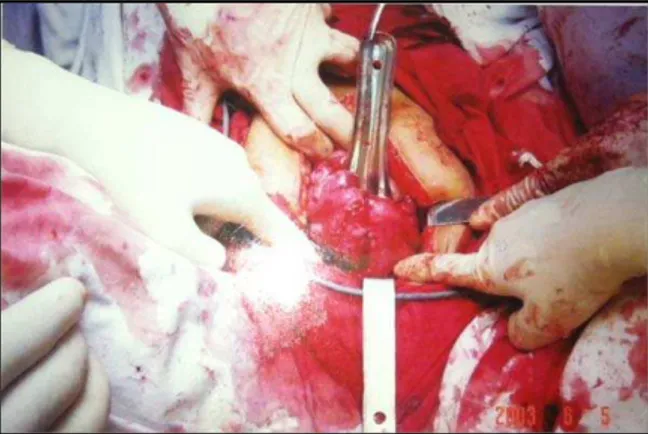
was done after due pre-operative preparation on 5th June 2010. On laprotomy two separate horns
of uterus were seen. Right horn was 5×3×2 cm and the left horn was 4×3×2 in size (Figure 1).
Both were incised at medial border, endometrium was exposed. Per vaginally, attempts to create passage between uterus and
cervix were done with the help of Hegar dilator no. 5.
Figure 1: Bicornuate Uterus with two separate
horns with intervening septum between two horns
Figure 2: Unification of uterus and creating of
communication between endocervical canal and the uterine cavity
pISSN: 0976 3325 eISSN: 2229 6816: 2229 6816
National Journal of Community Medicine Vol 2 Issue 2 July-Sept 2011 Page 315 and dilator passed through it. Red rubber
catheter was kept in situ to maintain the patency and myometrium was sutured around it in three layers with vicryl no.1 (Figure 2).
Chocolate cyst was incised. Hemostasis was achieved. Abdomen was closed. On 5th day red
rubber catheter was expelled by itself. Cu T 200 was inserted to maintain patency. Tablet Premarin (conjugated estrogen) (0.625mg) once daily was started for 21 days. Follow up ultrasound showed uterus of 5cm in length and cervix was 2.5cm. Triple lined endometrium of 11 mm size was seen with Cu T in situ. Patient was reassured and Tablet Meprate (medroxyprogesterone acetate) 10mg once daily for 10 days was started. On 47th day she
menstruated normally for 5 days.On follow up examination Cu T threads could be seen in situ.
DISCUSSION
The incidence of uterine congenital anomalies is approximately 1 in 200 out of which bicornuate uterus is the most common (about 45%).1-3
Congenital anomalies of lateral fusion (uterus) of mullerian duct normally do not co-exist along with anomalies of vertical fusion (cervix or vagina). So atresia of endocervical canal with bicornuate uterus is a rare entity.4, 5
In Indian setup such patients present at a late stage after many complications. They are psychologically immature and most of the times they are non compliant. Thus they require sympathetic handling by the gynaecologist. Patient may present with hematometra or endometrioses due to the functioning
endometrium and cryptomenorrhaea due to cervical lumen atresia.6 Confirmatory diagnosis
of such condition can only be done by combination of clinical examination, sonography as well as diagnostic laprascopy.4
In most of the cases the treating gynecologist is tempted to go for hysterectomy due to already existing complication like haematometra and endometriosis. But it is possible to go for unification operation (metroplasty) along with creation of passage through atretic lumen of cervix, thus restoring the normal menstrual function.6-8 This can be a great benefit for the
patient emotionally and psychologically.
REFERENCES
1. Gery WL, Weed JC. Congenital atresia of uterine cervix. Obstet Gynec 1973:42; 213
2. Farber M, Mitchell GW. Bicornuate uterus and partial atresia of fallopian tube. Am J Obstet Gynec. 1979; 134: 881.
3. Nahum GC: Uterine anomalies: how common are they and what is the distribution among subtypes? J Reprod Med 1998; 43:877-887
4. Valdes C, Mailini S, Malinak LR. Ultrasound evaluation of female genital tract anomalies: a review of 64 cases. Am J Obstet Gynecol. 1984; 149(no 3): 285-92
5. Markham SM, Waterhouse TB. Structural anomalies of the reproductive tract. Curr Opin Obstet Gynecol. 1992; 4(6): 867-73.
6. Bakri YN, Al Sugar A, Huggocon C. Bicronuate non fused rudimentary horns with functioning endometrium and complete cervico-vaginal agenesis. Fertil Steril 1992; 58 (No3): 620-1.
7. Musich JR, Behrman SJ. Obstetrical outcome before and after metroplasty in woman with uterine anomalies. Obstet and Gynec 1978; 52:63.